Chromatin remodeling as a molecular bridge between insulin resistance and hyperandrogenism in polycystic ovary syndrome: Current evidence and future perspectives

Maoxing Ran
1

,
Zirui Li
1
,
Yuanjun Liu
1
,
Xin Guo
1
,
Huimin Dang
2,*
*Correspondence to:
Huimin Dang, Department of Traditional Chinese Medicine, The Second Affiliated Hospital of Xi’an Jiaotong University, Xi’an 710004, Shaanxi, China.
E-mail: wbb.23@163.com
Comput Biomed. 2026;1:202623. 10.70401/cbm.2026.0024
Received: July 10, 2026Accepted: September 11, 2026Published: September 11, 2026
Abstract
Polycystic ovary syndrome (PCOS) is a heterogeneous endocrine and metabolic disorder, in which the interaction between insulin resistance (IR) and hyperandrogenism (HA) is a major driver of reproductive and metabolic complications. Accumulating evidence suggests that chromatin remodeling may represent a regulatory layer linking metabolic stress, inflammation, steroidogenic gene expression, and androgen receptor activity. Research indicates that chromatin remodeling may be a key regulatory factor linking metabolic stress, inflammation, steroidogenic gene expression, and androgen receptor activity. This review systematically summarizes current evidence on chromatin remodeling in PCOS-related IR and HA, focusing on histone modifications, DNA methylation, ATP-dependent remodeling complexes, transcription factors, and non-coding RNA-mediated regulation. We distinguish between direct evidence from PCOS tissues and cells and indirect evidence from metabolic, endocrine, and hormone-responsive disease models, proposing a hierarchy of evidence. Available data suggest that chromatin remodeling may influence insulin signaling genes, adipogenesis, ovarian steroidogenic enzymes, and co-transcriptional networks involving Forkhead box protein O1 (FOXO1), Peroxisome Proliferator-Activated Receptor γ (PPARγ), nuclear factor-kappa B (NF-κB), androgen receptor (AR) signaling, and SWI/SNF-related complexes. However, PCOS-specific functional validation remains limited, particularly in theca cells, granulosa cells, adipose tissue, and cell-type-resolved chromatin. Integrating multi-omics, single-cell epigenomics, and CRISPR-based epigenome editing may help identify causal chromatin regulators and clinically relevant PCOS subtypes. We further discuss how computational biomedical approaches, including bioinformatics, Artificial Intelligence (AI), machine learning, and multi-omics integration, can enhance mechanistic inference, biomarker discovery, and PCOS subtype stratification. A chromatin-centric framework may deepen our understanding of the IR-HA cycle and support future biomarker discovery and precision interventions in PCOS.
Keywords
Polycystic ovary syndrome, chromatin remodeling, insulin resistance, hyperandrogenism, epigenetics, steroidogenesis, multi-omics, bioinformatics, machine learning, artificial intelligence, multi-omics integration
1. Introduction
Polycystic ovary syndrome (PCOS) is one of the most common endocrine disorders in women of reproductive age, characterized by reproductive dysfunction, metabolic alterations, and significant clinical heterogeneity. IR and HA are two core pathophysiological components of PCOS. Hyperinsulinemia can stimulate ovarian androgen production and reduce sex hormone-binding globulin levels, thereby increasing free androgen exposure. Conversely, excess androgens may exacerbate adipose dysfunction, inflammatory responses, and impaired insulin sensitivity. This mutually reinforcing relationship can lead to ovulatory dysfunction, female infertility, cutaneous manifestations, and long-term cardiometabolic risks[1-6].
Previous studies on PCOS have primarily focused on circulating hormones, glucose metabolism, ovarian morphology, and clinical phenotypes. However, the molecular mechanisms linking metabolic and endocrine abnormalities remain incompletely elucidated. Growing evidence suggests that epigenetic regulation, particularly chromatin remodeling, is an important mechanism involved in transcriptional programs such as insulin signaling, adipogenesis, inflammation, steroidogenesis, and androgen receptor (AR) activity. Among these, the dynamic chromatin accessibility in adipose-derived stem cells from women with PCOS, along with whole-transcriptome associations involving chromatin-related genes, provides preliminary theoretical evidence for this concept[7,8].
Inflammation and oxidative stress further support the integrated model of IR-HA interaction. In PCOS, saturated fat intake can activate nuclear factor-kappa B (NF-κB)-related inflammatory responses in monocytes, which are associated with reduced insulin sensitivity and excessive androgen production[9]. Meanwhile, metabolomics analysis suggests the presence of distinct molecular features in the PCOS subgroup dominated by HA and IR[10-12]. These observations indicate that the IR-HA cycle is not only a hormonal interaction but also a transcriptional network that may be influenced by chromatin status.
This review critically elaborates on the existing evidence regarding chromatin remodeling as a molecular bridge between IR and HA in PCOS. We highlight the plausibility of the mechanisms, level of evidence, research techniques, and translational implications. Given the limited chromatin evidence directly targeting PCOS, we clearly distinguish disease-specific evidence from indirect evidence derived from metabolic diseases, endocrine models, and other hormone-responsive disorders.
2. Chromatin Remodeling and Epigenetic Regulation in Endocrine-Metabolic Disease
2.1 Core mechanisms of chromatin remodeling
Strictly speaking, “chromatin remodeling” refers specifically to ATP-dependent nucleosome rearrangement. In this review, we adopt a broader chromatin-centered regulatory network as the overarching concept, including histone covalent modifications, DNA methylation, and non-coding RNA mechanisms targeting chromatin. These distinct yet interconnected epigenetic layers operate through different molecular mechanisms but collectively regulate transcriptional output. Chromatin is organized around nucleosomes, each consisting of approximately 147 base pairs of DNA wrapped around a histone octamer. Transcription is strongly influenced by the accessibility of promoters, enhancers, insulators, and other regulatory elements. Chromatin remodeling encompasses histone post-translational modifications, DNA methylation, nucleosome positioning, histone variant exchange, and ATP-dependent remodeling. Together, these processes determine the access of transcription factors and RNA polymerase complexes to regulatory regions[13-18].
Histone acetylation is generally associated with open chromatin and active transcription, while histone methylation can either activate or repress transcription depending on the specific residue modified (e.g., H3K4me3, H3K27me3, or H3K9me3). ATP-dependent remodeling complexes, including the SWI/SNF, ISWI, CHD, and INO80/SWR families, utilize ATP hydrolysis to reposition, evict, or restructure nucleosomes. Through these actions, chromatin remodeling factors translate metabolic and signaling cues into transcriptional output.
2.2 Chromatin remodeling as a metabolic regulatory layer
Chromatin remodeling serves as an important link between nutrient availability and gene expression. The INO80 and SWI/SNF complexes coordinate metabolic gene programs, while tissue-specific BAF and PBAF subcomplexes regulate glycolysis and metabolic adaptation[19]. In obesity-related metabolic dysfunction, the SWI/SNF-associated factor BAF60a modulates inflammatory activation in adipose tissue macrophages, and its loss may enhance proinflammatory responses and exacerbate IR[20]. Environmental exposures may further reshape chromatin accessibility in insulin-sensitive tissues through remodeling factors such as SMARCA5, highlighting the context-dependent plasticity of chromatin-mediated metabolic regulation[21,22].
Chromatin regulatory mechanisms may contribute to PCOS pathogenesis across several tissues and cell types, including adipose tissue, ovarian granulosa and theca cells, skeletal muscle, liver, and immune cells. However, direct evidence in PCOS remains limited, and the relative contributions of individual tissues are not yet clear. These mechanisms should therefore be presented as a hierarchical model rather than as an established causal pathway (Table 1).
Table 1. Evidence hierarchy for chromatin remodeling in PCOS-related IR-HA crosstalk.
| Evidence level | Typical source | Main value | Main limitation |
| Direct PCOS evidence | Human PCOS-patient-derived tissues and primary cells | Supports disease-specific relevance | Limited sample size, tissue heterogeneity, few functional assays |
| Indirect endocrine-metabolic evidence | Obesity, diabetes, insulin-resistant tissues, steroidogenic cell models | Supports pathway plausibility for IR or steroidogenesis | May not capture PCOS-specific ovarian and metabolic contexts |
| Hormone-responsive disease evidence | AR-related cancer models and chromatin studies | Clarifies AR-chromatin interactions and co-regulator biology | Risk of overextension to PCOS without ovarian validation |
| Technology-driven hypotheses | ATAC-seq, ChIP-seq, single-cell multi-omics, epigenome editing | Identifies candidate regulators and cell states | Requires causal validation and clinical stratification |
IR: insulin resistance; HA: hyperandrogenism; PCOS: polycystic ovary syndrome; ATAC-seq: assay for transposase-accessible chromatin with high-throughput sequencing; ChIP-seq: chromatin immunoprecipitation sequencing.
3. Chromatin Remodeling in PCOS-Related Insulin Resistance
3.1 Chromatin accessibility and insulin signaling genes
Insulin signaling depends on the coordinated expression and phosphorylation of insulin receptor substrates, phosphoinositide 3-kinase (PI3K), protein kinase B (AKT), and glucose transporters. Chromatin accessibility at the promoters and enhancers of metabolic genes can determine whether these pathways are transcriptionally active. Studies of adipose tissue development and metabolic disorders have shown that open chromatin at energy metabolism-related loci is associated with increased transcription of metabolic genes[23]. These findings were mainly obtained from non-PCOS metabolic models and therefore provide only indirect mechanistic evidence, while direct evidence from human PCOS samples remains limited. Nevertheless, they provide a basis for further investigation of chromatin regulation involving IRS1, PI3K/AKT, SLC2A4 (GLUT4), and related metabolic nodes in PCOS.
Non-coding RNAs may also participate in chromatin-mediated regulation of insulin signaling. For example, the long non-coding RNA HULC has been reported to affect insulin-like growth factor 1 receptor (IGF1R) and downstream PI3K/AKT signaling in a non-PCOS disease context[24]. However, such chromatin-targeting functions are specific to certain lncRNAs. In contrast, classical microRNAs (miRNAs) mainly act at the post-transcriptional level by regulating mRNA stability and translation and generally do not directly remodel chromatin states. Vitamin D receptor-related signaling and sirtuin-mediated deacetylation may regulate AKT phosphorylation and insulin sensitivity, further supporting the interplay among nuclear receptor signaling, histone acetylation, and metabolic pathways[25,26].
3.2 Histone-modifying enzymes and ATP-dependent complexes in metabolic dysfunction
Histone deacetylases (HDACs) and histone acetyltransferases (HATs) regulate chromatin accessibility and the transcription of metabolic genes. In prenatal androgen exposure models, tissue-specific alterations in HDAC1, HDAC2, HDAC3, and EP300 were associated with metabolic dysfunction[27]. More broadly, HDAC dysregulation has been implicated in metabolic disorders such as type 2 diabetes[28]. Because the direction and consequences of epigenetic changes are strongly influenced by tissue type and developmental stage, these findings should be interpreted with caution.
SWI/SNF-related complexes cooperate with transcription factors that regulate adipogenesis and inflammatory responses. BAF60a mediates inflammatory activation in adipose tissue macrophages, whereas ARID1A is involved in IGF-1 signaling[20,29]. Histone and chromatin regulatory mechanisms have also been associated with insulin signaling genes and SLC2A4 expression in adipocytes[30,31]. Studies have shown that BAF complexes cooperate with transcription factors and RNA polymerase II during nucleosome remodeling[32]. However, direct evidence in PCOS remains limited, and these mechanisms need to be validated in insulin-sensitive cells under PCOS-related conditions.
4. Chromatin Remodeling in Hyperandrogenism and Steroidogenic Regulation
4.1 Steroidogenic enzyme genes and chromatin states
Hyperandrogenism in PCOS is driven by dysregulated steroidogenesis, particularly in theca cells, and may also involve altered aromatase activity in granulosa cells and adipose tissue. Key genes, including CYP17A1, CYP11A1, members of the HSD17B family, and CYP19A1, are transcriptionally regulated by steroidogenic factors and chromatin states. Open chromatin enriched in active histone marks, such as H3K27ac and H3K4me3, may facilitate transcription factor binding and RNA polymerase recruitment, whereas repressive marks, such as H3K27me3 and H3K9me3, may suppress transcriptional activity.
Evidence from steroidogenic cells and ovarian models indicates that DNA methylation, histone methylation, and microRNA-mediated regulation can influence the expression of steroidogenesis-related factors. For example, methylation of steroidogenic factor 1-related regions, H3K27me3-mediated repression of RUNX1 expression, and microRNAs targeting HSD17B1 have been shown to regulate steroidogenesis and hormonal balance[33-35]. These findings support a chromatin-centered hypothesis of PCOS. However, much of the evidence comes from non-PCOS human samples or non-human models and therefore requires validation in theca and granulosa cells from patients with PCOS. In the ovary, theca cells primarily synthesize androgens through CYP17A1, and their steroidogenic transcriptional program is tightly regulated by local chromatin accessibility. In granulosa cells, chromatin states regulate CYP19A1-mediated aromatization and estrogen production. However, high-quality chromatin profiling data from primary human PCOS theca cells remain scarce, representing a major technical limitation in this field.
In PCOS, different target cell types may involve distinct chromatin regulatory mechanisms. In theca cells, active histone marks at the CYP17A1 locus, such as H3K27ac and H3K4me3, may promote androgen biosynthesis, whereas in granulosa cells, the chromatin state of CYP19A1 may regulate aromatization. In adipose tissue and skeletal muscle, chromatin accessibility at the PPARγ and SLC2A4 (GLUT4) loci may affect insulin sensitivity. In immune cells, NF-κB-responsive loci may be potential targets of SWI/SNF chromatin-remodeling complexes. These proposed mechanisms remain hypothetical and require direct chromatin profiling in samples from patients with PCOS.
4.2 Androgen receptor signaling and chromatin remodeling
The AR is a nuclear receptor transcription factor whose activity depends on ligand availability, coregulatory factors, pioneer factors, and chromatin structure. In hormone-responsive diseases, AR recruits chromatin-remodeling complexes, including NuRD, SWI/SNF, and the BRD9-containing GBAF subcomplex, to regulate enhancer and promoter accessibility. Nuclear mTOR, SUMOylation, FOXA1, HOXB13, GATA2, ACK1, and histone phosphorylation may further regulate AR-dependent transcription[36-41].
The relevance of AR-chromatin interactions to PCOS should be regarded as a basis for hypothesis generation rather than as a definitive conclusion. PCOS is not an AR-driven malignancy, and its ovarian microenvironment and systemic endocrine milieu differ substantially from those of prostate cancer. Nevertheless, the AR literature remains informative because it shows how androgen signaling is regulated through chromatin accessibility and coregulator recruitment. Future studies should determine whether the distribution of AR-binding sites, enhancer activity, and chromatin-remodeler occupancy differs between HA-dominant and non-HA PCOS subtypes.
5. Chromatin-Mediated Crosstalk Between Insulin Resistance and Hyperandrogenism
As shown in Figure 1, this chromatin-mediated crosstalk model may integrate metabolic, inflammatory, and steroidogenic signaling in PCOS. Hyperinsulinemia may reshape metabolic transcription through PI3K/AKT, FOXO1, mechanistic target of rapamycin (mTOR), and inflammatory pathways. These signals may alter histone acetylation, histone methylation, and chromatin accessibility at metabolic and steroidogenic gene loci. Conversely, androgen excess may reprogram transcription in adipose tissue, immune cells, and ovarian cells through AR-dependent transcriptional complexes. The resulting chromatin landscape may maintain a pathological state in which IR and HA reinforce each other.
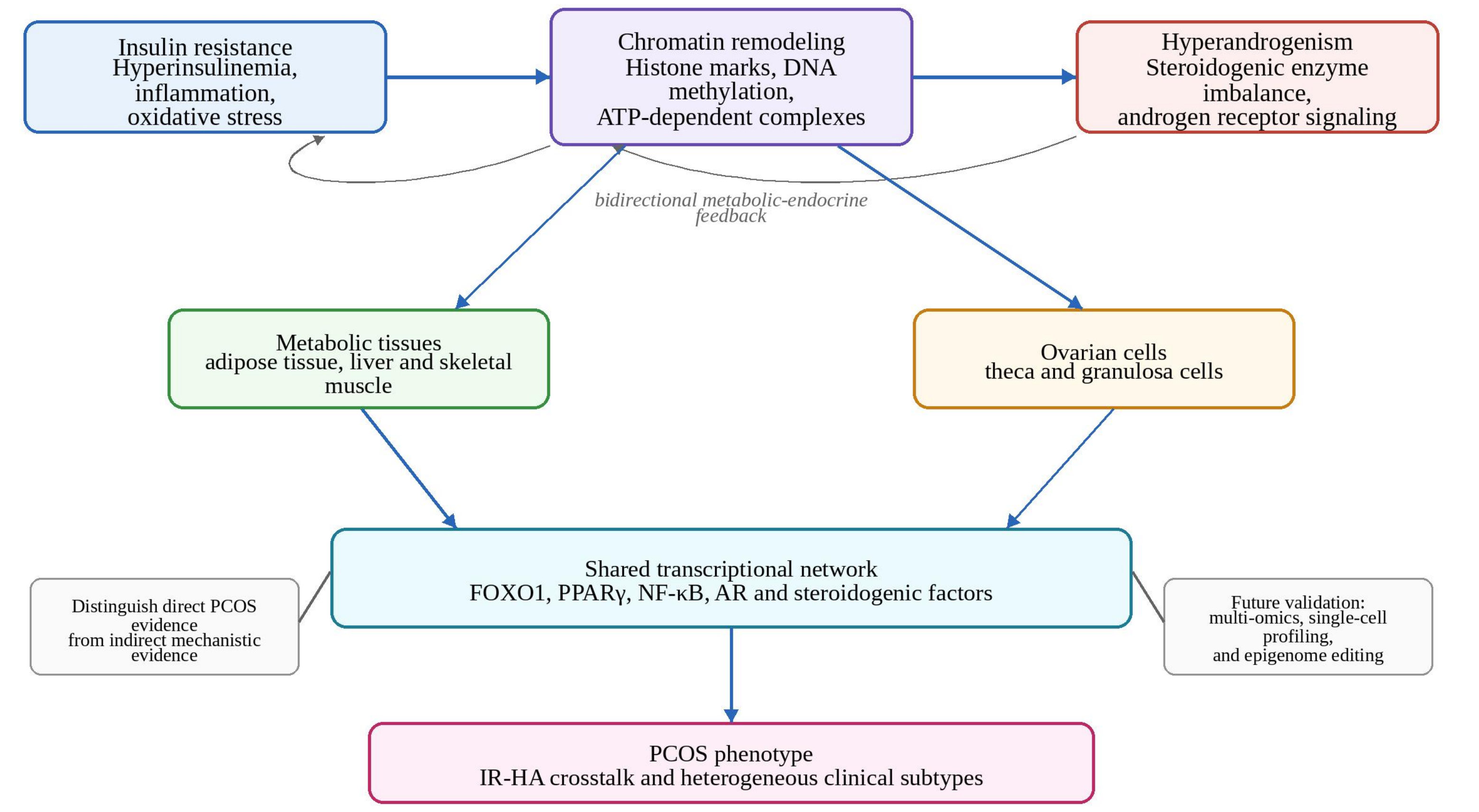
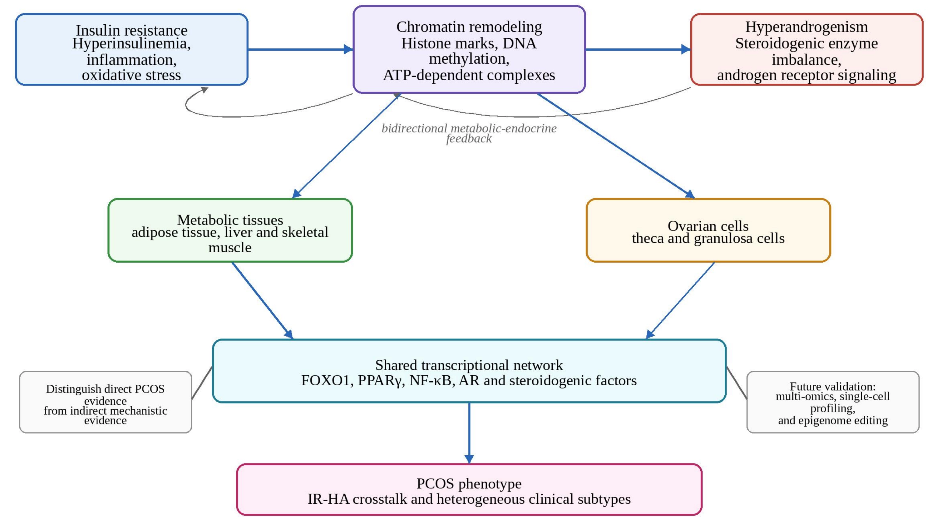
Figure 1. Proposed chromatin-mediated crosstalk between insulin resistance and hyperandrogenism in PCOS. The figure summarizes a working model in which chromatin remodeling integrates metabolic stress, inflammatory signaling, steroidogenic regulation, and androgen receptor-related transcriptional programs. IR: insulin resistance; HA: hyperandrogenism; PCOS: polycystic ovary syndrome; AR: androgen receptor.
Several shared transcriptional regulators may serve as network nodes. FOXO1 is a well-established insulin-responsive transcription factor that may interact with chromatin-remodeling mechanisms in a context-dependent manner. PPARγ regulates adipogenesis and insulin sensitivity and depends on chromatin accessibility at lipid metabolism-related loci. NF-κB links inflammation to metabolic dysfunction and may influence insulin sensitivity as well as the ovarian fibrotic and steroidogenic environment. The regulation of PPARγ/NF-κB/TGF-β1/Smad2/3 signaling by Wisp2 in ovarian granulosa cells further supports the interplay between inflammatory and fibrotic pathways, chromatin regulation, and endocrine function[42]. Given the broader evidence for ovarian histone modifications[43], PGR/RUNX-associated chromatin remodeling may also contribute to transcriptional regulation in the ovary[44]. Candidate chromatin-associated regulators and their potential roles in PCOS-related IR-HA crosstalk are summarized in Table 2.
Table 2. Candidate chromatin-related regulators and their possible roles in PCOS-related IR-HA crosstalk.
| Regulator or pathway | Possible role in IR | Possible role in HA | Evidence status |
| HDACs/HATs | Modify chromatin openness at metabolic genes and inflammatory loci | May affect steroidogenic gene transcription | Indirect evidence; tissue- and timing-dependent[27,30] |
| SWI/SNF/BAF complexes | Regulate adipose and inflammatory transcriptional programs | May cooperate with steroidogenic and AR-related transcription factors | Plausible but under-tested in PCOS[20,32] |
| FOXO1 | Insulin-responsive transcriptional node | May influence ovarian cell function indirectly | Requires PCOS-specific chromatin validation |
| PPARγ | Adipogenesis, lipid metabolism, insulin sensitivity | May interact with ovarian inflammatory/fibrotic signaling | Supported by metabolic and granulosa-cell models[42] |
| NF-κB | Inflammation-linked insulin resistance | May couple inflammatory stress to ovarian dysfunction | Supported by PCOS inflammation studies and indirect models[9,42] |
| AR and co-regulators | May affect adipose and metabolic phenotypes | Directly mediates androgen-responsive transcription | Strong in other hormone-responsive models; limited in PCOS[40] |
IR: insulin resistance; HA: hyperandrogenism; AR: androgen receptor; HDACs: histone deacetylases; HATs: histone acetyltransferases; PCOS: polycystic ovary syndrome; NF-κB: nuclear factor-kappa B.
6. Research Technologies for Defining Chromatin Regulation in PCOS
6.1 Bulk and single-cell epigenomic approaches
High-throughput epigenomic assays can help move research from association to mechanistic investigation. Assay for transposase-accessible chromatin with high-throughput sequencing (ATAC-seq) identifies open chromatin regions and enables promoter and enhancer mapping with relatively small amounts of starting material. Chromatin immunoprecipitation sequencing (ChIP-seq) profiles histone modifications and transcription factor binding but requires high-quality antibodies and generally more starting material. DNase I hypersensitive site sequencing (DNase-seq), Cleavage Under Targets and Tagmentation (CUT&Tag), Cleavage Under Targets and Release Using Nuclease (CUT&RUN), and Hi-C complement these methods by examining chromatin accessibility, protein-DNA interactions, and three-dimensional genome organization[45-51].
Single-cell multi-omics can reveal cellular heterogeneity that may be obscured by analyses of whole ovarian or adipose tissue. The integration of Single-cell RNA sequencing (scRNA-seq) and scATAC-seq, as well as trimodal approaches such as TEA-seq, can identify cell-specific regulatory elements, transcription factors, and lineage states. These approaches are particularly relevant to PCOS because theca cells, granulosa cells, immune cells, adipocytes, and stromal cells may contribute differently to insulin resistance and hyperandrogenism[52,53].
6.2 CRISPR-based epigenome editing and functional validation
CRISPR/Cas9-based systems provide tools for testing causal relationships. Nuclease-active Cas9 can knock out candidate genes, while catalytically inactive dCas9 fused with epigenetic effectors can specifically direct DNA methylation, histone acetylation, or transcriptional activation/repression to specific loci without altering the DNA sequence. Such methods can be used to test whether chromatin state changes at CYP17A1, CYP19A1, IRS1, IGF1R, SLC2A4, or inflammatory regulators are sufficient to affect insulin signaling or steroidogenesis[54,55].
A rigorous validation workflow should integrate patient stratification, multi-omics analyses, functional perturbation in relevant cell models, and confirmation in independent cohorts. Because the chromatin environment can affect editing efficiency and epigenetic drugs may alter chromatin accessibility at target sites, experimental designs should consider chromatin state, cell type, and disease subtype[56,57]. A stepwise framework is proposed to strengthen causal inference: first, candidate loci are prioritized through genetic and epigenetic analyses, including Mendelian randomization and colocalization; next, cell-type-specific epigenome editing, such as dCas9-mediated targeting of CYP17A1, CYP19A1, IRS1, and SLC2A4, is used to test whether locus-specific changes are sufficient to produce the relevant effects; complementary perturbation experiments and independent validation provide converging evidence; and organoid or in vivo models are used to assess systemic effects. Such functional validation is necessary to distinguish causal chromatin remodeling from secondary adaptation in PCOS (Table 3).
Table 3. Technologies for studying chromatin remodeling in PCOS.
| Technology | Primary readout | Main strength | Key limitation |
| ATAC-seq | Open chromatin regions | Low input, maps regulatory accessibility | Does not identify specific histone marks |
| ChIP-seq | Histone marks or transcription factor binding | Directly maps protein-DNA interactions | Requires high-quality antibodies and sufficient material |
| CUT&Tag/CUT&RUN | Histone marks or chromatin-bound proteins | Lower input and lower background than conventional ChIP | Protocol standardization in clinical samples is needed |
| Hi-C/3D genomics | Chromatin interactions and genome organization | Reveals enhancer-promoter and domain-level regulation | Data complexity and resolution constraints |
| Single-cell multi-omics | Cell-specific transcriptome and chromatin state | Resolves tissue heterogeneity and subtype-specific regulatory programs | Higher cost and demanding bioinformatics |
| CRISPR/dCas9 epigenome editing | Causal modulation of chromatin state | Tests regulatory causality at target loci | Delivery, off-target effects, and chromatin-context dependence |
PCOS: polycystic ovary syndrome; ATAC-seq: assay for transposase-accessible chromatin with high-throughput sequencing; ChIP-seq: chromatin immunoprecipitation sequencing; CUT&Tag: cleavage under targets and tagmentation; CUT&RUN: cleavage under targets and release using nuclease.
7. Computational Biomedical Approaches for Chromatin-Centered PCOS Research
As shown in Table 4, computational approaches can be incorporated into the existing chromatin-centered framework without altering its biological logic. High-throughput epigenomic technologies generate candidate loci, transcriptional profiles, and regulatory networks, but computational methods are needed to translate these datasets into interpretable mechanisms. In PCOS, computational biomedicine may help link heterogeneous clinical phenotypes with molecular and clinical features, including transcriptomic profiles, chromatin accessibility, DNA methylation, metabolomics, lipidomics, immune cell composition, and imaging characteristics[58].
Table 4. Computational biomedical approaches for PCOS chromatin remodeling research.
| Computational approach | Main data input | Potential contribution to IR-HA chromatin research | Key limitation |
| Bioinformatics and network analysis | Bulk RNA-seq, methylation arrays, ATAC-seq, ChIP-seq, public databases | Prioritizes pathways, transcription factors, chromatin regulators and immune-cell signatures for mechanistic testing | Generates associations rather than causal evidence[58] |
| Machine learning and AI | Clinical variables, EHR data, ultrasound images, transcriptomics, proteomics, lipidomics | Supports diagnosis, subtype classification, biomarker screening and feature prioritization | Risk of overfitting, limited external validation and inconsistent diagnostic criteria[59,60] |
| Multi-omics integration | Genomics, transcriptomics, epigenomics, metabolomics, lipidomics and hormone profiles | Connects endocrine-metabolic phenotypes with regulatory networks and candidate druggable genes | High dimensionality, missing data and batch effects require rigorous modeling[61,62] |
| Single-cell and spatial computation | scRNA-seq, scATAC-seq, spatial transcriptomics, cell-type annotation | Assigns chromatin-regulated programs to theca, granulosa, adipose, immune and stromal cell compartments | Cost, sample availability and computational complexity remain substantial[52,53] |
| Explainable and causal modeling | SHAP, feature importance, Mendelian randomization, colocalization, perturbation data | Improves biological interpretability and helps distinguish correlation from mechanistic hypotheses | Requires independent cohorts and experimental validation[27] |
PCOS: polycystic ovary syndrome; IR-HA: insulin resistance-hyperandrogenism; ATAC-seq: assay for transposase-accessible chromatin with high-throughput sequencing; ChIP-seq: chromatin immunoprecipitation sequencing; AI: artificial intelligence; scRNA-seq: single-cell RNA sequencing; scATAC-seq: single-cell assay for transposase-accessible chromatin with high-throughput sequencing.
7.1 Bioinformatics and network-based analyses
Bioinformatics provides an initial approach for identifying candidate regulators involved in IR-HA interactions. Differential expression analysis, pathway enrichment, protein–protein interaction networks, weighted gene co-expression network analysis, transcription factor motif analysis, and immune cell deconvolution can be used to prioritize genes and pathways altered in PCOS-related tissues. These methods are particularly useful when direct chromatin data are unavailable because transcriptomic features can be integrated with known chromatin regulators, histone-modifying enzymes, and transcription factor networks to generate testable hypotheses. For example, a computational workflow integrating RNA sequencing, public datasets, LASSO/SVM-RFE feature selection, XGBoost validation, and CIBERSORT-based immune cell inference identified candidate diagnostic genes and changes in immune cell composition in a PCOS granulosa cell dataset[58]. Although such studies cannot establish chromatin-mediated causality, they provide an important link between molecular screening and biological validation.
7.2 Artificial intelligence and machine learning for diagnosis, classification, and biomarker discovery
Artificial intelligence and machine learning are increasingly used to address the clinical heterogeneity of PCOS. A systematic review of AI and machine-learning applications in PCOS reported that published models have used clinical data, electronic health records, imaging data, and genetic or proteomic information. Common algorithms include support vector machines, k-nearest neighbors, and regression-based models[59]. These approaches may improve diagnosis and phenotypic classification, but their translational value depends on standardized diagnostic criteria, adequate sample sizes, external validation, and transparent reporting. In biomarker discovery, ensemble learning applied to untargeted lipidomics has identified lipid candidates with potential diagnostic value, illustrating how omics data and machine learning can be combined to screen molecular features[60]. In chromatin-centered PCOS research, machine learning could also be used to prioritize candidate enhancers, transcription factors, and chromatin-remodeling factors associated with insulin signaling, steroidogenesis, or inflammatory responses. However, these models should be considered and interpreted as hypothesis-generating unless they are validated in independent cohorts and experimentally tested in relevant ovarian or metabolic cell types.
7.3 Multi-omics integration and cell-type-resolved computational modeling
Multi-omics integration is particularly relevant to PCOS because insulin resistance and hyperandrogenism arise from interactions among endocrine, metabolic, inflammatory, and ovarian cellular processes. Common integration strategies include early integration through feature concatenation, intermediate integration through latent factors or shared embeddings, and late integration of model outputs. Advanced methods, including multi-view learning, graph-based integration, and deep generative models, can address high dimensionality, batch effects, missing data, and nonlinear relationships across data layers[61]. In PCOS research, integrative multi-omics analyses have begun to combine genome-wide association studies (GWAS), expression quantitative trait loci (eQTL), methylation quantitative trait loci (mQTL), Mendelian randomization, immune deconvolution, single-cell RNA sequencing, and molecular docking to prioritize druggable genes and candidate therapeutic targets. These approaches are consistent with a computational biomedicine perspective because they connect genetic susceptibility, epigenetic regulation, tissue-specific expression, immune context, and potential pharmacological modulation[62]. Several publicly available genetic and epigenetic resources can support such multi-omics integration, including the GWAS Catalog for summary-level GWAS results, GTEx for eQTL datasets, public mQTL databases, and the RAVAR database for rare variant-trait associations. These resources provide valuable data for future chromatin-focused studies of PCOS.
Modeling with cell-type resolution should be a priority for future research. Bulk ovarian, adipose tissue, and skeletal muscle datasets obscure cell-type-specific signals, whereas PCOS may involve distinct regulatory programs in theca cells, granulosa cells, adipocytes, immune cells, and stromal cells. Single-cell transcriptomics, single-cell ATAC-seq, and spatial transcriptomics can help localize chromatin-regulated transcriptional programs to specific cellular compartments. Computational integration of single-cell transcriptomic and epigenomic data can identify enhancer-gene links, transcription factor activity, and cell-state transitions that are often masked in bulk data. These approaches are particularly important for determining whether chromatin remodeling is a primary driver of dysregulated steroidogenesis, a consequence of metabolic stress, or an adaptive response to chronic inflammation.
7.4 Computational limitations, interpretability, and validation
Despite their potential, computational approaches have important limitations. PCOS datasets are often small and heterogeneous and may be affected by differences in diagnostic criteria, age, body mass index, ethnicity, medication exposure, tissue source, and menstrual status. High-dimensional omics data are particularly susceptible to overfitting, batch effects, and data leakage when feature selection, model training, and validation are not strictly separated. Computational studies should therefore report preprocessing steps, feature selection procedures, hyperparameter tuning, validation strategies, and model calibration. Ultimately, computational predictions should be validated experimentally using primary theca cells, granulosa cells, adipose-derived cells, organoids, or CRISPR-based epigenome editing.
8. Translational Implications, Limitations, and Future Priorities
Chromatin-based models may have several translational implications. First, chromatin features may help classify PCOS into biologically meaningful subtypes, such as insulin resistance-dominant, hyperandrogenism-dominant, inflammatory, or adipose dysfunction subtypes. Second, chromatin regulators may serve as biomarkers or therapeutic targets if their disease specificity, reversibility, and tissue distribution are clearly defined. Third, epigenomic profiles may help identify patients who are more likely to respond to lifestyle interventions, insulin-sensitizing agents, antiandrogen therapy, or emerging epigenetic approaches.
This field still faces important limitations. Many chromatin studies relevant to this topic are based on non-PCOS disease models, immortalized cell lines, or androgen-responsive cancers in males. Direct evidence from PCOS studies is limited to a small range of sample types and often lacks functional perturbation experiments. PCOS itself is also heterogeneous, and epigenomic patterns may be influenced by age, obesity status, ethnicity, medication exposure, menstrual status, and diagnostic criteria. Future studies should explicitly control for or stratify these variables.
Future research should prioritize: (i) constructing PCOS-specific chromatin maps in theca cells, granulosa cells, adipose tissue, skeletal muscle, liver, and immune cells; (ii) conducting paired analyses of transcriptomic, epigenomic, metabolomic, microbiome, imaging, and hormonal data; (iii) integrating multi-omics data using transparent and reproducible computational workflows; (iv) mapping tissue heterogeneity using single-cell and spatial omics; (v) functionally validating candidate regulatory elements using CRISPR-based approaches; and (vi) conducting longitudinal studies to determine whether chromatin alterations are a cause, consequence, or adaptive response within the IR-HA cycle. From a computational biomedicine perspective, the field should move beyond the identification of individual biomarkers toward interpretable models that integrate chromatin states, endocrine phenotypes, metabolic profiles, and treatment responses. This will require external validation, standardized diagnostic criteria, rigorous feature selection, explainable artificial intelligence, model calibration, and clinically meaningful decision thresholds.
A key unresolved question is how to distinguish causal chromatin changes that drive the IR-HA cycle from secondary molecular consequences. Most available observational data cannot determine whether alterations in chromatin states drive the development and progression of PCOS-related insulin resistance and hyperandrogenism or merely represent adaptive responses to persistent hyperinsulinemia or excessive androgen exposure. Addressing this causal evidence gap will require carefully designed longitudinal patient sampling and functional perturbation experiments using epigenome editing.
9. Conclusion
Chromatin remodeling provides a biologically plausible framework for understanding how insulin resistance and hyperandrogenism reinforce each other in PCOS. Mechanisms involving histone modifications, DNA methylation, ATP-dependent remodeling complexes, non-coding RNAs, and transcriptional coregulators may influence insulin signaling, adipogenic programming, inflammatory responses, steroidogenic enzyme expression, and AR-related transcription. However, PCOS-specific evidence remains limited, and many of the proposed mechanisms should currently be regarded as testable hypotheses rather than established causal pathways.
The field needs to move from broad epigenetic associations toward cell-type-specific, computationally integrated, and functionally validated chromatin mechanisms. Integrating multi-omics, single-cell epigenomics, targeted epigenome editing, interpretable machine learning, and well-annotated clinical data may help clarify how chromatin remodeling contributes to IR-HA interactions and ultimately support precision diagnosis and individualized treatment strategies for PCOS.
Acknowledgements
The figures in this manuscript were created using Microsoft PowerPoint (Microsoft Corp., Redmond, WA, USA). All authors read and approved the final manuscript.
Authors contribution
Ran M: Conceptualization, formal analysis, writing-original draft.
Li Z: Investigation, data curation.
Liu Y, Guo X: Writing-review & editing, resources.
Dang H: Conceptualization, supervision, project administration, funding acquisition, writing-review & editing.
Conflicts of interest
The authors declare no conflicts of interest.
Ethical approval
Not applicable.
Consent to participate
Not applicable.
Consent for publication
Not applicable.
Availability of data and materials
Not applicable.
Funding
This work was supported by the National Famous Traditional Chinese Medicine Experts Inheritance Studio Construction Project (National Administration of Traditional Chinese Medicine, Renjiao Letter [2019] No. 41); the Natural Science Basic Research Program of Shaanxi Province in 2025 (Project No. 2025JC-YBMS-882); and the 2022 National Famous Traditional Chinese Medicine Experts Inheritance Studio Construction Project (National Administration of Traditional Chinese Medicine, Renjiao Letter [2022] No. 75).
Copyright
© The Author(s) 2026.
References
-
2. O’Brien B, Dahiya R, Kimble R. Hyperandrogenism, insulin resistance and acanthosis nigricans (HAIR-AN syndrome): An extreme subphenotype of polycystic ovary syndrome. BMJ Case Rep. 2020;13(4):e231749.[DOI]
-
6. Li W, Liu C, Yang Q, Zhou Y, Liu M, Shan H. Oxidative stress and antioxidant imbalance in ovulation disorder in patients with polycystic ovary syndrome. Front Nutr. 2022;9:1018674.[DOI]
-
7. Leung KL, Sanchita S, Pham CT, Davis BA, Okhovat M, Ding X, et al. Dynamic changes in chromatin accessibility, altered adipogenic gene expression, and total versus de novo fatty acid synthesis in subcutaneous adipose stem cells of normal-weight polycystic ovary syndrome (PCOS) women during adipogenesis: Evidence of cellular programming. Clin Epigenet. 2020;12(1):181.[DOI]
-
12. Zhang Z, Hong Y, Chen M, Tan N, Liu S, Nie X, et al. Serum metabolomics reveals metabolic profiling for women with hyperandrogenism and insulin resistance in polycystic ovary syndrome. Metabolomics. 2020;16(2):20.[DOI]
-
15. Blessing C, Knobloch G, Ladurner AG. Restraining and unleashing chromatin remodelers–structural information guides chromatin plasticity. Curr Opin Struct Biol. 2020;65:130-138.[DOI]
-
16. Sundaramoorthy R, Owen-Hughes T. Chromatin remodelling comes into focus. F1000Res. 2020;9:1011.[DOI]
-
17. Clapier CR. Sophisticated conversations between chromatin and chromatin remodelers, and dissonances in cancer. Int J Mol Sci. 2021;22(11):5578.[DOI]
-
22. Palanivel R, Dazard JE, Park B, Costantino S, Moorthy ST, Vergara-Martel A, et al. Air pollution modulates brown adipose tissue function through epigenetic regulation by HDAC9 and KDM2B. JCI Insight. 2025;10(18):e187023.[DOI]
-
23. Du K, Shi Y, Bai X, Chen L, Sun W, Chen S, et al. Integrated analysis of transcriptome, microRNAs, and chromatin accessibility revealed potential early B-cell Factor1-regulated transcriptional networks during the early development of fetal brown adipose tissues in rabbits. Cells. 2022;11(17):2675.[DOI]
-
25. Yuan Q, Zhang R, Sun M, Guo X, Yang J, Bian W, et al. Sirt1 mediates vitamin D deficiency-driven gluconeogenesis in the liver via mTorc2/Akt signaling. J Diabetes Res. 2022;2022:1755563.[DOI]
-
26. Charidemou E, Tsiarli MA, Theophanous A, Yilmaz V, Pitsouli C, Strati K, et al. Histone acetyltransferase NAA40 modulates acetyl-CoA levels and lipid synthesis. BMC Biol. 2022;20(1):22.[DOI]
-
28. Kumar KK, Aburawi EH, Ljubisavljevic M, Leow MKS, Feng X, Ansari SA, et al. Exploring histone deacetylases in type 2 diabetes mellitus: Pathophysiological insights and therapeutic avenues. Clin E pigenetics. 2024;16(1):78.[DOI]
-
29. Jdeed S, Erdős E, Bálint BL, Uray IP. The role of ARID1A in the nonestrogenic modulation of IGF-1 signaling. Mol Cancer Res. 2022;20(7):1071-1082.[DOI]
-
32. Brahma S, Henikoff S. The BAF chromatin remodeler synergizes with RNA polymerase II and transcription factors to evict nucleosomes. Nat Genet. 2024;56(1):100-111.[DOI]
-
33. Hong Z, Xu Y, Xu Y, Ding M, Zhao Z, Wang C, et al. Prenatal BPA exposure disrupts androgen synthesis in testicular Leydig cells via epigenetic modifications of steroidogenic factor 1. Ecotoxicol Environ Saf. 2025;307:119409.[DOI]
-
34. Zhong Y, Li L, He Y, He B, Li Z, Zhang Z, et al. Activation of steroidogenesis, anti-apoptotic activity, and proliferation in porcine granulosa cells by RUNX1 is negatively regulated by H3K27me3 transcriptional repression. Genes. 2020;11(5):495.[DOI]
-
35. Bhandary P, Shetty PK, Shetty P, Manjeera L, Patil P. microRNA mediated downregulation of HSD17B1 impairs estrone-to-estradiol conversion in polycystic ovarian syndrome. Reprod Biol. 2025;25(4):101085.[DOI]
-
36. Dufour CR, Scholtes C, Yan M, Chen Y, Han L, Li T, et al. The mTOR chromatin-bound interactome in prostate cancer. Cell Rep. 2022;38(12):110534.[DOI]
-
39. Nguyen DT, Mahajan U, Angappulige DH, Doshi A, Mahajan NP, Mahajan K. Amino terminal acetylation of HOXB13 regulates the DNA damage response in prostate cancer. Cancers. 2024;16(9):1622.[DOI]
-
43. Le ML, Zeng LJ, Luo T, Zheng LP. Role of histone posttranslational modifications in the regulation of ovarian function. Sheng Li Xue Bao. 2023;75(1):91-98. Chinese.[PubMed]
-
45. Ma S, Zhang Y. Profiling chromatin regulatory landscape: Insights into the development of ChIP-seq and ATAC-seq. Mol Biomed. 2020;1(1):9.[DOI]
-
46. Zhang J, Tong S, Liang S, Li F, Zhang C, Ding N, et al. ATAC-seq library preparation of murine bone marrow-derived neutrophils. J Vis Exp. 2025(215):e67490. Chinese.[DOI]
-
48. Cheng K, McCarrey JR. Profiling the epigenetic landscape of the spermatogonial stem cell-Part 1: Epigenomics assays. In: Oatley JM, Hermann BP, editors. Spermatogonial stem cells: Methods in Molecular Biology. New York: Humana; 2023. p. 71-108.[DOI]
-
50. Imoto Y. Comprehensive noise reduction in single-cell data with the RECODE platform. Cell Rep Methods. 2025;5(10):101178.[DOI]
-
53. Zeng P, Ma Y, Lin Z. scAWMV: An adaptively weighted multi-view learning framework for the integrative analysis of parallel scRNA-seq and scATAC-seq data. Bioinformatics. 2023;39:btac739.[DOI]
-
61. Baião AR, Cai Z, Poulos RC, Robinson PJ, Reddel RR, Zhong Q, et al. A technical review of multi-omics data integration methods: From classical statistical to deep generative approaches. Brief Bioinform. 2025;26(4):bbaf355.[DOI]
Copyright
© The Author(s) 2026. This is an Open Access article licensed under a Creative Commons Attribution 4.0 International License (https://creativecommons.org/licenses/by/4.0/), which permits unrestricted use, sharing, adaptation, distribution and reproduction in any medium or format, for any purpose, even commercially, as long as you give appropriate credit to the original author(s) and the source, provide a link to the Creative Commons license, and indicate if changes were made.
Publisher’s Note
Science Exploration remains a neutral stance on jurisdictional claims in published
maps
and institutional affiliations. The views expressed in this article are solely those
of
the author(s) and do not reflect the opinions of the Editors or the publisher.
Share And Cite


Science Exploration Style
Ran M, Li Z, Liu Y, Guo X, Dang H. Chromatin remodeling as a molecular bridge between insulin resistance and hyperandrogenism in polycystic ovary syndrome: Current evidence and future perspectives. Comput Biomed. 2026;1:202623. https://doi.org/10.70401/cbm.2026.0024
Tips
Copy completed.
Submit a Manuscript
Author Instructions
Cite this Article
Article Metrics
0
View
0
Download
Cited
Article Updates

- Abstract
- Keywords
- 1. Introduction
- 2. Chromatin Remodeling and Epigenetic Regulation in Endocrine-Metabolic Disease
- 3. Chromatin Remodeling in PCOS-Related Insulin Resistance
- 4. Chromatin Remodeling in Hyperandrogenism and Steroidogenic Regulation
- 5. Chromatin-Mediated Crosstalk Between Insulin Resistance and Hyperandrogenism
- 6. Research Technologies for Defining Chromatin Regulation in PCOS
- 7. Computational Biomedical Approaches for Chromatin-Centered PCOS Research
- 8. Translational Implications, Limitations, and Future Priorities
- 9. Conclusion
- Acknowledgements
- Authors contribution
- Conflicts of interest
- Ethical approval
- Consent to participate
- Consent for publication
- Availability of data and materials
- Funding
- References
- Copyright
 Get citation
Science Exploration Style

Ran M, Li Z, Liu Y, Guo X, Dang H. Chromatin remodeling as a molecular bridge between insulin resistance and hyperandrogenism in polycystic ovary syndrome: Current evidence and future perspectives. Comput Biomed. 2026;1:202623. https://doi.org/10.70401/cbm.2026.0024
copy
Share Link
copy