The evolution of extracellular vesicles: From passive transporters to active architects of microenvironmental homeostasis

Jingjun Zhou
1,2

,
Yuxin Xiong
1,2
,
Ruolang Pan
3
,
Jianzhong Shao
1,2
,
Ye Chen
1,2,*
*Correspondence to:
Ye Chen, College of Life Sciences, and Department of Genetic and Metabolic Disease, Children’s Hospital, Zhejiang University School of Medicine, National Clinical Research Center for Children and Adolescents’ Health and Diseases, Hangzhou 310058, Zhejiang, China; Key Laboratory of Cell and Molecular Intelligent Design and Development of Zhejiang Province, Hangzhou 310058, Zhejiang, China.
E-mail: yechency@zju.edu.cn
EXO. 2026;1:202621. 10.70401/EXO.2026.0018
Received: May 29, 2026Accepted: August 12, 2026Published: August 12, 2026
Abstract
Once dismissed as mere cellular waste, extracellular vesicles (EVs) have undergone a conceptual redefinition, emerging as programmable therapeutic scaffolds with broad biomedical applications. Modern EVs design has progressed past the conventional framework of localized cargo delivery to isolated recipient cells; instead, the focus has shifted toward systemic, multi-cellular niche remodeling aimed at restoring tissue-level homeostasis. This review provides a comprehensive analysis of the engineering strategies to overcome the biological bottlenecks of naive EVs, specifically rapid systemic clearance and inefficient cytosolic delivery. We detail current strategies for active loading and for bypassing endolysosomal entrapment to facilitate in-situ translation of therapeutic mRNA. Furthermore, we discuss how the synergy between engineered EVs and responsive biomaterial scaffolds provides the spatiotemporal control necessary for localized reprogramming of diseased microenvironments. Finally, by examining application paradigms across oncology, regenerative medicine, and neurodegeneration alongside existing regulatory classification frameworks, this review provides a roadmap for transitioning intelligent vesicle platforms from benchtop discovery to clinical-grade compliance.
Keywords
Extracellular vesicles, engineered extracellular vesicles, mRNA delivery, biomaterial-assisted delivery, in-situ translation, niche remodeling, microenvironmental homeostasis
1. Introduction: From Natural Messengers to Engineered Therapeutic Platforms
Extracellular vesicles (EVs) were formerly regarded as “cellular dust” or “metabolic waste”. In 2007, Valadi et al. discovered that EVs can transfer functional mRNA and miRNA between cells, helping to establish their role in intercellular communication[1]. For years, the field largely viewed EVs through a “Postman Model”, in which their therapeutic value was judged primarily by their ability to deliver a defined cargo to a recipient cell. More recent work, however, has expanded this view and positioned EVs as versatile platforms for therapeutic engineering.
Naturally secreted by nearly all cell types, these lipid bilayer nanoparticles carry a diverse array of bioactive proteins, lipids, and nucleic acids that are essential for maintaining physiological homeostasis, but also contribute to pathological processes[1-3]. Their inherent biocompatibility, stability, and ability to traverse biological barriers, such as the blood-brain barrier (BBB), make them attractive candidates for drug delivery[4]. Nevertheless, the clinical translation of naive EVs remains hindered by significant technical bottlenecks, including low drug loading efficiency, poor tissue specificity, rapid systemic clearance, and substantial heterogeneity. These limitations become even clearer when EVs are compared with more established delivery platforms. Lipid nanoparticles (LNPs) have achieved greater clinical maturity for nucleic acid delivery[5,6], whereas viral vectors generally provide high gene-transfer efficiency and more established manufacturing frameworks[7,8]. By contrast, engineered EVs may offer complementary advantages in biological interfacing, multimodal cargo presentation, and local microenvironmental modulation, but they still lag behind these established platforms in scalable manufacturing, potency assessment, batch-to-batch reproducibility, and regulatory precedent[9] (Table S1). Mechanistically, engineered EVs work differently from synthetic LNPs or viral capsids. Synthetic LNPs often rely on apolipoprotein E (ApoE) opsonization for hepatic uptake, and viral capsids are frequently constrained by their fixed packaging volumes. In contrast, engineered EVs take advantage of their endogenous surface properties to engage in intercellular signaling within complex tissue niches. These challenges have prompted the field to move beyond the exploitation of natural EV biology towards the deliberate engineering of EVs as programmable platforms for in-situ translation and functional niche remodeling. This strategic evolution aims to expand the role of EVs from passive messengers into active, precision therapeutic platforms. In this form, engineered EVs are capable of dynamically reshaping diseased microenvironments, such as the tumor immune niche or regenerative tissue milieu[10] (Figure 1).
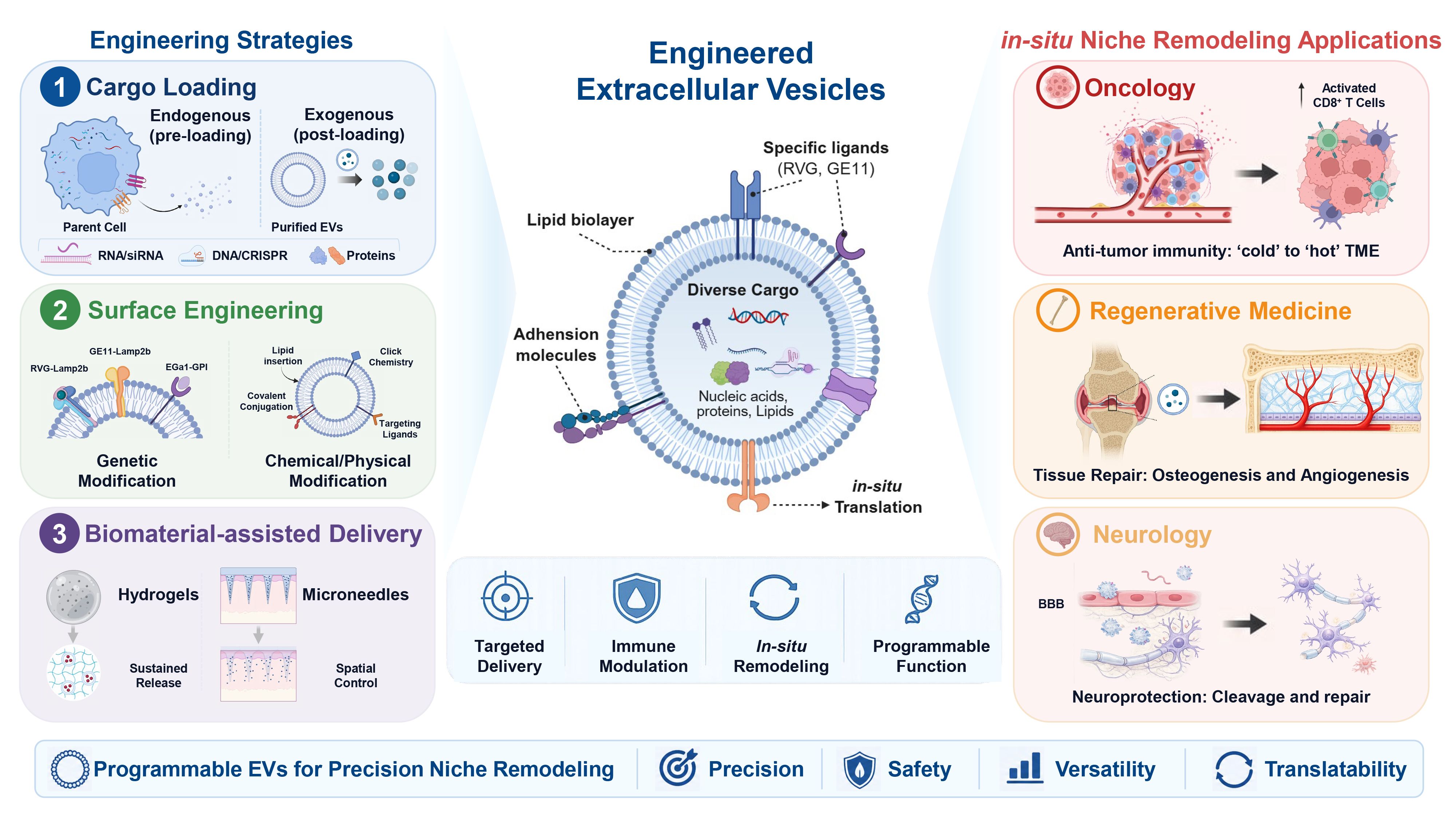
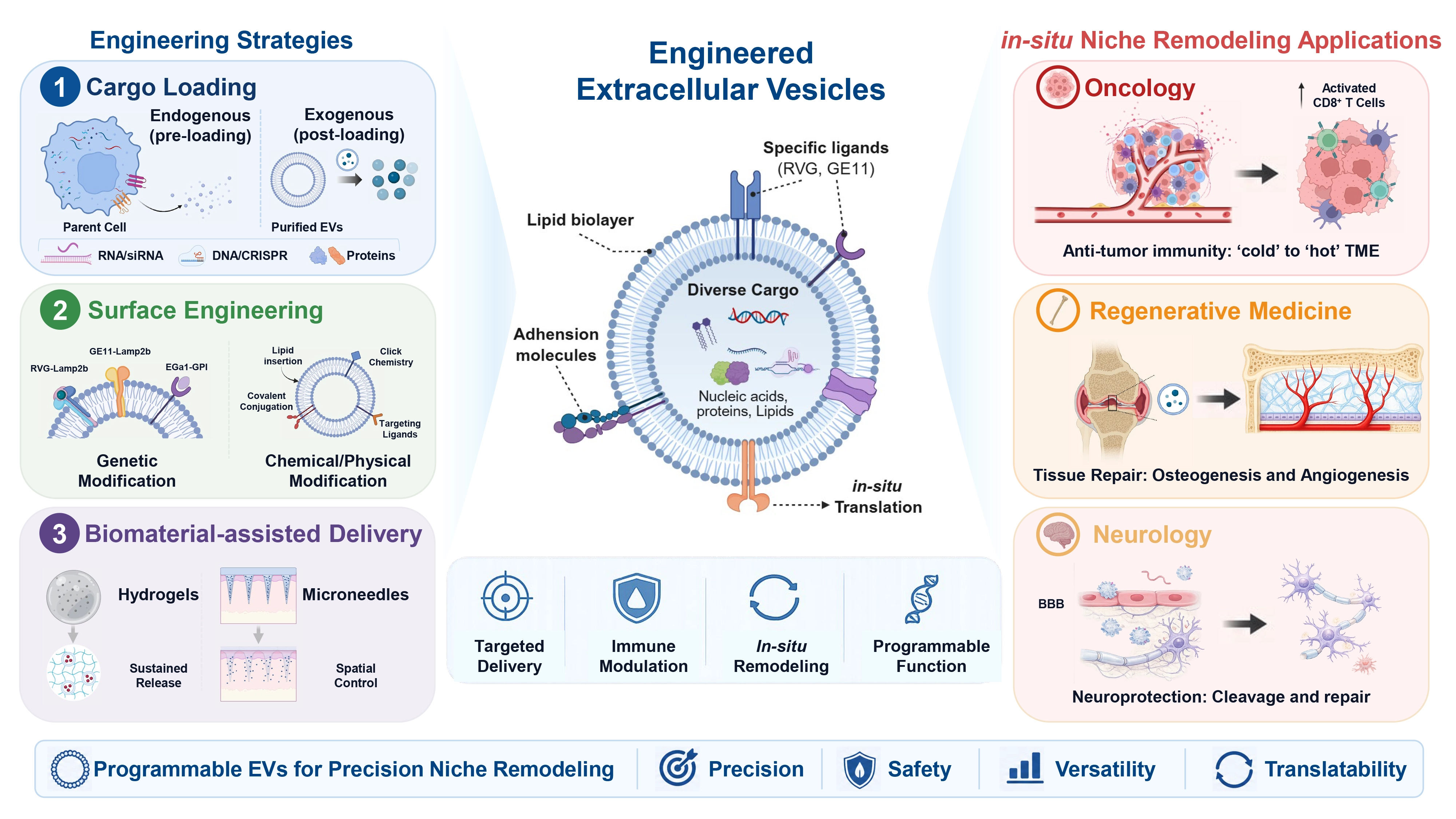
Figure 1. Advanced bioengineering toolkit for EVs and their application paradigms in in-situ niche remodeling. A conceptual overview of transforming naive EVs into programmable therapeutic platforms via a modular engineering framework. Modular assembly and processing: The framework categorizes EV modification into three distinct technological dimensions: cargo loading, surface functionalization, and biomaterial-assisted delivery. Cargo loading involves the internal packaging of mRNAs, functional proteins, or gene-editing components. Surface functionalization includes the display of brain-homing or cell-specific ligands, as well as chemical conjugation methods such as click chemistry. Biomaterial-assisted delivery uses hydrogels or responsive formulations to improve local retention and enable controlled, time-dependent release. Downstream applications: Successfully engineered EV platforms can be designed to reshape diseased microenvironments across three major therapeutic niches: the oncological niche (reversal of immunosuppressive signals and checkpoint disruption); the regenerative niche (orchestration of neovascularization, cell proliferation, and osteochondral defect repair); and the neural niche (traversing the BBB via targeting peptides to mitigate neuroinflammation and accelerate protein aggregate clearance). EVs: extracellular vesicles; BBB: blood brain barrier.
1.1 The biology of EVs in intercellular communication
EVs are heterogeneous and may be classified according to their presumed biogenesis, physical properties, or molecular composition. Exosomes arise through the endosomal multivesicular-body pathway, whereas ectosomes or microvesicles bud directly from the plasma membrane[11]. Because these biogenetic origins are difficult to determine experimentally, minimal information for studies of extracellular vesicles (MISEV2023) recommends using the generic term “EV” or operational descriptions such as “small EV” and “large EV” unless the vesicle origin has been demonstrated[11]. Accordingly, throughout this Review, we use “EV” as the default term and retain more specific terminology only when supported by the original study or when it forms part of an established platform or regulatory product description. EV cargo, including proteins, lipids, and nucleic acids, can be selectively sorted during vesicle biogenesis[12]. Early studies demonstrated that EV-associated mRNAs can be transferred to recipient cells and translated into proteins, providing early evidence for functional RNA transfer between cells[1,13]. These biological properties have also inspired the development of engineered EV platforms for therapeutic cargo delivery.
1.2 From cargo delivery to microenvironmental remodeling
Traditionally, EV-based therapeutic strategies have primarily been conceptualized as a form of targeted delivery. In this model, EVs function as biological nanocarriers, transporting functional cargo such as proteins, nucleic acids, or gene-editing systems to specific recipient cells. This effect is often framed as a linear, single-cell event, where an EV alters only the autonomous behavior of the immediate recipient cell. However, an emerging perspective proposes that the ultimate therapeutic relevance of engineered EVs may extend beyond single-cell targeting to the coordinated reprogramming of functional niches and pathological microenvironments. In this Review, building on the general concept of a functional tissue niche[14], we use the term “niche remodeling” to describe coordinated changes in more than one cellular or extracellular component of a local tissue microenvironment, rather than an isolated response in a single recipient cell. Evidence for niche remodeling should therefore include changes in the composition, state, or interactions of multiple niche components, together with a measurable tissue-level functional outcome. In many pathologies, these niches often become dysregulated, supporting rather than inhibiting disease progression. A prime example is the tumor microenvironment (TME), where cancer cell-derived EVs transfer oncogenic proteins and nucleic acids to immune cells, fibroblasts, and endothelial cells to promote immunosuppression, angiogenesis, and metastasis[15-18]. Similarly, in degenerative conditions like osteoarthritis, the joint microenvironment becomes increasingly pro-inflammatory and catabolic, which can actively impair tissue repair[19]. The emerging therapeutic strategy of “in-situ niche remodeling” aims to reverse this dysregulation by delivering specific molecular signals directly to the diseased sites. Engineered EVs can carry therapeutic mRNAs, including those encoding immunomodulatory cytokines, and thereby alter the cellular and molecular composition of diseased microenvironments[20,21]. Rather than acting only on individual recipient cells, EVs can influence the local tissue microenvironment by coordinating responses across multiple cell types. In disease, this process often supports pathological niche formation, including malignant and pre-metastatic microenvironments[17,18]. Engineering strategies aim to exploit the same biological properties to drive tissue repair and functional recovery[20,22].
1.3 The imperative for engineering: Overcoming the limitations of natural EVs
Despite their inherent biological advantages, naive EVs face significant challenges that limit their efficacy as standardized therapeutic agents. These limitations highlight the need for advanced engineering strategies to convert EVs into more reliable and powerful therapeutic platforms.
The core limitations of natural EVs and the engineering strategies designed to address them are summarized in Table S2. Collectively, these technological components facilitate a transition from viewing EVs as simple “messengers” to using them as “programmable platforms”. This conceptual evolution is exemplified by the emphasis on in-situ translation. Engineered EVs deliver mRNA to recipient cells within a targeted niche to produce their own therapeutic proteins. This strategy offers significant advantages over the delivery of recombinant proteins, including more sustained expression and the ability to generate complex, multi-domain proteins that are typically difficult to manufacture or stabilize externally. Ultimately, by systematically addressing the inherent deficits of natural vesicles, bioengineering also helps turn EVs into more precise and controllable tools for remodeling the pathological microenvironment.
2. Engineering Strategies for Cargo Loading and Enabling Efficient In-Situ Translation
The therapeutic utility of engineered EVs is currently limited by three closely related challenges: limited loading of large nucleic acid cargoes, the potential loss of biological activity during forced loading procedures, and endosomal entrapment, which prevents delivered mRNA from accessing the ribosomal machinery for in-situ translation[23,24].
2.1 Endogenous loading via parental cell engineering
Endogenous loading, often referred to as “pre-loading”, utilizes a cell’s innate biosynthetic machinery to encapsulate therapeutic cargo during vesicle biogenesis. By genetically modifying the producer cell, this approach enables cargo incorporation during EV biogenesis without requiring post-isolation membrane-disruptive loading procedures (Table S3). This strategy preserves the structural integrity and bioactivity of the EV membrane while facilitating the efficient loading of complex macromolecules. A primary method involves constructing chimeric fusion proteins that link a therapeutic payload or an RNA-binding domain (RBD) to EV-enriched transmembrane anchors such as CD63, CD9, and lysosome-associated membrane glycoprotein 2b (Lamp2b) or to endogenous RNA-binding proteins like heterogeneous nuclear ribonucleoprotein A2B1 (hnRNPA2B1). This molecular linkage promotes the sorting of target molecules into nascent multivesicular bodies (MVBs) for eventual secretion within EVs[12,25,26]. A notable example includes fusing viral capsid components, such as the human immunodeficiency virus (HIV) Gag protein, with EV-associated domains to create engineered chimeric vesicles that successfully package and protect full-length mRNA for targeted systemic delivery to neurons[27].
Research over the past decade has significantly expanded the capacity of endogenous loading. For instance, the Targeted and Modular EV Loading (TAMEL) system utilizes MS2 coat protein (MCP) fusions to recruit MS2 stem-loop-tagged RNA, achieving up to a 40-fold increase in enrichment[28]. More recently, the vesicular stomatitis virus G glycoprotein (VSV-G)-Foldon-Intein-Cre (VFIC) system achieved nearly 80% genome editing efficiency in recipient cells by integrating the membrane-fusion capabilities of VSV-G with the cargo-loading precision of inteins[29]. Similarly, platforms employing CD63-RBD fusions have enabled the efficient loading of tumor-targeted mRNA and produced a significant antitumor immune response in aggressive melanoma models[30]. Beyond functional proof-of-concept studies, Erana-Perez et al. reported greater protein-loading capacity in the large EV fraction than in the corresponding small EV fraction, together with differences in membrane stability and production characteristics[31]. These findings highlight EV size and isolation strategy as practical considerations when selecting an EV preparation for a particular therapeutic application.
2.2 Post-isolation loading techniques
In contrast to endogenous loading, exogenous loading adds therapeutic cargo to EVs after their isolation and purification from parental cells. This approach offers high experimental flexibility, because the EV population can be characterized and standardized before loading (Table S4). However, these techniques typically rely on physical or chemical perturbation and may affect EV membrane properties or promote cargo aggregation, depending on the loading method[32,33].
Exogenous loading techniques are broadly categorized into passive and active loading. Passive loading strategies, such as simple incubation, rely on concentration gradients and are generally suitable only for small hydrophobic drugs, exhibiting extremely low efficiency for macromolecules such as nucleic acids or proteins. Consequently, for macromolecules like mRNA, active loading strategies are required, which involve the transient disruption of EV membrane integrity to permit cargo entry. Although active methods significantly improve loading efficiency, they must be optimized to load cargo effectively without compromising vesicle structure or biological activity[34,35].
2.3 Enhancing cytosolic delivery and translation efficiency
The functional efficacy of an engineered extracellular vesicle (EV) is strongly influenced by its capacity to bypass the endolysosomal pathway, a process known as endosomal escape (EE)[36]. Because a substantial proportion of internalized EVs become sequestered and subsequently degraded within endolysosomes, only a limited fraction of the delivered mRNA payload typically reaches the cytoplasm intact[24]. Therefore, enhancing EE capacity represents an important consideration for improving the efficiency of in-situ translation (Table S5). VSV-G is widely used for this purpose, leveraging the acidic endosomal environment to mediate vesicle-endosome membrane fusion and release genetic cargo into the cytosol. Recent studies demonstrated that VSV-G integration can facilitate efficient cytosolic delivery of functional macromolecules, such as Cre recombinase[29]. Beyond viral proteins, synthetic or microbe-derived fusogenic peptides have also been employed for this purpose. Peptides such as aurein 1.2 and peptides composed of repeating glutamic acid-alanine-leucine-alanine sequences (GALA) have been shown to enhance cytosolic protein delivery by promoting EE[37,38].
Alongside vehicle engineering, the mRNA payload also needs to be optimized to support efficient translation once it reaches the cytosol. Key modifications include the substitution of uridine with pseudouridine to diminish immunogenicity while enhancing mRNA stability and translation kinetics[39]. Furthermore, untranslated region (UTR) engineering, including the optimization of 5’ UTR sequences, can enhance protein expression[40,41]. Finally, maintaining terminal structure integrity, specifically a complete 5’ cap and an adequate 3’ poly(A) tail, is critical to protect the mRNA from premature degradation and facilitate efficient ribosomal recruitment[42].
2.4 Biophysical trade-offs and scalability constraints in cargo loading and EE
Although advanced endogenous platforms, such as the TAMEL, CD63-RBD, and VSV-G plus EV-sorting domain-intein-cargo/VSV-G-foldon-intein-cargo (VEDIC/VFIC) systems[28-30], have improved nucleic acid packaging or enabled genome editing applications, scaling these processes to meet clinical demand introduces considerable biophysical and manufacturing challenges. Exogenous methods such as sonication, extrusion, and freeze-thaw cycling may compromise EV integrity and function, and their implementation at scale requires careful control of processing conditions, including those used during tangential flow filtration (TFF)[43,44]. Endogenous loading also presents trade-offs. Genetic modification of producer cells may influence the properties of the resulting EVs and therefore requires careful optimization. How these engineering strategies influence intracellular delivery and the in vivo fate of engineered EVs remains unclear. Consequently, achieving optimal cargo loading and efficient EE requires balancing maximum technical performance against the scalable preservation of vesicle structure and function.
3. Precision Targeting: Directing Engineered EVs to Specific Niches
The therapeutic efficacy of engineered EVs depends largely on the precision of their targeting capacity, which can be enhanced through modification of their membrane surface. Native EVs typically exhibit non-specific accumulation in the liver, spleen, and lungs, which substantially constrains their therapeutic window[45,46]. To address this limitation, surface engineering strategies have been developed to display targeting moieties and redirect EV tropism toward specific cell types within pathological niches. These strategies can be broadly grouped into pre-isolation genetic engineering of producer cells and post-isolation modification of purified EVs[4,47]. Selecting the optimal approach requires balancing the robustness and scalability of genetic methods against the modularity and precise control provided by chemical conjugation. As the field advances, the quantitative assessment of ligand density and conjugation efficiency has become increasingly important to ensure clinical reproducibility and potency.
3.1 Genetic engineering for ligand display
Genetic engineering of parental cells can confer targeting properties on EVs by displaying specific ligands on their surface. Targeting peptides, nanobodies, or protein domains are genetically fused to EV-enriched membrane proteins so that they are incorporated into the vesicle membrane during biogenesis (Table S6). Among the available scaffolds, Lamp2b has been widely utilized due to its natural abundance on EV surfaces. Tetraspanins, including CD9, CD63, and CD81, together with membrane-associated proteins such as prostaglandin F2 receptor negative regulator (PTGFRN), have been investigated as scaffolds for EV surface display[48,49].
This approach has been used to redirect EVs toward specific tissues and pathological microenvironments and to improve their delivery across biological barriers. In an early example of brain-targeted delivery, the rabies virus glycoprotein (RVG) peptide was fused to the N-terminus of Lamp2b. This modification targets neuronal nicotinic acetylcholine receptors, facilitating BBB penetration and achieving a greater than 1.5-fold increase in cerebral cargo accumulation in vivo[4]. Beyond the central nervous system (CNS), this strategy significantly enhances tumor-specific delivery. For instance, GE11 peptide-modified EVs drastically improve the delivery of chemotherapeutics like doxorubicin to epidermal growth factor receptor (EGFR)-positive cancer cells, maximizing cytotoxicity while mitigating off-target effects[50]. Similarly, surface display of the integrin-binding internalizing RGD (iRGD) peptide suppresses pulmonary metastasis and extends survival in breast cancer models. This strategy has also been applied to cardiovascular disease, where fusing cardiac-targeting peptides (CTPs) to EV membranes selectively enhances myocardial uptake and promotes functional tissue repair in models of myocardial infarction[51,52].
3.2 Chemical and physical surface modifications
Post-isolation modification provides a flexible alternative to genetic engineering, allowing EVs to be conjugated with various targeting molecules, including synthetic peptides, antibodies, carbohydrates, and polymers, without genetically modifying the producer cells. These methods are especially beneficial for rapid screening of different targeting ligands and for functionalizing EVs from primary cells or hard-to-transfect sources[47,53,54]. However, translating these physical and chemical methods requires careful optimization. The central challenge is to maximize conjugation efficiency without compromising the structural integrity, morphology, and intrinsic biological activity of the vesicles.
Precise surface functionalization can be achieved through a flexible set of chemical methods, mainly covalent and non-covalent modification. Among covalent approaches, bioorthogonal click chemistry, such as strain-promoted azide-alkyne cycloaddition (SPAAC), has become widely used due to its high specificity, rapid reaction kinetics, and minimal disruption of the EV membrane. A typical example is the use of hyaluronic acid-functionalized EVs to target CD44-expressing tissues[53]. Alternatively, traditional covalent conjugation via 1-ethyl-3-(3-dimethylaminopropyl)carbodiimide/N-hydroxysuccinimide (EDC/NHS) chemistry or thiol-maleimide reactions facilitates stable bonds with surface-exposed amines or thiols to achieve high ligand densities[55,56]. However, this density must be carefully tuned, because excessive crowding may paradoxically reduce targeting specificity. Lipid insertion provides a non-covalent alternative to these covalent methods. In this approach, amphiphilic molecules such as 1,2-distearoyl-sn-glycero-3-phosphoethanolamine-poly (ethylene glycol) (DSPE-PEG) conjugates spontaneously integrate into the EV lipid bilayer[48,57]. Although lipid insertion is valued for its simplicity and ability to preserve vesicle structure, its efficiency can vary depending on the lipid composition of the host membrane. Collectively, these chemical and physical modification strategies have broadened the options for directing EVs toward selected tissues and cell types across preclinical disease models (Table S7). However, enhanced target-tissue uptake does not necessarily imply exclusive targeting, and whole-body biodistribution remains highly dependent on the EV platform and experimental model.
4. Biomaterial-Assisted Delivery for Spatiotemporal Control within Niches
The therapeutic performance of engineered EVs is strongly influenced by their in vivo pharmacokinetics, particularly rapid clearance and limited retention at target sites. After intravenous administration, EVs are rapidly cleared by the mononuclear phagocyte system and preferentially accumulate in organs such as the liver and spleen, whereas locally administered EVs can diffuse away from the injection site[58]. These limitations are particularly relevant to niche remodeling, which depends on sustained signaling within a defined tissue environment rather than transient exposure. Biomaterial-assisted delivery can improve local EV retention and support sustained release from the delivery site[59,60]. By extending local exposure, biomaterial-assisted delivery may better support tissue regeneration and sustained remodeling of the local tissue niche.
4.1 Hydrogel-based EV delivery systems
Hydrogels, characterized by highly hydrated polymer networks, are widely applied to EV delivery due to their tunable physical properties, injectability, and ability to encapsulate biologics without denaturation[59,61] (Table S8). Their therapeutic value is largely based on the adjustable diffusion and degradation kinetics of the polymer matrix, which allow EV release rates to be precisely controlled[59]. Hydrogel formulation can extend EV release over days to weeks compared with free EV delivery[59,61]. Mechanistically, this prolonged retention is achieved by balancing passive diffusion and active matrix degradation. In supramolecular hydrogels, for instance, polymer chains can interact non-covalently with the EV lipid bilayer, forming dynamic crosslinks that retard passive diffusion. This strategy has been demonstrated by hydroxypropyl methylcellulose (HPMC) modified with hydrophobic carbon chains, which insert directly into the EV membrane. In this system, release kinetics are controlled by the crosslinking density[59]. Covalently crosslinked networks, such as gelatin methacrylate (GelMA), provide a complementary strategy. In GelMA systems, crosslinking conditions can modulate EV release and reduce the magnitude of initial burst release[61].
The primary benefit of this sustained release is a marked enhancement of therapeutic outcomes, particularly in regenerative medicine. For example, GelMA hydrogels delivering epidermal stem cell-derived EVs accelerate diabetic wound closure by promoting angiogenesis and re-epithelialization through the hypoxia-inducible factor 1/vascular endothelial growth factor A (HIF-1α/VEGFA) pathway[62]. Similarly, PEG-based hydrogels have been used for the systemic delivery of mesenchymal stem/stromal cell (MSC)-derived EVs in chronic liver failure models. These hydrogels produced enhanced anti-fibrotic and anti-apoptotic effects compared to bolus injections by prolonging EV retention[60]. Beyond general tissue repair, hydrogels have been engineered to recapitulate the native cartilage extracellular matrix (ECM) to create tissue-specific niches. A recent study demonstrated that a hydrogel scaffold combining decellularized cartilage ECM (dECM) with chondrocyte-derived EVs enhanced osteochondral repair by activating eukaryotic translation initiation factor 4E-binding protein (4E-BP) and mothers against decapentaplegic homolog 2 (Smad2) signaling to promote matrix synthesis[63]. From the perspective of practical translation and application, however, the clinical translation of these hydrogel reservoirs faces significant bottlenecks. This is mainly due to the difficulty of ensuring consistent EV release across batches and preserving vesicular structural integrity. For example, in covalently cross-linked networks like GelMA, free radicals produced during photopolymerization carry substantial risks. They can lead to the oxidation of signaling lipids on the surface of EVs and result in the cross-linking of exposed structural proteins.
4.2 Advanced formulations: Microneedles (MNs) and responsive systems
In addition to sustained release, advanced biomaterial formulations can also improve delivery precision by supporting minimally invasive administration and microenvironment-responsive “smart” release. MN technology offers a powerful solution for intradermal and transdermal EV delivery, bypassing the skin’s formidable stratum corneum barrier in a painless and localized manner. These platforms are typically based on dissolving or swellable polymers and can encapsulate EVs within the needle matrix or present them as a surface coating. Upon skin insertion, the matrix hydrates and releases the EV payload directly into dermal tissues, promoting local EV retention while reducing systemic exposure[64-66]. This localized delivery strategy offers potential for dermatological interventions, vaccination, and localized regenerative therapies.
Parallel to physical delivery, stimuli-responsive systems can coordinate EV release with local pathological cues, including oxidative or acidic microenvironments[67-69]. In oxidative microenvironments, such as chronic wounds or intervertebral disc degeneration, hydrogels can be engineered to degrade selectively in the presence of reactive oxygen species (ROS). For example, a ROS-cleavable hydrogel loaded with quercetin-enriched EVs enables targeted release, neutralizes ROS, and promotes M1-to-M2 macrophage polarization, significantly enhancing tissue repair[67]. Similarly, pH‑responsive systems take advantage of the acidic nature of the TME to improve EV delivery. For example, EVs functionalized with pH-sensitive adjuvants demonstrate greater cellular uptake and a better therapeutic index under acidic stressors[68,69]. Composite hydrogels offer a more advanced form of spatiotemporal control by coordinating sequential therapeutic cascades, such as the early release of anti-inflammatory EVs in response to primary inflammatory signals[70]. However, the reproducibility of these responsive release profiles across heterogeneous disease microenvironments remains to be established.
5. Application Paradigms: In-Situ Translation for Niche Remodeling in Disease Contexts
Advanced EV engineering strategies are shifting the field from systemic drug delivery toward the localized reprogramming of pathological microenvironments across oncology, regenerative medicine, and neurology.
5.1 Remodeling the tumor immune niche
This approach extends cancer therapy beyond conventional systemic administration toward localized modulation of the tumor immune niche, with the potential to improve therapeutic specificity and reduce systemic toxicity[20,71] (Table 1). Engineered EVs combine their biocompatibility with engineered targeting features to deliver therapeutic payloads within the TME, thereby helping to address some of the biological barriers that limit conventional therapies[56]. A primary axis of this immunomodulation framework involves engineering EVs to deliver immunostimulatory mRNAs that reprogram immunosuppressive microenvironments into immunogenic niches. Recent advances in surface modification and cargo loading techniques have facilitated the localized translation of therapeutic proteins, thereby enhancing immune activation and driving antitumor responses. For instance, the localized delivery of cytokine-encoding mRNAs, such as interleukin-12 (IL-12), via engineered EVs has demonstrated robust immune stimulation in preclinical models. Specifically, EV-encapsulated IL-12 mRNA administered via inhalation induces local expression, leading to elevated interferon-γ production, expansion of cytotoxic T cells, and enhanced antitumor activity, while mitigating systemic toxicity[20].
Table 1. Representative applications of engineered EVs in cancer immunotherapy.
| Therapeutic Strategy | Engineered EV Cargo | Target Cells/Components | Expected Immunotherapeutic Effects and Mechanism | Representative Evidence |
| Editing immune-evasive or pro-tumorigenic pathways | CRISPR/Cas9 plasmids, Cas9/sgRNA RNPs, or modular CRISPR components | Tumor cells or TME-associated stromal/immune cells | Genetic disruption of tumor survival, DNA repair, oncogenic, or immune-evasive pathways to increase tumor vulnerability and improve antitumor immunity | EV-mediated CRISPR/Cas9 delivery has been explored for editing cancer-relevant genes such as KRASG12D and PARP-1[72,73]. |
| Cytokine mRNA immunotherapy | IL-12 mRNA | Lung TME; immune cells including CD8+ T cells, NK cells, and antigen-presenting cells | Local IL-12 expression promotes IFN-γ production, cytotoxic immune activation, systemic antitumor immunity, and reduced systemic cytokine toxicity | Inhalable EVs carrying IL-12 mRNA induced local IL-12 expression and suppressed lung tumor growth[20]. |
| Tumor immunogenicity enhancement | mRNA-loaded EVs or mRNAs encoding immune-modulatory factors | Tumor cells or antigen-presenting cells | IFN-γ mRNA delivery increases MHC-I expression and promotes an immune-stimulatory TME. | mRNA-loaded EV platforms provide a basis for delivering immunomodulatory mRNAs to enhance antigen presentation[71]. |
| TAM repolarization by EVs | M1 macrophage-derived EV cargo; engineered M1 macrophage-derived EVs carrying NF-κB p50 siRNA and miR-511-3p | M2-like TAMs | Reprograms immunosuppressive M2-like TAMs toward pro-inflammatory, antitumor M1-like macrophages | M1 macrophage-derived or engineered EVs repolarized M2-like TAMs toward an antitumor M1-like phenotype[74,75]. |
| CRISPR/Cas9-mediated TAM re-education | Cas9-sgRNA RNP targeting Pik3cg plus CpG-rich DNA fragments | TAMs | Disrupts a macrophage-polarization regulator while providing TLR9 stimulation, stabilizing TAMs in an M1-like antitumor state | Pik3cg-targeting Cas9 RNP nanovesicles re-educated TAMs and remodeled the immunosuppressive TME[76]. |
EVs: extracellular vesicles; CRISPR/Cas: clustered regularly interspaced short palindromic repeats/CRISPR-associated; sgRNA: single-guide RNA; RNPs: ribonucleoproteins; TME: tumor microenvironment; PARP-1: poly (ADP-ribose) polymerase 1; IL-12: interleukin-12; NK: natural killer; IFN-γ: interferon-γ; MHC-I: major histocompatibility complex class I; TME: tumor microenvironment; TAM: targeted and modular; NF-κB: nuclear factor κB; siRNA: small interfering RNA; miR-511-3p: microRNA-511-3p; Pik3cg: phosphatidylinositol-4,5-bisphosphate 3-kinase catalytic subunit gamma; CpG: cytosine-phosphate-guanine; TLR9: toll-like receptor 9.
Beyond mRNA-mediated transient expression, EVs have also been investigated as vehicles for clustered regularly interspaced short palindromic repeats/CRISPR-associated (CRISPR/Cas)9 delivery and gene editing[72,73,77]. Recent modular EV-engineering approaches have enabled active loading of CRISPR-Cas9 ribonucleoproteins through MS2 aptamer-MCP interactions and UV-cleavable photocleavable domain (PhoCl)-mediated cargo release, supporting functional delivery of wild-type Cas9, dCas9 transcriptional activators, and adenine base editor 8e (ABE8e) base editors in reporter and endogenous gene-editing models, although these strategies remain largely preclinical and require further optimization for in vivo translation[77]. Notably, vesicle-mediated CRISPR/Cas9 delivery has also been extended to immune-cell reprogramming within the TME. Zhao et al. developed TAM-targeted bacterial protoplast-derived nanovesicles that co-deliver Cas9-singleguide RNA (sgRNA) ribonucleoproteins targeting phosphatidylinositol-4,5-bisphosphate 3-kinase catalytic subunit gamma (Pik3cg), a key regulator of macrophage polarization, together with cytosine-phosphate-guanine (CpG)-rich DNA fragments as toll-like receptor 9 (TLR9) agonists. This strategy stabilized TAMs in an M1-like antitumor state, remodeled the immunosuppressive TME, and inhibited tumor growth in vivo, highlighting the potential of vesicular CRISPR/Cas9 platforms to edit not only tumor cells but also stromal and immune components of the TME[76]. Despite these encouraging findings, the durability of therapeutic responses and the long-term safety of immune modulation and genome editing within the TME require further evaluation.
5.2 Functional niche reconstruction in regenerative medicine
Beyond oncology, engineered EVs serve as programmable scaffolds in regenerative medicine by delivering functional payloads, including mRNAs encoding growth factors and transcriptional regulators involved in tissue repair. By encapsulating specific mRNAs, these vesicles mimic or amplify endogenous pro-regenerative pathways, thereby modulating recipient cell behaviors to enhance proliferation, differentiation, and localized microenvironmental remodeling. Stem cell-derived EVs, in particular, promote healing through complex paracrine mechanisms, immune regulation, pro-regenerative signaling, and remodeling of damaged microenvironments[78-82]. This approach is highly effective in treating ischemic diseases, such as diabetic wounds and myocardial infarction, where the induction of angiogenesis is a prerequisite for repair. For instance, engineered EVs loaded with pro-angiogenic VEGF-A mRNA significantly accelerate neovascularization and tissue perfusion in preclinical models. Notably, vesicles generated via cellular nanoporation to deliver VEGF-A mRNA have been shown to induce transient local expression and improve functional recovery in mouse models of hindlimb and myocardial ischemia while maintaining a low immunogenic profile[78].
Parallel to soft-tissue and vascular engineering, programmable EVs provide a cell-free platform for supporting bone regeneration. EVs harvested from genetically modified MSCs with enhanced osteogenic factor expression, such as bone morphogenetic protein 2 (BMP2), exhibit increased osteoinductive activity and have been investigated as cell-free platforms to stimulate osteogenic differentiation and bone repair. EVs produced by BMP2-engineered MSCs have shown enhanced osteoinductive activity and improved bone-regenerative outcomes in preclinical models[79,80,83]. Crucially, the utility of engineered EVs extends to regulating ECM dynamics within fibrotic and catabolic disease microenvironments. By delivering regulatory RNAs and other bioactive cargo, engineered EVs can modulate hepatic stellate-cell activity and signaling within fibrotic microenvironments[81,82]. Collectively, these studies support the potential of engineered EVs to reshape pathological microenvironments toward conditions that favor tissue repair and regeneration, although the durability of these effects remains to be established.
5.3 Targeting the neural niche in neurodegenerative disorders
In neurodegenerative diseases, delivering therapeutics across the BBB into the CNS remains a major challenge. EVs have emerged as promising carriers due to their biocompatibility and intrinsic capacity to transport diverse molecular payloads, from small molecules to gene-editing machinery. However, several studies indicate that EV transport across the BBB is heterogeneous across vesicle sources and experimental conditions and can involve distinct transcytotic mechanisms[84,85]. Functionalizing the EV membrane topology with brain-homing ligands, most notably the RVG peptide that targets neuronal nicotinic acetylcholine receptors, has been reported to enhance BBB penetration in certain preclinical settings, although the degree of enhancement remains highly dependent on the specific model and dosing regimen. In preclinical models, systemically administered RVG-engineered EVs have achieved functional gene silencing, demonstrating enhanced brain targeting[4]. RVG-modified EVs carrying brain-derived neurotrophic factor (BDNF) have also been shown to promote neurogenesis and alleviate neuroinflammation[86], highlighting the therapeutic potential of engineered surface ligands for neural targeting.
Beyond neurotrophic support, engineered EVs serve as useful platforms for delivering CRISPR-Cas9 gene editing components and other macromolecules within the CNS. Recent platforms, such as VEDIC and VFIC, combine EV-sorting domains, self-cleaving inteins, and fusogenic VSV-G to enhance cargo loading, cargo release, and EE. Intracerebroventricular administration of these engineered vesicles has achieved functional recombination within the hippocampus and cortex, primarily targeting astrocytes and microglia, confirming the feasibility of genome editing in CNS-resident cells[29]. Furthermore, EVs derived from mammalian cell factories can restore intracellular proteostasis in lysosomal storage disorders by delivering catalytically active enzymes, such as alpha-galactosidase A (GLA), to lysosomes more efficiently than soluble recombinant alternatives[87]. Taken together, these studies support the application of engineered EVs as a modular platform for neural niche targeting (Table 2); nevertheless, most evidence remains preclinical, and a substantial proportion of efficacy data still derives from prophylactic or early-intervention paradigms in young animals. Further studies are therefore needed to clarify BBB transport mechanisms, optimize brain-region and cell-specific targeting, and validate long-term safety and therapeutic efficacy in disease-relevant neurodegenerative models that better reflect age, disease stage, and chronic pathology.
Table 2. Representative applications of engineered EVs in neurodegenerative disease therapy.
| Disease | Engineering strategy/targeting | Engineered EV cargo | Expected therapeutic effects and mechanisms | Representative Evidence |
| AD | RVG peptide modification for BBB-crossing delivery; mannose modification for microglia targeting | BACE1 siRNA; Gemfibrozil-loaded EVs; MSC-derived neuroprotective EV cargo | Reduce Aβ production, enhance microglial Aβ clearance, modulate neuroinflammation and autophagy, and protect neurons from AD-related toxicity | RVG-EVs enabled brain delivery of BACE1 siRNA and reduced BACE1 expression[4]; mannose- modified EVs promoted microglial Aβ clearance[88]; RVG-modified MSC-derived EVs reduced Aβ deposition and improved cognition in APP/PS1 mice[89]. |
| PD | Ex vivo cargo loading; designer EXOtic EV engineering | Catalase protein; catalase mRNA | Reduces oxidative stress, neuroinflammation, and neurotoxicity, thereby protecting dopaminergic neurons in PD models | Catalase-loaded EVs and EXOtic-engineered EVs delivering catalase mRNA attenuated neuroinflammation and neurotoxicity in preclinical PD models[43,90]. |
| ALS | Synthetic-biology-enabled self-assembled therapeutic small EVs with CNS/spinal neuron delivery capability | SOD1 siRNA | Silences mutant SOD1 in spinal neurons, reduces toxic protein burden, mitigates muscle atrophy, and improves ALS-related motor phenotypes | Self-assembled small EVs carrying SOD1 siRNA targeted spinal neurons, reduced mutant SOD1 expression, alleviated muscle atrophy, and improved disease phenotypes in SOD1G93A mice[91]. |
| HD | Ex vivo loading of hsiRNA into EVs; RVG-Lamp2b-based neuron-targeted EV engineering | Huntingtin- or mHTT-targeting siRNA | Reduces Huntingtin/mHTT expression, decreases toxic protein burden, and may alleviate neuronal dysfunction and motor deficits | hsiRNA-loaded EVs reduced Huntingtin mRNA/protein in primary neurons[92]; RVG-targeted self-assembled EVs delivered mHTT-silencing siRNA to the cortex and striatum and improved HD-related phenotypes in mice[93]. |
| Stroke/cerebral ischemia | RVG-mediated ischemic brain targeting; biomimetic EV loading | FGF20; brain-targeted heptapeptide | Reduces infarct volume, protects neurons from mitochondrial damage, enhances neuroplasticity, and promotes functional recovery after ischemic injury | RVG-FGF20-EVs improved ischemic brain delivery, reduced infarct volume, and enhanced recovery in MCAO mice[94]; heptapeptide-loaded EVs targeted the brain and reduced mitochondria-mediated neuronal injury[95]. |
EVs: extracellular vesicles; AD: Alzheimer’s disease; RVG: rabies virus glycoprotein; BBB: blood-brain barrier; APP: amyloid precursor protein; BACE1: beta-site APP cleaving enzyme 1; siRNA: small interfering RNA; MSC: mesenchymal stem/stromal cell; PS1: presenilin; PD: Parkinson’s disease; ALS: amyotrophic lateral sclerosis; CNS: central nervous system; SOD1: superoxide dismutase type 1; HD: Huntington’s disease; mHTT: mutant huntingtin; FGF20: fibroblast growth factor 20; MCAO: mouse middle cerebral artery occlusion; hsiRNA: hydrophobically modified siRNA.
6. Regulatory Science, Good Manufacturing Practice (GMP) Manufacturing, and Clinical Translation of Engineered EV Therapeutics
While advanced bioengineering strategies underscore the transformative potential of EVs, translating these programmable scaffolds from laboratory research to clinical implementation requires overcoming significant manufacturing and regulatory hurdles. The field must move from discovery-oriented, small-scale workflows toward robust, GMP-compliant production. This transition demands standardized quality control metrics and adherence to evolving regulatory frameworks to guarantee the safety, efficacy, and batch-to-batch consistency of EVs.
6.1 GMP manufacturing workflows: The path to scalability
Producing engineered EVs for clinical application requires scalable manufacturing processes that preserve product quality. Conventional isolation techniques, particularly ultracentrifugation, are limited by low yields, poor reproducibility, and the risk of structural damage during large-scale operations[11,96]. To meet GMP standards, industrial workflows are shifting toward integrated bioprocessing technologies, focusing on strict process control rather than simple isolation.
This involves implementing standardized cell culture systems, such as 3D hollow-fiber or stirred-tank bioreactors. These platforms support more controlled and reproducible EV production than conventional flask-based culture[97,98]. Furthermore, downstream processing has evolved to prioritize purification efficiency and the removal of process-related impurities. TFF is widely adopted for volume reduction and buffer exchange, offering high product recovery and superior scalability compared with ultracentrifugation[99]. TFF can be combined with downstream chromatographic polishing steps, including size-exclusion and multimodal chromatography, to further improve EV purity. These steps help to eliminate residual soluble proteins, co-isolated lipoproteins, and nucleic acid contaminants, thereby improving the purity of the EV product[99-102]. More broadly, Process Analytical Technology (PAT) approaches used in bioprocessing can support real-time monitoring of critical process parameters and quality attributes, although their implementation in EV manufacturing remains at an early stage[103].
6.2 Critical quality attributes (CQAs) for engineered EV therapeutics
As engineered EVs transition into clinical development, establishing CQAs is a regulatory imperative. Unlike traditional small molecules, engineered EVs are complex biological products characterized by heterogeneity in size, density, and molecular composition. This complexity necessitates an orthogonal, multi-dimensional analytical matrix to ensure consistent safety and efficacy across manufacturing batches[104,105].
Identity CQAs should include EV-associated proteins from complementary categories. Commonly assessed proteins include the transmembrane proteins CD9, CD63, and CD81, together with cytosolic EV-associated proteins such as tumor susceptibility gene 101 (TSG101) and ALG-2-interacting protein X (ALIX). Engineered EV products also require assessment of product-specific attributes, including targeting-ligand density and therapeutic mRNA content[11,106]. Purity CQAs are equally critical; they must address process-related impurities, particularly host-cell proteins (HCPs) and host-cell DNA (HCD), which present significant safety liabilities regarding immunogenicity and genotoxicity[9,107,108]. Moreover, because therapeutic payloads exist in dynamic equilibrium between intra-vesicular and free states during manufacturing and storage, protection assays must be integrated into quality control protocols. These methods, such as RNase and proteinase digestion assays or physical separation followed by quantification, help verify that the therapeutic cargo remains encapsulated within the vesicles rather than co-purifying as free, soluble molecules[30,32].
6.3 Potency assay development and comparability studies
Developing robust potency assays remains a major analytical hurdle because no single test can capture the multifactorial mechanism of action (MoA) of engineered EVs[109]. Instead, a matrixed panel of orthogonal, stability-indicating assays is required to evaluate distinct functional steps[9,107]. For RNA-loaded EVs, this matrix must resolve the delivery cascade into separate, quantifiable readouts: target-cell binding, functional cytosolic delivery (best measured via reporter systems such as Cre/loxP due to the technical challenges of quantifying direct EE), and the final pharmacodynamic response, such as protein expression or gene knockdown[20,29,110-113]. These functional assays must control for co-isolated, non-encapsulated cargo to avoid false-positive signals and should be calibrated against qualified reference standards[11,32,114].
When manufacturing processes are optimized or scaled up, risk-based comparability assessments are essential to ensure pre- and post-change materials remain analytically and functionally similar[115,116]. Potency assays serve as the core of this evaluation, providing a functional measure of whether process alterations compromise biological activity[107]. However, potency data must be interpreted alongside complementary physicochemical and structural readouts to confirm overall product consistency[11,115]. When differences are detected across potency or other CQAs, their potential biological relevance should be assessed in the context of the product’s MoA, dose metric, and intended clinical use.
6.4 Regulatory expectations and classification frameworks
The regulatory framework for engineered EVs remains complex and continues to evolve. In the absence of dedicated Food and Drug Administration (FDA) or European Medicines Agency (EMA) guidance, therapeutic EVs are evaluated within existing frameworks for drugs, biological products, and gene therapies based on their cellular origin, manufacturing, cargo, and MoA[107]. The central regulatory question concerns not their vesicular structure, but rather how they are generated, modified, formulated, and intended to function.
In the United States, exosome products intended to treat diseases are generally regulated as drugs and biological products and are subject to premarket review[117,118]. Clinical investigation generally requires an Investigational New Drug (IND) application, whereas marketing of products regulated as biological products requires an approved biologics license application (BLA). Engineered EVs carrying nucleic acids or genome-editing components may fall within the FDA’s human gene therapy framework when their therapeutic effects are mediated by the transcription or translation of transferred genetic material or by genome editing[107,119].
In the European Union, engineered EV products are assessed case by case against the definitions applicable to Advanced Therapy Medicinal Products (ATMPs). The EMA/Committee for Advanced Therapies (CAT) has classified exosomes carrying recombinant cystic fibrosis transmembrane conductance regulator (CFTR) mRNA and microRNA-17 as a gene therapy medicinal product. Developers may request a scientific recommendation from the CAT to clarify whether a product meets the definition of an ATMP[120,121].
Across jurisdictions, regulatory evaluations remain risk-based and product-specific. Critical focus areas include preparation purity, manufacturing reproducibility, residual host contaminants, adventitious-agent safety, and payload stability. These considerations become particularly important for engineered platforms that combine parental cell modification, surface ligand display, and biomaterial formulation[122,123]. Consequently, early engagement with regulatory authorities is essential to align on product classification, CQAs, and nonclinical study designs prior to pivotal development.
7. Conclusion and Future Perspectives
The intersection of advanced bioengineering and synthetic biology is transforming EVs from simple biological messengers into a programmable therapeutic platform. By addressing the inherent limitations of native vesicles, such as poor loading efficiency and low tissue specificity, engineering strategies for in-situ translation and niche remodeling expand the therapeutic scope of precision medicine. These advances have been driven by a multi-pronged approach encompassing high-efficiency cargo loading, precision surface modification, and spatiotemporal control through biomaterial integration. These strategies have supported diverse applications, ranging from the localized delivery of immunomodulatory mRNAs to reshape the TME to the transport of neurotrophic factors across the BBB.
Despite substantial preclinical progress, the trajectory toward clinical adoption remains constrained by several major bottlenecks that require interdisciplinary solutions (Table 3). These challenges span the entire developmental pipeline. Key challenges include the low yields of traditional cell culture, a lack of standardized GMP protocols, and the inherent heterogeneity of EV populations. To overcome these challenges, the field needs scalable production processes, including large-scale bioreactors, together with broadly accepted approaches to EV characterization informed by guidelines such as MISEV. Lessons from the clinical evolution of other complex biologics, notably adeno-associated virus (AAV) vectors, will offer valuable insights for navigating this regulatory landscape.
Table 3. Key challenges in the clinical translation of engineered EVs.
| Challenge Category | Specific Hurdles | Potential Consequences | Current Mitigation Strategies |
| Manufacturing & Scalability | Low yield from traditional cell cultures; lack of standardized GMP protocols; high production costs. | Inability to meet clinical-grade demand; batch-to-batch variability impedes reproducibility and complicates dosing. | Development of large-scale bioreactors and hollow-fiber cell culture systems; optimization of serum-free media; cell stimulation to boost EV secretion per cell. |
| Characterization & Standardization | Heterogeneity of EV populations; lack of universally accepted potency assays; evolving MISEV guidelines. | Difficulty in defining a consistent product; challenges in correlating dose with biological effect; regulatory uncertainty. | Implementation of single-EV analysis technologies; adoption of orthogonal characterization methods (NTA, SEC, nanoFCM); adherence to updated MISEV2023 recommendations. |
| Safety & Immunogenicity | Potential for off-target effects; unintended immune activation or suppression; tumorigenic risk associated with certain producer cells (e.g., MSCs). | Adverse events in clinical trials; long-term safety concerns; limitations in repeat dosing. | Rigorous profiling of EV surface cargo and tropism; selection of low-immunogenicity source cells (e.g., HEK293); comprehensive in vivo toxicology studies. |
| Regulatory & Pharmacokinetic | Short plasma half-life (minutes) and rapid clearance by the mononuclear phagocyte system, leading to accumulation in the liver and spleen. | Suboptimal therapeutic concentrations at the target site; necessitating frequent or high-dose administration, increasing cost and potential toxicity. | Surface modification with “self” markers (e.g., CD47) to evade phagocytosis; formulation within controlled-release biomaterial scaffolds. |
EVs: extracellular vesicles; GMP: good manufacturing practice; MISEV: minimal information for studies of extracellular vesicles; NTA: nanoparticle tracking analysis; SEC: size-exclusion chromatography; nanoFCM: nano flow cytometry; MSCs: mesenchymal stem/stromal cells.
In parallel, data-driven methodologies are opening new avenues for characterizing EV heterogeneity and informing the rational design of engineered variants. Explainable machine-learning models have been used to predict EV-associated proteins from sequence features and identify determinants associated with EV protein sorting[124]. More broadly, AI-based approaches may support the integration of complex EV datasets and the development of more predictive engineering strategies[125]. More immediately, high-throughput multi-omics profiling, EV subpopulation analysis, and single-vesicle characterization technologies are advancing our understanding of vesicle heterogeneity, offering refined tools to delineate molecular identity, purity, and potency. Recent plasma EV multi-omics studies have begun to define characteristic protein and lipid features of circulating EV preparations[3]. In parallel, advances in single-vesicle imaging[126] and open-access single-EV omics resources[127] are providing additional tools to resolve EV heterogeneity. Ultimately, these analytical strategies will be central to meeting evolving regulatory expectations for comprehensive product characterization.
Building on these analytical and data-driven advances, the next generation of EV-based therapeutics may increasingly integrate materials science and genetic engineering to yield synthetic and responsive delivery platforms. A major direction is the development of fully synthetic EV mimetics, assembled from the bottom up or generated through cell-free systems. By reducing some of the scalability and heterogeneity issues associated with cell culture, these mimetics may offer a more reproducible platform for therapeutic delivery. Hybrid systems combine synthetic lipid frameworks with natural EV components, with the aim of enhancing loading capacity while retaining biological recognition. Next-generation platforms may move beyond static targeting by incorporating dynamic, stimuli-responsive functionalities. For instance, logic-gated designs could trigger cargo release only when they encounter specific combinations of microenvironmental cues like pH shifts, enzymatic activity, or elevated ROS. Coupling these systems with cell-free protein synthesis (CFPS) may further enable on-demand production of personalized payloads within vesicles, bridging the gap between the laboratory bench and the clinical bedside, and offering revolutionary therapeutic modalities for complex diseases.
In conclusion, while significant challenges remain, the strategic engineering of EVs for in-situ niche remodeling holds considerable promise for advancing the treatment of complex diseases. Continued advances in manufacturing, characterization, and intelligent design, together with efforts to address remaining translational barriers, may ultimately enable engineered EVs to progress from preclinical development toward clinical application and open new possibilities for therapeutic intervention.
Supplementary materials
The supplementary material for this article is available at: Supplementary materials.
Acknowledgments
The authors declare that ChatGPT 5.6 was used solely for linguistic polishing and language editing during the manuscript preparation process. All core research content, including study design, data synthesis, mechanistic interpretations, conceptual figure design, and structured tables, is original and was not generated using AI tools. The authors are responsible for the accuracy and scientific content of the article.
Authors contribution
Zhou J, Xiong Y: Writing-original draft, writing-review & editing, visualization.
Pan R: Writing-review & editing.
Shao J: Writing-review & editing, supervision.
Chen Y: Conceptualization, writing-review & editing, supervision.
Conflicts of interest
The authors declare no conflicts of interest.
Ethical approval
Not applicable.
Consent to participate
Not applicable.
Consent for publication
Not applicable.
Availability of data and materials
Not applicable.
Funding
This work was supported by the Natural Science Foundation of Zhejiang Province (Z23C070002).
Copyright
© The Author(s) 2026.
References
-
6. Polack FP, Thomas SJ, Kitchin N, Absalon J, Gurtman A, Lockhart S, et al. Safety and efficacy of the BNT162b2 mRNA covid-19 vaccine. N Engl J Med. 2020;383(27):2603-2615.[DOI]
-
8. Stibbs DJ, Silva Couto P, Takeuchi Y, Rafiq QA, Jackson NB, Rayat AC. Continuous manufacturing of lentiviral vectors using a stable producer cell line in a fixed-bed bioreactor. Mol Ther Methods Clin Dev. 2024;32(1):101209.[DOI]
-
10. Cheng L, Peng D, Liu Z, Tang J, Zhang P, Li M, et al. Engineered bacterial outer membrane vesicles enhanced tumor immunotherapy through remodeling tumor stroma and targeted delivery of CD73 siRNA. Biomaterials. 2026;327:123725.[DOI]
-
13. Skog J, Würdinger T, van Rijn S, Meijer DH, Gainche L, Curry Jr WT, et al. Glioblastoma microvesicles transport RNA and proteins that promote tumour growth and provide diagnostic biomarkers. Nat Cell Biol. 2008;10(12):1470-1476.[DOI]
-
14. Lander AD, Kimble J, Clevers H, Fuchs E, Montarras D, Buckingham M, et al. What does the concept of the stem cell niche really mean today? BMC Biol. 2012;10(1):19.[DOI]
-
26. Wu Q, Liu P, Liu X, Li G, Huang L, Ying F, et al. hnRNPA2B1 facilitates ovarian carcinoma metastasis by sorting cargoes into small extracellular vesicles driving myofibroblasts activation. J Nanobiotechnology. 2025;23(1):273.[DOI]
-
28. Hung ME, Leonard JN. A platform for actively loading cargo RNA to elucidate limiting steps in EV-mediated delivery. J Extracell Vesicles. 2016;5:31027.[DOI]
-
31. Erana-Perez Z, Igartua M, Santos-Vizcaino E, Hernandez RM. Differential protein and mRNA cargo loading into engineered large and small extracellular vesicles reveals differences in in vitro and in vivo assays. J Control Release. 2025;379:951-966.[DOI]
-
33. Chen C, Li Y, Wang Q, Cai N, Wu L, Yan X. Single-particle assessment of six different drug-loading strategies for incorporating doxorubicin into small extracellular vesicles. Anal Bioanal Chem. 2023;415(7):1287-1298.[DOI]
-
35. Lamichhane TN, Jay SM. Production of extracellular vesicles loaded with therapeutic cargo. In: Sirianni RW, Behkam B, editors. Targeted drug delivery: Methods and protocols. New York: Humana; 2018. p. 37-47.[DOI]
-
40. Leppek K, Das R, Barna M. Functional 5’ UTR mRNA structures in eukaryotic translation regulation and how to find them. Nat Rev Mol Cell Biol. 2018;19(3):158-174.[DOI]
-
41. Sample PJ, Wang B, Reid DW, Presnyak V, McFadyen IJ, Morris DR, et al. Human 5’ UTR design and variant effect prediction from a massively parallel translation assay. Nat Biotechnol. 2019;37(7):803-809.[DOI]
-
47. Geng L, Matsumoto M, Yao F, Umino M, Kamiya M, Mukai H, et al. Microfluidic post-insertion of polyethylene glycol lipids and KK or RGD high functionality and quality lipids in milk-derived extracellular vesicles. Eur J Pharm Sci. 2024;203:106929.[DOI]
-
48. Zhang J, Brown A, Johnson B, Diebold D, Asano K, Marriott G, et al. Genetically engineered extracellular vesicles harboring transmembrane scaffolds exhibit differences in their size, expression levels of specific surface markers and cell-uptake. Pharmaceutics. 2022;14(12):2564.
-
50. Yang Y, Wang F, Li Y, Chen R, Wang X, Chen J, et al. Engineered extracellular vesicles with polypeptide for targeted delivery of doxorubicin against EGFR-positive tumors. Oncol Rep. 2024;52(5):154.[DOI]
-
53. Lim GT, You DG, Han HS, Lee H, Shin S, Oh BH, et al. Bioorthogonally surface-edited extracellular vesicles based on metabolic glycoengineering for CD44-mediated targeting of inflammatory diseases. J Extracell Vesicles. 2021;10(5):e12077.[DOI]
-
54. Geng T, Leung E, Chamley LW, Wu Z. Functionalisation of extracellular vesicles with cyclic-RGDyC potentially for glioblastoma targeted intracellular drug delivery. Biomater Adv. 2023;149:213388.[DOI]
-
58. Zhao S, Di Y, Fan H, Xu C, Li H, Wang Y, et al. Targeted delivery of extracellular vesicles: The mechanisms, techniques and therapeutic applications. Mol Biomed. 2024;5(1):60.[DOI]
-
59. Patel N, Avery E, Chung EJ. Supramolecular hydrogels for sustained extracellular vesicle delivery. MRS Commun. 2024;14(5):1037-1044.[DOI]
-
60. Mardpour S, Ghanian MH, Sadeghi-Abandansari H, Mardpour S, Nazari A, Shekari F, et al. Hydrogel-mediated sustained systemic delivery of mesenchymal stem cell-derived extracellular vesicles improves hepatic regeneration in chronic liver failure. ACS Appl Mater Interfaces. 2019;11(41):37421-37433.
-
62. Wang Y, Cao Z, Wei Q, Ma K, Hu W, Huang Q, et al. VH298-loaded extracellular vesicles released from gelatin methacryloyl hydrogel facilitate diabetic wound healing by HIF-1α-mediated enhancement of angiogenesis. Acta Biomater. 2022;147:342-355.[DOI]
-
64. Zheng M, Sheng T, Yu J, Gu Z, Xu C. Microneedle biomedical devices. Nat Rev Bioeng. 2024;2(4):324-342.[DOI]
-
66. Bui VD, Son S, Xavier W, Nguyen VQ, Jung JM, Lee J, et al. Dissolving microneedles for long-term storage and transdermal delivery of extracellular vesicles. Biomaterials. 2022;287:121644.[DOI]
-
70. Li S, Zheng W, Deng W, Li Z, Yang J, Zhang H, et al. Logic-based strategy for spatiotemporal release of dual extracellular vesicles in osteoarthritis treatment. Adv Sci. 2024;11(26):2403227.[DOI]
-
71. Dong S, Liu X, Bi Y, Wang Y, Antony A, Lee D, et al. Adaptive design of mRNA-loaded extracellular vesicles for targeted immunotherapy of cancer. Nat Commun. 2023;14:6610.[DOI]
-
73. Kim SM, Yang Y, Oh SJ, Hong Y, Seo M, Jang M. Cancer-derived exosomes as a delivery platform of CRISPR/Cas9 confer cancer cell tropism-dependent targeting. J Control Release. 2017;266:8-16.[DOI]
-
78. You Y, Tian Y, Guo R, Shi J, Kwak KJ, Tong Y, et al. Extracellular vesicle-mediated VEGF-A mRNA delivery rescues ischaemic injury with low immunogenicity. Eur Heart J. 2025;46(17):1662-1676.[DOI]
-
79. Huang CC, Kang M, Lu Y, Shirazi S, Diaz JI, Cooper LF, et al. Functionally engineered extracellular vesicles improve bone regeneration. Acta Biomater. 2020;109:182-194.[DOI]
-
80. Li F, Wu J, Li D, Hao L, Li Y, Yi D, et al. Engineering stem cells to produce exosomes with enhanced bone regeneration effects: An alternative strategy for gene therapy. J Nanobiotechnology. 2022;20(1):135.[DOI]
-
87. Seras-Franzoso J, Díaz-Riascos ZV, Corchero JL, González P, García-Aranda N, Mandaña M, et al. Extracellular vesicles from recombinant cell factories improve the activity and efficacy of enzymes defective in lysosomal storage disorders. J Extracell Vesicles. 2021;10(5):e12058.
-
92. Didiot MC, Hall LM, Coles AH, Haraszti RA, Godinho BM, Chase K, et al. Exosome-mediated delivery of hydrophobically modified siRNA for huntingtin mRNA silencing. Mol Ther. 2016;24(10):1836-1847.[DOI]
-
98. Gobin J, Muradia G, Mehic J, Westwood C, Couvrette L, Stalker A, et al. Hollow-fiber bioreactor production of extracellular vesicles from human bone marrow mesenchymal stromal cells yields nanovesicles that mirrors the immuno-modulatory antigenic signature of the producer cell. Stem Cell Res Ther. 2021;12(1):127.
-
99. Visan KS, Lobb RJ, Ham S, Lima LG, Palma C, Edna CPZ, et al. Comparative analysis of tangential flow filtration and ultracentrifugation, both combined with subsequent size exclusion chromatography, for the isolation of small extracellular vesicles. J Extracell Vesicles. 2022;11(9):e12266.
-
100. Gurriaran-Rodriguez U, De Repentigny Y, Kothary R, Rudnicki MA. Isolation of small extracellular vesicles from regenerating muscle tissue using tangential flow filtration and size exclusion chromatography. Skelet Muscle. 2024;14(1):22.[DOI]
-
101. Keysberg C, Schneider H, Otte K. Production cell analysis and compound-based boosting of small extracellular vesicle secretion using a generic and scalable production platform. Biotechnol Bioeng. 2023;120(4):987-999.[DOI]
-
106. Corso G, Heusermann W, Trojer D, Görgens A, Steib E, Voshol J, et al. Systematic characterization of extracellular vesicle sorting domains and quantification at the single molecule-single vesicle level by fluorescence correlation spectroscopy and single particle imaging. J Extracell Vesicles. 2019;8:1663043.[DOI]
-
107. Takakura Y, Hanayama R, Akiyoshi K, Futaki S, Hida K, Ichiki T, et al. Quality and safety considerations for therapeutic products based on extracellular vesicles. Pharm Res. 2024;41(8):1573-1594.[DOI]
-
109. Gimona M, Brizzi MF, Choo ABH, Dominici M, Davidson SM, Grillari J, et al. Critical considerations for the development of potency tests for therapeutic applications of mesenchymal stromal cell-derived small extracellular vesicles. Cytotherapy. 2021;23(5):373-380.[DOI]
-
114. Adamo G, Picciotto S, Gargano P, Paterna A, Raccosta S, Rao E, et al. DetectEV: A functional enzymatic assay to assess integrity and bioactivity of extracellular vesicles. J Extracell Vesicles. 2025;14:e70030.[DOI]
-
115. Food and Drug Administration, HHS . International conference on harmonisation; guidance on Q5E comparability of biotechnological/biological products subject to changes in their manufacturing process; availability. Notice. Fed Regist. 2005;70(125):37861-37862.[PubMed]
-
116. Clément N, Kassim SH, Leblanc D, Spink K, Tomtishen J. The comparability tales: A phase-appropriate roadmap for CGT drug product development. Mol Ther Methods Clin Dev. 2024;32(1):101170.[DOI]
-
117. US Food and Drug Administration. Public safety notification on exosome products [Internet]. 2019. Available from: https://www.fda.gov/vaccines-blood-biologics/safety-availability-biologics/public-safety-notification-exosome-products
-
118. US Food and Drug Administration. Human gene therapy products incorporating human genome editing: Guidance for industry [Internet]. 2024. Available from: https://www.fda.gov/media/156894/download
-
120. European Medicines Agency. Advanced therapy medicinal products: Overview [Internet]. Available from: https://www.ema.europa.eu/en/human-regulatory-overview/advanced-therapy-medicinal-products-overview
-
121. European Medicines Agency. Scientific recommendation on classification of advanced therapy medicinal products: Exosomes carrying recombinant mRNA encoding for the cystic fibrosis transmembrane conductance regulator protein and microRNA-17 [Internet]. 2018. Available from: https://www.ema.europa.eu/en/documents/report/scientific-recommendation-classification-advanced-therapy-medicinal-products-exosomes-carrying-recombinant-mrna-encoding-cystic-fibrosis-transmembrane-conductance-regulator-protein-and-microrna-17_en.pdf
-
122. Jay SM. Addressing barriers to clinical translation of extracellular vesicle therapeutics. Mol Ther. 2025;33(5):1879-1880.[DOI]
-
126. Wu J, Dou Q, Mao M, Wan X, Wu M, Hu TY, et al. Single extracellular vesicle imaging via rolling circle amplification-expansion microscopy. Nat Commun. 2025;16:7498.[DOI]
Copyright
© The Author(s) 2026. This is an Open Access article licensed under a Creative Commons Attribution 4.0 International License (https://creativecommons.org/licenses/by/4.0/), which permits unrestricted use, sharing, adaptation, distribution and reproduction in any medium or format, for any purpose, even commercially, as long as you give appropriate credit to the original author(s) and the source, provide a link to the Creative Commons license, and indicate if changes were made.
Publisher’s Note
Science Exploration remains a neutral stance on jurisdictional claims in published
maps
and institutional affiliations. The views expressed in this article are solely those
of
the author(s) and do not reflect the opinions of the Editors or the publisher.
Share And Cite


Science Exploration Style
Zhou J, Xiong Y, Pan R, Shao J, Chen Y. The evolution of extracellular vesicles: From passive transporters to active architects of microenvironmental homeostasis. EXO. 2026;1:202621. https://doi.org/10.70401/EXO.2026.0018
Tips
Copy completed.
Submit a Manuscript
Author Instructions
Cite this Article
Article Metrics
0
View
0
Download
Cited
Article Updates

- Abstract
- Keywords
- 1. Introduction: From Natural Messengers to Engineered Therapeutic Platforms
- 2. Engineering Strategies for Cargo Loading and Enabling Efficient In-Situ Translation
- 3. Precision Targeting: Directing Engineered EVs to Specific Niches
- 4. Biomaterial-Assisted Delivery for Spatiotemporal Control within Niches
- 5. Application Paradigms: In-Situ Translation for Niche Remodeling in Disease Contexts
- 6. Regulatory Science, Good Manufacturing Practice (GMP) Manufacturing, and Clinical Translation of Engineered EV Therapeutics
- 7. Conclusion and Future Perspectives
- Supplementary materials
- Acknowledgments
- Authors contribution
- Conflicts of interest
- Ethical approval
- Consent to participate
- Consent for publication
- Availability of data and materials
- Funding
- References
- Copyright
 Get citation
Science Exploration Style

Zhou J, Xiong Y, Pan R, Shao J, Chen Y. The evolution of extracellular vesicles: From passive transporters to active architects of microenvironmental homeostasis. EXO. 2026;1:202621. https://doi.org/10.70401/EXO.2026.0018
copy
Share Link
copy