Multi-level regulation of CtIP, BRCA1, and RAD51 expression: From transcription to post-translational modification

Yuheon Chung
1

,
Minyoung Kim
1,2
,
Seula Jeong
1
,
Enkhzul Amarsanaa
1,3
,
Yoonsung Lee
4
,
Kyungjae Myung
1,3,*
*Correspondence to:
Kyungjae Myung, Center for Genomic Integrity, Institute for Basic Science, Ulsan 44919, Republic of Korea; Department of Biological Sciences, Ulsan National Institute of Science and Technology, Ulsan 44919, Republic of Korea.
E-mail: kmyung@ibs.re.krkmyung@ibs.re.kr
Ageing Cancer Res Treat. 2026;3:202613. 10.70401/acrt.2026.0029
Received: March 30, 2026Accepted: June 26, 2026Published: July 01, 2026
Abstract
Homologous recombination (HR) is a high-fidelity DNA double-strand break repair pathway that plays a central role in preserving genome stability and suppressing tumorigenesis. Core HR proteins, including CtIP, BRCA1, and RAD51, function in a tightly coordinated manner to mediate DNA end resection, pathway choice, and homology-directed strand exchange. Increasing evidence indicates that the abundance, activity, and chromatin engagement of these HR regulators are not constitutive but are dynamically controlled across multiple regulatory layers, including transcriptional programs, post-transcriptional RNA regulation, and post-translational proteostasis mechanisms. These regulatory systems integrate cell-cycle, stress signaling, and metabolic context to regulate HR capacity. In this review, we present an integrated framework describing how CtIP, BRCA1, and RAD51 are regulated across transcriptional, post-transcriptional, and post-translational levels, with an emphasis on mechanisms that jointly control multiple HR proteins as well as gene-specific regulatory modules.
Keywords
Homologous recombination, CtIP, BRCA1, RAD51, post-translational regulation
1. Introduction
DNA double-strand breaks (DSBs) are repaired in mammalian cells primarily through non-homologous end joining (NHEJ) and homologous recombination (HR). HR is distinguished by template-directed repair and contributes to the preservation of sequence integrity during DNA replication and chromosome maintenance[1-4]. Canonical HR proceeds through a defined sequence of mechanistic stages including DNA end resection, recombination pathway coordination, recombinase filament assembly, homology search, and strand invasion[2,5-7]. The biochemical reactions underlying these steps and the core proteins involved have been extensively characterized through genetic, structural, and reconstitution studies[8-12].
Although HR is often described as a pathway defined by its repair proteins, cellular HR capacity is not determined solely by pathway composition[13,14]. Measurements of recombination efficiency, pathway usage, and HR reporter activity show substantial variability across proliferative states and stress conditions, including contexts in which core HR genes are neither mutated nor deleted[15-20]. These observations indicate that HR output is strongly shaped by regulatory control acting on key execution factors. Regulatory effects are especially consequential at mechanistic transition points governing pathway entry, progression, and completion, because perturbation at these steps constrains downstream HR reactions regardless of the status of other components.
HR progression can be viewed as passing through three principal control transitions: commitment to recombination through DNA end resection, coordination of recombination with chromatin and checkpoint signaling, and execution of homologous strand pairing and exchange (Figure 1)[21-23]. C-terminal binding protein 1 interacting protein (CtIP), breast cancer gene 1 (BRCA1), and RAD51 occupy central and non-redundant positions at these respective stages. CtIP promotes resection initiation together with the MRE11-RAD50-NBS1 complex and associated nucleases, thereby contributing to pathway commitment[24-27]. BRCA1 functions in pathway coordination by linking end processing, checkpoint signaling, and chromatin-associated repair regulation[28-30]. RAD51 forms the recombinase filament that mediates homology search and strand exchange as the core catalytic step of HR[31-33]. Because these proteins define entry, coordination, and execution phases of recombination, their regulation produces pathway level effects and provides a structured basis for examining how HR activity is modulated.
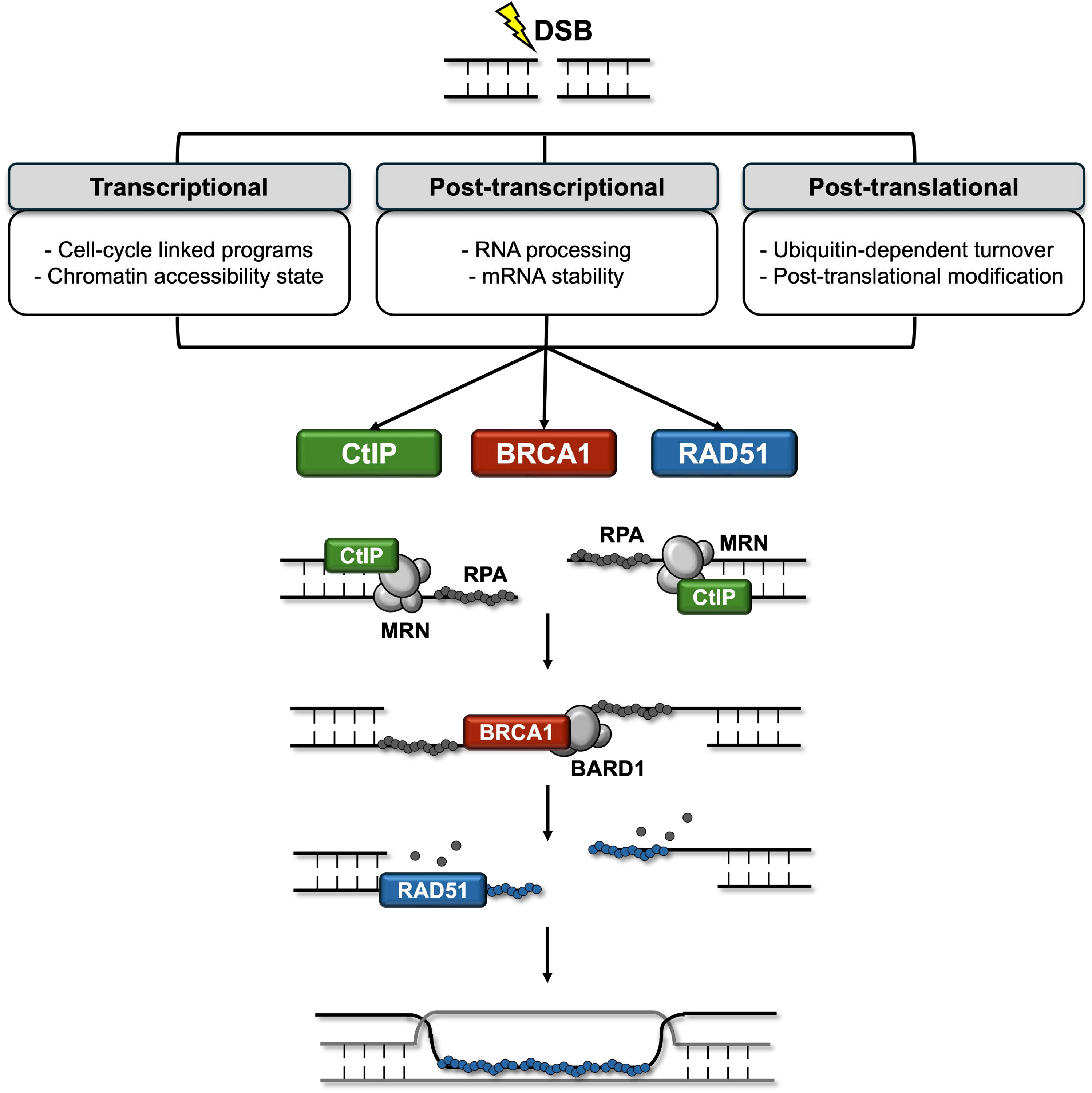
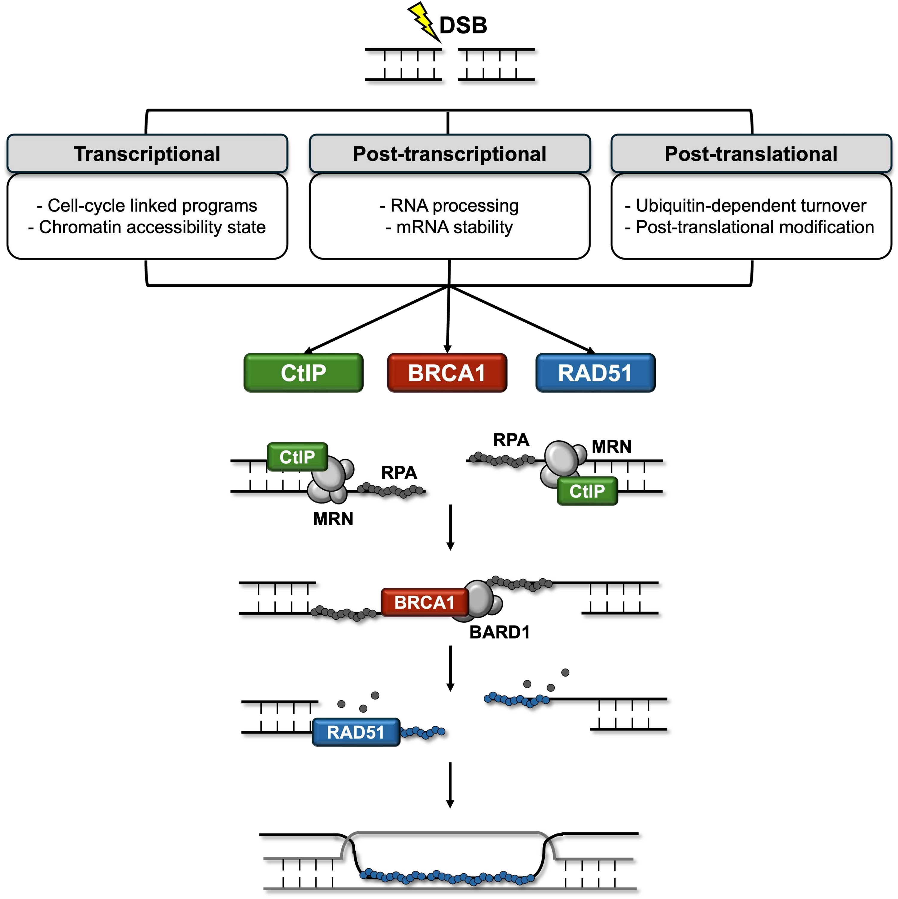
Figure 1. This schematic illustrates the hierarchical stages of the HR repair process and the convergence of multi-layered regulatory inputs on its core execution proteins: CtIP, BRCA1, and RAD51. Canonical HR is depicted as a three-stage mechanical transition: (1) Commitment, initiated by CtIP-mediated DNA end resection; (2) Coordination, governed by BRCA1 to integrate checkpoint signaling and chromatin remodeling; and (3) Execution, centered on RAD51-dependent homology search and strand exchange. The diagram highlights how cellular HR capacity is not constitutive but is dynamically fine-tuned by transcriptional programs, post-transcriptional RNA regulation, and post-translational proteostasis mechanisms. These regulatory layers integrate extrinsic and intrinsic cues, including cell-cycle progression, genotoxic stress, and metabolic context, to ensure genome stability and suppress tumorigenesis. HR: homologous recombination; CtIP: C-terminal binding protein 1 interacting protein; BRCA1: breast cancer gene 1.
Each of these proteins is subject to regulation across multiple biological layers. Their transcription is controlled by cell cycle–linked transcription programs and chromatin accessibility states, their transcripts are shaped by RNA processing and stability mechanisms, and their protein abundance and activity are dynamically influenced by ubiquitin-dependent turnover and post-translational modification. Convergence of regulatory inputs on these stage-defining nodes makes them practical integration points for organizing HR regulatory mechanisms that are otherwise distributed across factor-specific studies.
This review is organized around regulatory mechanisms that directly influence CtIP, BRCA1, and RAD51 expression and thereby modulate HR execution at distinct mechanistic stages. We examine transcriptional, post-transcriptional, and post-translational regulatory pathways affecting the expression of these proteins, with an emphasis on mechanisms that have been linked to measurable effects on HR progression or pathway usage.
2. Transcriptional Regulation of CtIP, BRCA1, and RAD51 in HR
2.1 E2F transcription factors and cell-cycle–coupled activation of HR genes
E2F transcription factors constitute a well-defined regulatory module controlling expression of HR genes, including CtIP, BRCA1, and RAD51 (Figure 2A)[34-37]. Because HR activity is largely restricted to S and G2 phases, transcription of these genes is coupled to DNA replication programs[23,38]. Promoter-mapping and reporter analyses have identified functional E2F-responsive elements within the proximal promoter regions of CtIP, BRCA1, and RAD51, and chromatin immunoprecipitation[39] studies demonstrate cell-cycle–dependent occupancy by activating E2F members such as E2F1 and E2F3, with maximal binding during late G1 and S phase[37,39,40].
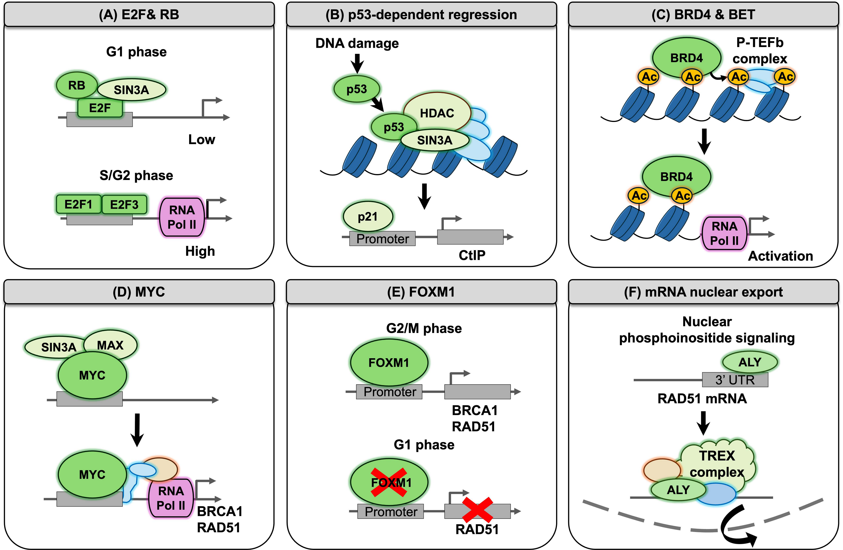
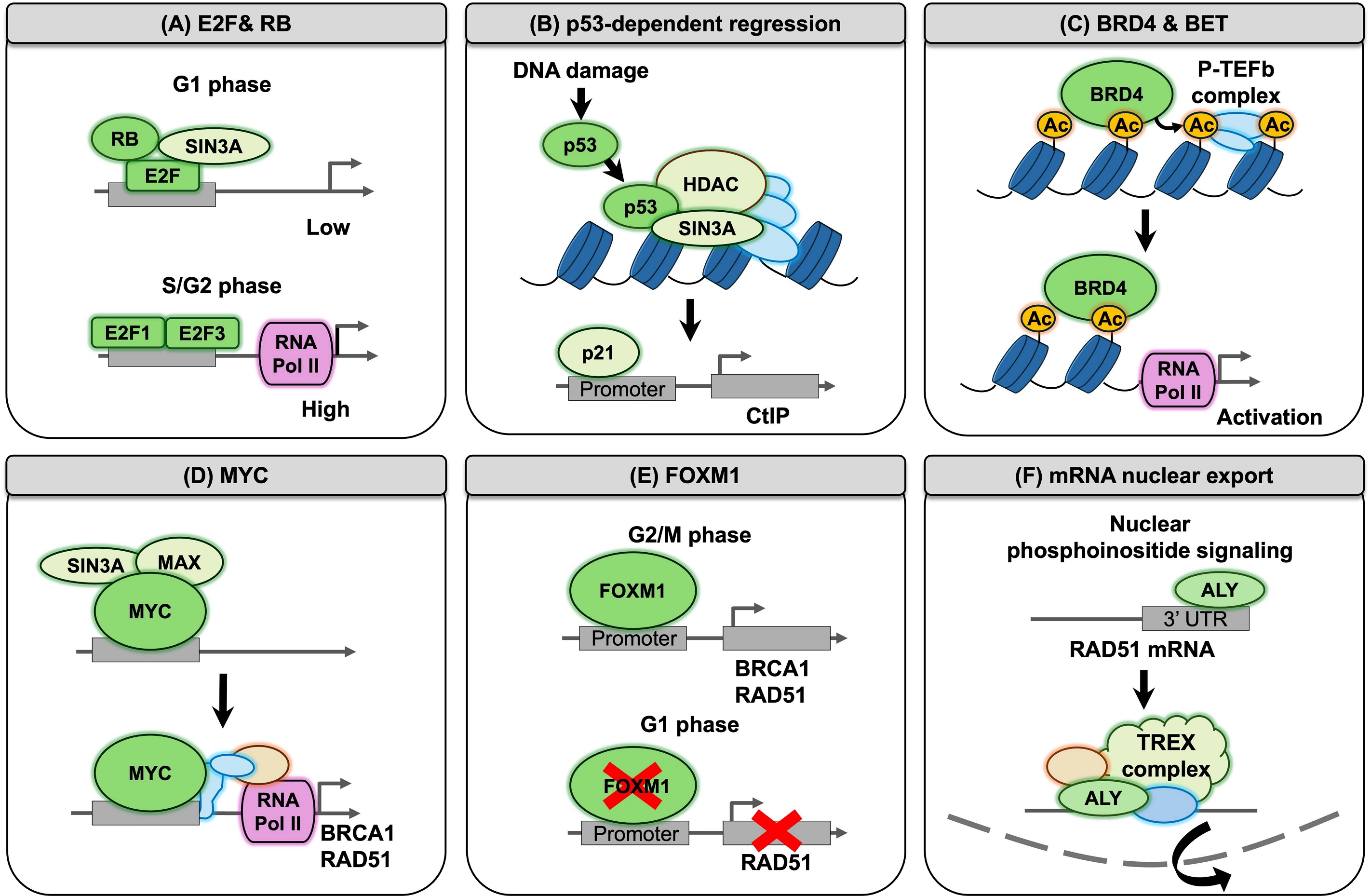
Figure 2. HR gene expression is regulated by multiple signaling modules that couple DNA repair capacity to cellular proliferative and stress-responsive states. (A) RB–E2F complexes recruit co-repressors such as SIN3A to maintain low transcriptional activity of HR genes, whereas during S/G2 phases activating E2F members promote RNA polymerase II-mediated transcription; (B) Under genotoxic stress, p53 recruits chromatin-modifying co-repressors including HDACs and SIN3A to HR gene promoters, resulting in chromatin compaction and transcriptional repression; (C) BRD4/BET proteins function as epigenetic coactivators by recruiting the P-TEFb complex, which promotes RNA polymerase II pause release and transcriptional elongation; (D) MYC amplifies transcription of HR genes such as BRCA1 and RAD51 through enhancer activation and increased RNA polymerase II loading; (E) FOXM1 drives proliferation-associated HR gene expression during G2/M phases, while HR gene transcription is reduced during G1; (F) RAD51 expression can also be regulated post-transcriptionally through selective nuclear export of RAD51 mRNA mediated by IPMK-dependent phosphoinositide signaling and the TREX export pathway. HR: homologous recombination; RB: retinoblastoma protein; HDACs: histone deacetylases; BRD4: bromodomain-containing protein 4; BET: bromodomain and extra-terminal; P-TEFb: positive transcription elongation factor b; BRCA1: breast cancer gene 1; FOXM1: forkhead box M1; IPMK: inositol polyphosphate multikinase; TREX: transcription-export complex.
Ectopic E2F1 expression increases BRCA1 and RAD51 transcription, whereas retinoblastoma protein (RB) pathway activity suppresses their expression by limiting E2F availability[41]. Under quiescent conditions, RB family proteins bind E2F and recruit chromatin repressors[42,43]. Cyclin-dependent kinase (CDK)-mediated RB phosphorylation releases E2F and permits HR gene transcription[44,45]. RB pathway disruption or CDK hyperactivation therefore produces sustained E2F activity and elevated HR gene expression[46,47]. Functional assays link E2F-driven transcriptional activation to increased RAD51 foci formation and enhanced recombination reporter activity, indicating that transcriptional regulation directly influences HR output[35].
2.2 p53-dependent transcriptional repression of HR genes under genotoxic stress
The tumor suppressor p53 exerts context-dependent transcriptional repression of HR genes, particularly RAD51 and BRCA1 (Figure 2B)[48-50]. Promoter-reporter and chromatin immunoprecipitation studies demonstrate p53 binding at RAD51 regulatory regions and reduced promoter activity in the presence of wild-type p53[48].
Mechanistically, repression involves recruitment of co-repressor complexes including histone deacetylases (HDACs) and SIN3A-associated chromatin remodelers, resulting in localized chromatin compaction[51-53]. HDAC inhibition partially reverses p53-dependent RAD51 repression, supporting a chromatin-mediated mechanism. p53 also indirectly suppresses HR gene transcription through p21 induction, which inhibits CDK activity and maintains RB in an E2F-repressive state[54,55]. Consistent with these mechanisms, p53 activation reduces RAD51 transcript levels, RAD51 foci formation, and HR reporter efficiency, whereas p53 loss is frequently associated with elevated RAD51 expression[48,56]. DNA damage–induced phosphorylation of p53 at residues including Ser15 and Ser20 contributes to stabilization and transcriptional activation of p53-dependent programs[57-59], although the specific phosphorylation events responsible for RAD51 repression remain incompletely defined[48,60]. Because suppression of RAD51 limits HR activity, p53-dependent repression of HR genes may increase cellular sensitivity to DNA-damaging agents[48,61]. However, the therapeutic consequences are context dependent, as p53 activation can simultaneously promote cell-cycle arrest, DNA repair coordination, senescence, or apoptosis[62,63].
2.3 Bromodomain-containing protein 4 (BRD4) and bromodomain and extra-terminal (BET) proteins as epigenetic coactivators of HR gene transcription
BRD4 functions as a chromatin-associated transcriptional coactivator linking histone acetylation to productive transcriptional elongation[64,65]. Genome-wide and locus-specific studies show BRD4 enrichment at regulatory regions of HR genes, including CtIP and RAD51[66]. BRD4 recruits the positive transcription elongation factor b complex, promoting CDK9 activation and RNA polymerase II pause release (Figure 2C)[64].
BET inhibition using compounds such as JQ1 disrupts BRD4 chromatin occupancy at HR gene loci, reducing nascent transcription and steady-state mRNA levels and decreasing HR efficiency in functional assays[67]. BET inhibitor treatment can therefore produce HR-deficient phenotypes without genetic mutation and increase sensitivity to DNA damaging agents and Poly(ADP-ribose) polymerase (PARP) inhibition[68,69]. Recent studies using UNI66, which inhibits BRD4, similarly demonstrate reduced CtIP and RAD51 transcription and impaired HR capacity[70].
2.4 MYC-driven transcriptional amplification of HR programs
The oncogenic transcription factor MYC broadly amplifies transcriptional output and contributes to activation of DNA repair gene programs[71,72]. MYC occupancy has been detected at HR gene regulatory regions, and MYC overexpression correlates with increased BRCA1 and RAD51 transcription in multiple tumor contexts (Figure 2D)[72]. MYC promotes transcription through enhancer activation and recruitment of elongation-supporting cofactors, increasing RNA polymerase II loading and activity[73,74]. MYC-associated HR gene upregulation is frequently observed in proliferative and replication-stress conditions[72,75,76].
2.5 Forkhead box protein M1 (FOXM1) and proliferation-linked activation of HR gene expression
FOXM1 regulates genes involved in DNA replication and mitosis and directly activates HR gene transcription. FOXM1 binds promoter regions of BRCA1 and RAD51 and increases their transcription, whereas FOXM1 suppression reduces RAD51 expression and HR efficiency (Figure 2E)[77,78]. Because FOXM1 expression is closely linked to the proliferative state, its regulatory effect on HR genes provides a transcriptional connection between cell-cycle progression and recombination capacity[79].
2.6 Nuclear export control of HR gene transcripts
Beyond transcriptional regulation, expression of HR genes can also be controlled at the level of mRNA nuclear export. Nuclear export of RAD51 transcripts is regulated by inositol polyphosphate multikinase (IPMK), which links nuclear phosphoinositide signaling to transcript-selective mRNA export. IPMK-dependent signaling promotes recognition of sequence motifs within the 3′ untranslated region of RAD51 mRNA by the mRNA export adaptor ALY, thereby facilitating export through the transcription-export complex pathway. Depletion or catalytic inactivation of IPMK results in nuclear retention of RAD51 mRNA, reduced RAD51 protein abundance, and impaired HR efficiency (Figure 2F)[80]. Under genotoxic stress conditions, nuclear export of DNA repair transcripts can also be dynamically regulated. Ribonucleoprotein complexes containing THOC4 and EIF4E associate with mRNAs encoding DNA damage response proteins, and the nuclear export receptor exportin-1 (XPO1) facilitates their transport to the cytoplasm. Inhibition of XPO1 impairs nuclear export of these transcripts and reduces the synthesis of repair proteins required for efficient DNA damage repair[81].
Collectively, transcriptional regulation of HR genes is tightly coordinated with cell cycle progression and the cellular stress response. Proliferation-associated transcription factors, including E2F, MYC, and FOXM1, activate transcription of CtIP, BRCA1, and RAD51 to establish HR competence during DNA replication, while chromatin-associated regulators such as BRD4 sustain transcriptional output. In contrast, genotoxic stress engages p53-dependent repression, limiting HR gene expression to prevent inappropriate recombination. In parallel, mRNA nuclear export provides an additional layer of regulation by controlling the availability of HR transcripts for translation. Together, these regulatory mechanisms ensure that HR capacity is dynamically modulated, activated during proliferation, restrained under stress, and attenuated following repair, thereby maintaining genomic stability across changing cellular contexts.
3. Post-Transcriptional Regulation of CtIP, BRCA1, and RAD51 in HR
3.1 Cell-cycle–dependent restriction of HR by microRNAs (miRNAs)
Post-transcriptional regulation mediated by miRNAs constitutes a critical regulatory layer governing HR. Rather than acting on individual targets in isolation, miRNA networks frequently modulate multiple HR components simultaneously, thereby modulating HR capacity in a context-dependent manner. These regulatory circuits can be broadly categorized into functional modules, including cell-cycle-dependent HR restriction, control of DNA end resection, direct suppression of core HR factors, and DNA damage–responsive adaptive regulation.
HR is tightly restricted during the G1 phase, when sister chromatids are unavailable. A defined group of miRNAs, including miR-1255b, miR-148b*, and miR-193b*, suppresses the expression of BRCA1, BRCA2, and RAD51 specifically during G1, thereby preventing aberrant HR activation (Figure 3)[82]. Inhibition of these miRNAs results in ectopic HR activity in G1 and increased genomic instability, demonstrating that miRNA-mediated repression contributes to cell-cycle gating of HR.
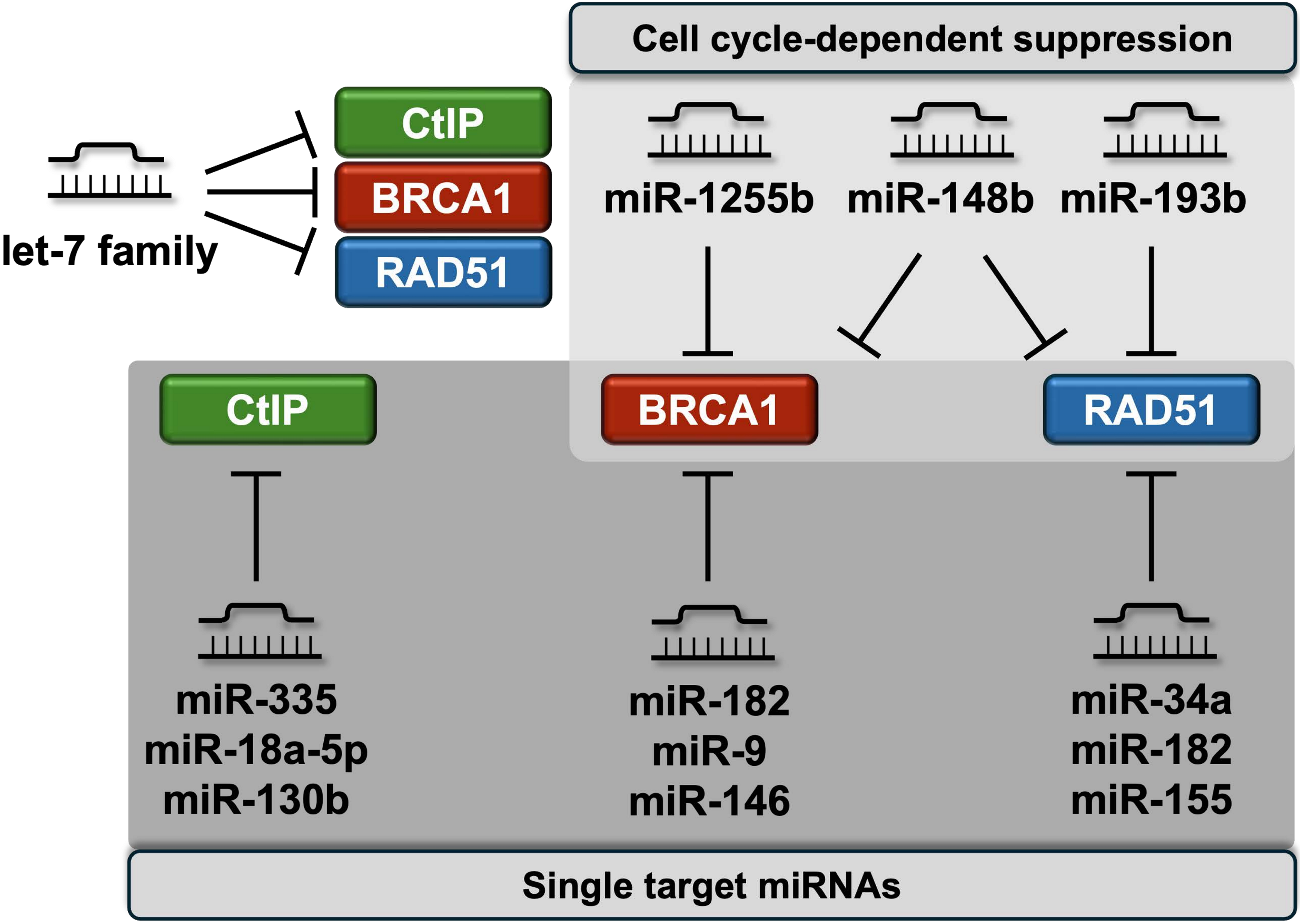
Figure 3. This figure delineates the complex miRNA-dependent networks that regulate the stability and translation of HR factor transcripts. A specific cohort of miRNAs, including miR-1255b, miR-148b, and miR-193b*, enforces G1-phase restriction of HR by suppressing BRCA1 and RAD51, thereby preventing inappropriate recombination when sister chromatids are absent. Multi-target suppressors, such as the let-7 family and the miR-17-92 cluster (e.g., miR-19a/b), coordinately downregulate multiple HR nodes to shift overall pathway efficiency. Furthermore, damage-responsive circuits are highlighted, such as the ATM-dependent repression of miR-335, which relieves CtIP inhibition following irradiation. These miRNA programs provide a rapid, non-genetic mechanism to attenuate HR activity in various physiological and pathological contexts, including oncogenic signaling. miRNA: microRNA; HR: homologous recombination; BRCA1: breast cancer gene 1; ATM: ataxia telangiectasia mutated; CtIP: C-terminal binding protein 1 interacting protein.
In addition, members of the let-7 family function as multi-target HR suppressors by coordinately downregulating BRCA1, BRCA2, and RAD51 transcripts[83,84]. This coordinated targeting highlights how a single miRNA family can regulate multiple nodes within the HR pathway and impose global constraints on HR capacity[82].
3.2 miRNA-mediated control of DNA end resection through CtIP regulation
DNA end resection represents a key commitment step in HR initiation and is tightly regulated at the post-transcriptional level[24]. Several miRNAs directly target CtIP, a key mediator of DNA end resection, thereby limiting HR efficiency[85-87].
Members of the miR-17-92 cluster, particularly miR-19a and miR-19b, suppress CtIP expression and impair resection activity, resulting in reduced HR efficiency and increased sensitivity to genotoxic stress (Figure 3)[85]. Given the frequent amplification of this cluster in cancer, miR-19-mediated CtIP repression provides a mechanistic link between oncogenic miRNA activation and attenuation of HR initiation.
Additional oncogenic miRNAs, including miR-18a-5p and miR-130b, similarly downregulate CtIP expression in solid tumors[86]. Functional rescue experiments demonstrate that restoration of CtIP reverses downstream phenotypes, confirming CtIP as a primary target. These findings indicate that miRNA-driven suppression of CtIP can generate functional HR deficiency in the absence of genetic mutations.
3.3 DNA damage-responsive miRNA regulation of HR
miRNA-mediated regulation of HR is not static but is dynamically regulated by DNA damage signaling pathways. A representative example is miR-335, which forms a regulatory circuit linked to ataxia telangiectasia mutated (ATM) signaling.
Under basal conditions, miR-335 suppresses CtIP expression. Following ionizing radiation (IR), activation of ATM leads to phosphorylation of cyclic adenosine monophosphate response element-binding protein, resulting in repression of miR-335 transcription. This relieves CtIP suppression and preserves HR competence under genotoxic stress (Figure 3)[87]. Perturbation of this axis alters CtIP levels, BRCA1 foci formation, and cellular survival after irradiation, supporting its role as a damage-responsive regulatory module. Although this pathway was originally defined as an ATM-dependent response to IR-induced DSBs[87], whether ataxia telangiectasia and Rad3-related (ATR) also contributes to the miR-335-CtIP regulation axis under replication stress conditions remains unexplored.
3.4 Direct targeting of core HR factors by miRNAs
In addition to regulating HR initiation, miRNAs can directly suppress core HR machinery components, including BRCA1, BRCA2, and RAD51. Multiple miRNAs, such as miR-182, miR-9, and members of the miR-146 family, directly target BRCA1 or BRCA2 transcripts, leading to reduced HR capacity (Figure 3)[88]. In some cases, polymorphisms within miRNA binding sites contribute to inter-individual variability in BRCA expression.
RAD51 is also subject to miRNA-mediated repression. miR-182 and miR-155 reduce RAD51 expression and impair HR efficiency in several tumor models[89]. Suppression of RAD51 is often accompanied by compensatory activation of alternative repair pathways, indicating that miRNAs can influence overall DNA repair pathway choice.
Furthermore, p53-induced miRNAs such as miR-34a directly target RAD51, linking transcriptional stress responses with post-transcriptional regulation of HR[90]. This illustrates convergence between signaling networks and miRNA-mediated control at key HR nodes.
Collectively, miRNA-mediated regulation operates across multiple functional layers of HR, including cell-cycle gating, control of DNA end resection, and direct suppression of core HR factors. Through coordinated targeting of multiple HR components, miRNA networks enable dynamic and reversible modulation of recombination capacity without requiring genetic alterations in HR genes.
4. Post-Translational Regulation of HR Proteins Through Proteasomal Degradation and SUMO–Ubiquitin Signaling
4.1 Proteasome-dependent degradation and ubiquitin-driven control of HR proteins
Proteasome-dependent degradation is a major post-translational mechanism controlling the abundance, chromatin residence time, and functional turnover of HR proteins[91-93]. The ubiquitin–proteasome system operates as a selective regulatory network that determines when and how long HR proteins remain active (Figure 4A)[91,94-96]. This control is particularly important in HR, where both insufficient activity and excessive persistence of repair proteins can compromise genome stability[97]. Multiple E3 ligase complexes ubiquitinate HR regulators under basal and damage-induced conditions, distinguishing steady-state turnover from repair remodeling[98-102].
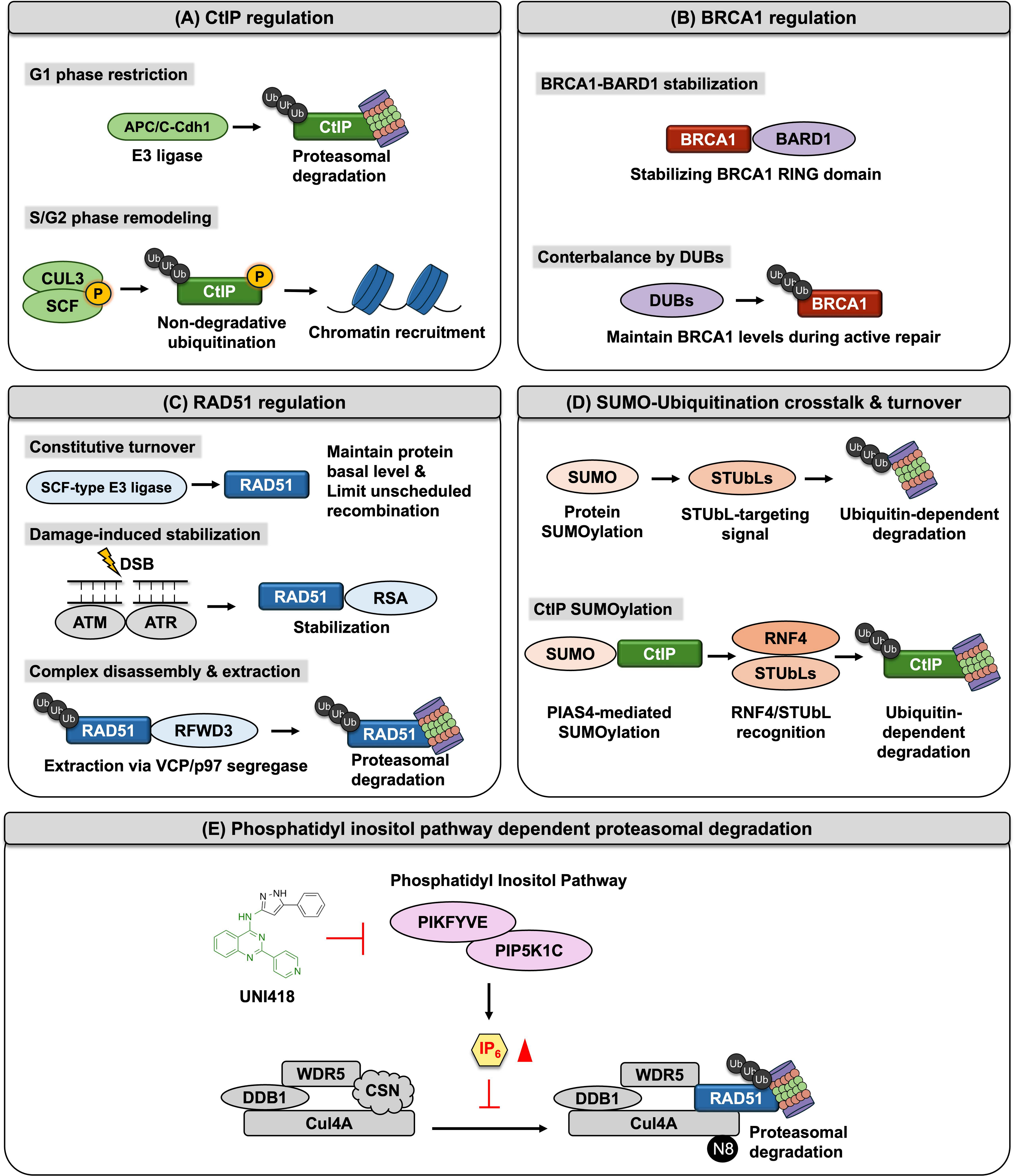
Figure 4. CtIP, BRCA1, and RAD51 are regulated by distinct but interconnected proteostasis mechanisms that control their abundance, chromatin association, and timely removal during HR. (A) CtIP regulation includes APC/C–Cdh1-mediated proteasomal degradation during G1 phase, which restricts CtIP accumulation outside the HR-permissive cell-cycle window, and CUL3- and SCF-associated ubiquitin signaling during S/G2, which promotes non-degradative ubiquitination and chromatin recruitment; (B) BRCA1 regulation is maintained through stabilization by BARD1, which protects the BRCA1 RING domain, while DUBs counterbalance turnover to preserve BRCA1 levels during active repair; (C) RAD51 regulation involves constitutive SCF-type E3 ligase-dependent turnover that limits unscheduled recombination, damage-induced stabilization downstream of ATM/ATR signaling after double-strand breaks, and subsequent complex disassembly through RFWD3-mediated ubiquitination, VCP/p97-dependent extraction, and proteasomal degradation; (D) SUMO–ubiquitin crosstalk further regulates HR factor turnover, in which SUMOylation of CtIP and RAD51 promotes recognition by STUbLs, leading to ubiquitin-dependent degradation; (E) The lower panel illustrates an additional phosphatidylinositol pathway-dependent proteasomal degradation mechanism, in which phosphatidylinositol pathway components, including PIPKFYVE and PIP5K1C, are linked to DDB1–WDR5–CUL4A-associated RAD51 degradation, whereas UNI418 is shown as an inhibitory compound affecting this pathway. CtIP: C-terminal binding protein 1 interacting protein; BRCA1: breast cancer gene 1; HR: homologous recombination; APC: anaphase-promoting complex; SCF: Skp1–Cullin1–F-box complex; BARD1: BRCA1-associated RING domain protein 1; RING: really interesting new gene; DUBs: deubiquitinating enzymes; ATM: ataxia telangiectasia mutated; ATR: ataxia telangiectasia and Rad3-related; RFWD3: RING finger and WD repeat domain 3; VCP: valosin-containing protein; SUMO: small ubiquitin-like modifier; STUbLs: SUMO-targeted ubiquitin ligases; CUL3: Cullin 3; CUL4A: Cullin 4A; WDR5: WD repeat domain 5.
CtIP stability is regulated by cell-cycle–linked ubiquitination that restricts DNA end resection competence. Anaphase-promoting complex/cyclosome–Cdh1-mediated ubiquitination targets CtIP for degradation during G1 phase and after resection completion, thereby limiting end processing to appropriate cell-cycle windows[99]. Additional E3 ubiquitin ligases, including Cullin 3 and Skp1–Cullin1–F-box complex (SCF) family complexes, further modulate CtIP half-life in response to phosphorylation state and checkpoint signaling[98,103]. Not all CtIP ubiquitination is degradative; specific ubiquitin chain architectures also support CtIP recruitment and chromatin retention at break sites, indicating context-dependent functional outcomes[104].
BRCA1 protein abundance is likewise governed by ubiquitin-dependent proteostasis. The BRCA1–BRCA1-associated RING domain protein 1 heterodimer stabilizes the BRCA1 RING domain, whereas alternative ligases promote turnover of unbound or mislocalized BRCA1 (Figure 4B)[102,105-111]. Deubiquitinating enzymes counterbalance degradative ubiquitination and maintain BRCA1 levels during active repair[112-117]. Perturbation of this balance alters HR efficiency without requiring BRCA1 mutation.
RAD51 levels are maintained through constitutive ubiquitin-dependent turnover that limits unscheduled recombination[100,118]. SCF-type ligase complexes regulate basal RAD51 abundance, whereas DNA damage signaling through ATR and ATM pathways promotes RAD51 stabilization and mediator association (Figure 4C)[101,119]. RAD51 removal after recombination intermediate resolution requires ubiquitination by E3 ligases such as RING finger and WD repeat domain 3 and extraction through valosin-containing protein (VCP/p97)-dependent segregase activity, followed by proteasomal degradation[100]. Loss of this extraction pathway results in prolonged RAD51 retention and incomplete recombination resolution, demonstrating that regulated turnover contributes to both suppression and completion of HR reactions.
Recent evidence suggests that metabolic signaling can also regulate RAD51 proteostasis[120]. Inhibition of the lipid kinases PIKfyve and PIP5K1C reduces intracellular levels of inositol hexaphosphate (IP6), promoting neddylation and activation of the Cullin 4A (Cul4A) ubiquitin ligase complex (Figure 4E). IP6 is known to regulate Cullin-RING ligase activity through the COP9 signalosome-mediated deneddylation cycle, thereby controlling CRL activity and substrate ubiquitination[121-123]. Activated Cul4A, together with the DDB1- and CUL4-associated factor protein, WD repeat domain 5, drives proteasome-dependent degradation of RAD51[120,124], linking phosphoinositide metabolism to post-translational control of RAD51 stability and HR activity.
4.2 SUMO–ubiquitin signaling crosstalk in HR pathway and protein turnover
SUMOylation provides an additional regulatory layer that frequently operates in coordination with ubiquitin signaling to control HR protein dynamics (Figure 4D)[125,126]. SUMO conjugation can function as a targeting signal for SUMO-targeted ubiquitin ligases, linking SUMO modification to ubiquitin-dependent processing[127,128]. This SUMO–ubiquitin relay contributes to ordered assembly and disassembly of HR complexes at DNA damage sites[129].
CtIP undergoes SUMOylation that promotes chromatin association and DNA end resection activity[130]. Following DSB induction, ATM-dependent hyperphosphorylation of CtIP further promotes PIAS4-dependent SUMOylation at Lys578, enabling recognition by the SUMO-targeted ubiquitin ligase RNF4. RNF4-mediated polyubiquitination and proteasomal degradation of SUMO-modified CtIP establish a negative feedback mechanism that limits CtIP accumulation at DSBs and prevents excessive end resection[131]. Thus, CtIP SUMOylation coordinates resection activity with the timely turnover of activated CtIP during HR. SUMO-modified repair proteins also contribute to DNA damage–associated chromatin compartments that concentrate HR machinery, while STUbL-dependent ubiquitination supports repair-complex remodeling and disassembly following repair.
RAD51 is also SUMOylated after DNA damage, and this modification promotes BRCA2-dependent RAD51 recruitment, chromatin loading, and efficient HR repair[132,133]. Direct STUbL-mediated degradation of SUMOylated RAD51 has not been clearly established[134]. Instead, STUbL-dependent turnover of SUMO-modified repair factors such as MDC1 and RPA indirectly facilitates RPA-to-RAD51 exchange and proper RAD51 loading during HR[135-137]. Disruption of STUbL function produces persistent retention of repair factors at damage sites and unresolved DNA damage signaling, supporting a role for SUMO–ubiquitin relay signaling in HR protein clearance[137-139].
Collectively, post-translational regulation of HR proteins is governed by coordinated ubiquitin–proteasome-mediated turnover and SUMO–ubiquitin relay signaling, which together define the temporal dynamics of repair factor activity. Ubiquitin-dependent proteostasis controls the abundance and chromatin association of CtIP, BRCA1, and RAD51, thereby preventing unscheduled recombination, while enabling efficient repair initiation. In parallel, SUMOylation and subsequent recognition by STUbL enzymes couple repair complex assembly to timely disassembly, ensuring proper resolution of recombination intermediates. These pathways dynamically integrate cell-cycle cues, DNA damage signaling, and metabolic inputs to fine-tune HR protein stability and function. Through the balanced control of stabilization, chromatin retention, and proteasome-dependent removal, post-translational mechanisms establish a regulatory framework that enables the initiation, progression, and termination of HR.
5. Therapeutic and Biomarker Implications of HR Regulatory Networks
5.1 Regulatory control of HR and PARP-inhibitor synthetic lethality
The regulatory nodes that govern CtIP, BRCA1, and RAD51 are not merely of academic interest, they directly influence how tumors respond to DNA-damaging agents such as PARP inhibitors[140-143]. HR-deficient cells rely on PARP-mediated base excision repair to maintain viability[140,141,144]. Pharmacological inhibition of PARP therefore induces synthetic lethality in cells lacking HR capacity[141-143]. Recent work demonstrates that disrupting the transcriptional or proteostatic regulation of CtIP and RAD51 can convert HR-proficient tumors into an HR-deficient state and dramatically enhance PARP inhibitor sensitivity[38,68,98,120]. For example, BET proteins such as BRD4 act as epigenetic co-activators for CtIP and RAD51 transcription[69,70]. Treatment with the BRD4 inhibitor UNI66 or classical BET inhibitors like JQ1 suppresses CtIP and RAD51 transcription, inducing an HR-deficient phenotype and producing synergistic cytotoxicity when combined with PARP inhibitors[67,68,70]. Similar synergy is observed when ATR or CHK1 inhibitors reduce RAD51 expression and disrupt HR[140,145], or when WEE1 inhibition lowers CtIP and RAD51 levels and heightens replication stress[146]. Loss of CtIP itself sensitizes breast cancer cells and xenograft models to PARP inhibitors[143,147], while depletion of the deubiquitinase USP52 destabilizes CtIP and promotes PARP-inhibitor sensitivity[148]. Together, these findings reveal a mechanistic link between the regulation of HR execution factors and the synthetic lethality exploited by PARP inhibitors, highlighting CtIP, BRCA1, and RAD51 as actionable nodes to induce HR deficiency and overcome drug resistance. The p53 pathway may also influence therapeutic responses through regulation of HR gene expression. Wild-type p53 represses RAD51 and BRCA1 transcription under genotoxic stress, potentially reducing HR capacity and increasing sensitivity to DNA-damaging agents[48,50,149]. However, whether p53 wild-type tumors are uniformly more responsive than p53-mutant tumors remains unclear, because p53 also activates cell-cycle checkpoints, apoptosis, senescence, and genome surveillance programs that can independently affect treatment outcomes[62,150]. Consequently, the relationship between p53 status, HR activity, and therapeutic response is likely to be highly context dependent.
5.2 Therapeutic strategies targeting HR pathways
The insights above have catalyzed the development of therapeutic strategies that deliberately target HR regulatory pathways. Several classes of agents are being tested in pre-clinical and clinical settings[146,151,152]. Small molecules such as UNI66, JQ1, and I-BET762 block BRD4 recruitment to HR gene promoters, reducing CtIP and RAD51 transcription[67,70]. These compounds induce HR deficiency and exhibit strong synergy with PARP inhibitors across diverse tumor models[68,69]. ATR inhibitors (e.g., ceralasertib) and CHK1/2 inhibitors downregulate RAD51 and disrupt HR[153]. Combined administration with PARP inhibitors results in enhanced DNA damage and synthetic lethality in BRCA-wild-type tumors[145,146]. Agents such as CAM833 block the BRCA2–RAD51 interaction and prevent recombinase filament assembly[154]. In HR-proficient models, RAD51 inhibitors sensitize tumors to PARP inhibition[151,155]. WEE1 kinase controls S-phase progression and indirectly regulates CtIP and RAD51 levels[156]. WEE1 inhibition reduces HR gene expression and, when combined with PARP inhibitors, induces replication stress and immune activation[157,158]. CDK inhibitors similarly mimic BRCA1 deficiency by downregulating HR genes and augmenting PARP-inhibitor efficacy[159]. Targeting deubiquitinases and E3 ligases can alter the stability of HR proteins[148,160]. For instance, USP52 stabilizes CtIP; its inhibition decreases CtIP abundance and sensitizes cells to PARP inhibition[148]. Strategies that promote RAD51 degradation through ubiquitin ligases or neddylation pathways are also being explored.
These therapeutic approaches demonstrate that HR pathway modulation is a viable strategy to induce synthetic lethality in tumors that are otherwise HR-proficient. Ongoing clinical trials combining PARP inhibitors with ATR, CHK1, WEE1, or BET inhibitors aim to translate these findings into effective combination therapies[161,162].
5.3 HR regulatory status as biomarkers
Because regulation of CtIP, BRCA1, and RAD51 can determine HR competence, measuring their regulatory status may predict responses to PARP inhibitors better than genomic HRD scores alone[163,164]. CtIP is frequently downregulated in breast cancer, and low CtIP expression correlates with reduced RAD51 foci formation and heightened sensitivity to PARP inhibitors[147]. More broadly, HR-deficient breast tumors may also exhibit increased sensitivity to DNA-damaging chemotherapy[163,164], particularly platinum-based agents[165], and potentially to anthracycline/cyclophosphamide-containing regimens[166], although the magnitude of benefit varies according to tumor subtype, treatment context, and the degree of functional HR deficiency.
Functional assays that quantify nuclear RAD51 foci provide dynamic biomarkers of HR activity: patients with low RAD51 immunohistochemistry scores experience longer progression-free survival on PARP inhibitors than those with high scores, and RAD51 levels increase upon acquisition of drug resistance[163,164,167-169]. A related metric, the RAD51 score derived from RAD51 focus formation, discriminates PARP-sensitive from PARP-resistant tumors and outperforms genomic HRD testing[170]. Multi-layer biomarkers that integrate CtIP, BRCA1, and RAD51 mRNA abundance, miRNA regulators, and post-translational modifications may further enhance predictive accuracy[85,171,172]. For example, assessing miR-19a/b levels alongside CtIP protein could identify tumors with miRNA-mediated HR suppression, while measuring ubiquitination or SUMOylation of RAD51 could indicate defective proteostasis[85,172]. Development of such composite biomarkers will aid stratification of patients for PARP inhibitor therapy and guide combination strategies.
5.4 Relevance to cancer and aging
The efficiency of HR impacts not only therapeutic response but also fundamental biological processes such as tumorigenesis and aging[173,174]. Age-associated increases in genomic rearrangements and loss of heterozygosity (LOH) correlate with declining HR activity[173,174]. In replicative senescent human cells, HR is virtually absent because RAD51 expression becomes undetectable; instead, cells rely on NHEJ, which is more error-prone[175,176]. A p53-dependent decrease in RAD51 during senescence promotes the formation of persistent DNA-damage foci and reinforces the senescent phenotype[48]. These changes contribute to accumulation of DNA damage, genomic instability, and an increased incidence of cancer with advancing age[174]. Mechanisms that suppress mitotic recombination or RAD51 recruitment may fail in aging cells, leading to greater reliance on error-prone repair pathways and higher frequencies of genomic rearrangements and LOH[176,177]. HR regulators may therefore be perturbed in aged tissues through decreased transcription, altered miRNA expression, or defective proteasomal turnover[176,178]. Understanding how CtIP, BRCA1, and RAD51 regulation changes over the lifespan could reveal why HR deficiency and genome instability rise with age and may offer opportunities to preserve genome integrity or mitigate cancer risk in the elderly.
6. Conclusion
HR capacity emerges from coordinated regulatory processes operating across transcriptional, post-transcriptional, and post-translational layers rather than from the presence of individual repair proteins alone[179]. The transcriptional programs controlling CtIP, BRCA1, and RAD51 link recombination competence to fundamental cellular states, including cell-cycle progression, chromatin accessibility, and stress-responsive signaling[97]. Regulatory factors such as E2F, MYC, FOXM1, and BRD4 collectively establish transcriptional environments that promote HR gene expression during proliferative conditions[35,64,71,77], whereas stress-responsive pathways such as p53 signaling restrain recombination activity under genotoxic stress[48]. These transcriptional circuits therefore function as gatekeeping mechanisms that couple HR activation to appropriate cellular contexts.
Beyond transcriptional control, miRNA-mediated post-transcriptional regulation provides an additional regulatory layer that can simultaneously modulate multiple HR proteins[82,180,181]. miRNA-mediated suppression of CtIP, BRCA1, and RAD51 illustrates how recombination capacity can be rapidly attenuated without genetic alterations to HR genes themselves[85,88-90]. Such regulation is particularly important for restricting HR to appropriate cell-cycle phases[82] and for dynamically adapting repair activity following DNA damage[180]. Importantly, several oncogenic miRNAs target HR regulators, suggesting that dysregulated miRNA expression in cancer may reshape recombination capacity through coordinated repression of multiple HR nodes.
Post-translational control further defines HR activity by regulating protein stability, chromatin residency, and the timely removal of repair proteins[91,95]. Ubiquitin-dependent proteasomal degradation and SUMO–ubiquitin relay signaling coordinate the assembly, activation, and disassembly of HR complexes[91,125,129]. These processes ensure that CtIP-mediated DNA end resection, BRCA1 dependent repair complex formation, and RAD51 filament dynamics occur within tightly controlled temporal windows[98-100,118]. Disruption of these proteostasis pathways can lead to either insufficient HR activity or persistent recombination intermediates, both of which threaten genome stability[94,96].
Together, these multilayer regulatory mechanisms emphasize that HR competence represents a dynamic network property shaped by interactions among transcriptional programs, RNA regulatory circuits, and protein turnover systems[91,97,182]. From a broader perspective, this network architecture provides flexibility that allows cells to rapidly adjust recombination activity in response to proliferative cues, chromatin state changes, and DNA damage signals. However, it also creates multiple points at which regulatory dysregulation can alter HR capacity without direct mutations in core repair genes. This concept may help explain why HR-deficient phenotypes are frequently observed in tumors lacking genetic alterations in canonical HR proteins[183,184].
Despite this multilayered regulatory framework, several important questions remain unresolved. First, it is not yet clear whether increased transcription or protein abundance of HR factors is always sufficient to enhance productive HR activity. For example, elevated RAD51 expression may increase recombination potential in some settings, but excessive or improperly regulated RAD51 accumulation can also interfere with repair completion and promote aberrant recombination intermediates[185-187]. Similarly, transcriptional programs driven by E2F, MYC, FOXM1, or BRD4 may support HR gene expression during proliferation, but their contribution to functional HR capacity is likely influenced by chromatin context, replication stress, and post-translational control of repair proteins[72,79,188-190].
Second, the regulatory effects of stress-responsive pathways such as p53 signaling remain context-dependent. Although p53-dependent cell-cycle arrest can restrain HR gene expression by limiting proliferation-associated transcriptional programs[79,149], p53 may also contribute to genome maintenance by preventing inappropriate repair, promoting repair pathway choice, or eliminating heavily damaged cells[62,191,192]. Thus, whether p53 signaling suppresses HR directly, indirectly through cell-cycle control, or through broader genome surveillance mechanisms remains an important issue[193].
Third, the physiological and pathological roles of miRNA-mediated HR regulation require further clarification. Several miRNAs can repress CtIP, BRCA1, RAD51, or multiple HR-associated factors, but it remains unclear whether these interactions primarily serve as normal mechanisms for fine-tuning repair activity or whether they become functionally significant mainly in cancer, where dysregulated miRNA expression may mimic or reinforce HR-deficient states[82]. Likewise, post-translational mechanisms that remove HR proteins from chromatin may either promote repair completion or prematurely terminate repair, depending on timing and cellular context[100,194]. Distinguishing these opposing outcomes will be essential for understanding how regulatory dysregulation alters HR capacity without direct mutation of core HR genes.
The regulatory control of HR gene expression also has important implications for therapeutic responses to DNA damage-targeting agents. In particular, HR activity is a key determinant of cellular sensitivity to PARP inhibitors, which exploit synthetic lethality in HR-deficient cells[141,142,151]. Tumors with impaired HR function, such as those harboring BRCA1 or BRCA2 mutations, exhibit pronounced sensitivity to PARP inhibition, whereas restoration or upregulation of HR activity can confer drug resistance[195,196]. In this context, regulatory mechanisms controlling the expression and activity of HR proteins, including transcriptional programs, miRNA-mediated regulation, and post-translational protein turnover, may influence the functional HR state of cancer cells and thereby affect the efficacy of PARP inhibitor–based therapies[143,144,146,151]. Consequently, understanding how these regulatory networks modulate CtIP, BRCA1, and RAD51 expression may provide important insights into variability in PARP inhibitor responses and may help identify new strategies to enhance therapeutic sensitivity in HR-proficient tumors.
However, an important unresolved question is whether regulatory suppression of HR factors produces the same therapeutic vulnerabilities as genetic inactivation of BRCA1, BRCA2, or other canonical HR genes[68,141,142]. Regulatory HR deficiency may be partial, reversible, or highly context-dependent, which could explain why HR gene expression signatures do not always correlate with PARP inhibitor responses[197]. Therefore, future biomarker strategies may need to integrate transcriptional, post-transcriptional, and proteostasis-based measurements with direct functional assays of HR activity[164,198].
Future studies integrating transcriptional profiling, RNA regulatory analysis, and proteostasis measurements with functional HR assays will be important for defining how these regulatory layers interact under physiological and pathological conditions. Such integrative approaches may reveal regulatory vulnerabilities that modulate recombination capacity and could inform therapeutic strategies targeting HR regulation in cancer and other genome instability-associated diseases.
Acknowledgements
ChatGPT was used solely for language polishing during the manuscript preparation process. All research content, including study design, interpretations, and figures, is original and was not generated using AI tools. The authors take full responsibility for the integrity, originality, and accuracy of the work.
Authors contribution
Chung Y, Kim M, Jeong S, Amarsanaa E: Conceptualization, methodology, writing-original draft, writing-review & editing.
Lee Y, Myung K: Conceptualization, methodology, writing-review & editing.
Conflicts of interest
The authors declare no conflicts of interest.
Ethical approval
Not applicable.
Consent to participate
Not applicable.
Consent for publication
Not applicable.
Availability of data and materials
Not applicable.
Funding
This work was supported by the Institute for Basic Science (Grant No. IBS-R022-D1).
Copyright
© The Author(s) 2026.
References
-
1. Wright WD, Shah SS, Heyer WD. Homologous recombination and the repair of DNA double-strand breaks. J Biol Chem. 2018;293(27):10524-10535.[DOI]
-
2. Li X, Heyer WD. Homologous recombination in DNA repair and DNA damage tolerance. Cell Res. 2008;18(1):99-113.[DOI]
-
3. Trenner A, Sartori AA. Harnessing DNA double-strand break repair for cancer treatment. Front Oncol. 2019;9:1388.[DOI]
-
4. Yin Y, Lin J, Cai M. Phase separation as a dynamic regulatory mechanism of DNA end resection. Ageing Cancer Res Treat. 2025;2(2):202515.[DOI]
-
6. Kowalczykowski SC. An overview of the molecular mechanisms of recombinational DNA repair. Cold Spring Harb Perspect Biol. 2015;7(11):a016410.[DOI]
-
7. Jasin M, Rothstein R. Repair of strand breaks by homologous recombination. Cold Spring Harb Perspect Biol. 2013;5(11):a012740.[DOI]
-
8. Sung P. Catalysis of ATP-dependent homologous DNA pairing and strand exchange by yeast RAD51 protein. Science. 1994;265(5176):1241-1243.[DOI]
-
10. Conway AB, Lynch TW, Zhang Y, Fortin GS, Fung CW, Symington LS, et al. Crystal structure of a Rad51 filament. Nat Struct Mol Biol. 2004;11(8):791-796.[DOI]
-
17. Karanam K, Kafri R, Loewer A, Lahav G. Quantitative live cell imaging reveals a gradual shift between DNA repair mechanisms and a maximal use of HR in mid S phase. Mol Cell. 2012;47(2):320-329.[DOI]
-
18. Bindra RS, Schaffer PJ, Meng A, Woo J, Måseide K, Roth ME, et al. Down-regulation of Rad51 and decreased homologous recombination in hypoxic cancer cells. Mol Cell Biol. 2004;24(19):8504-8518.[DOI]
-
19. Bindra RS, Glazer PM. Repression of RAD51 gene expression by E2F4/p130 complexes in hypoxia. Oncogene. 2007;26(14):2048-2057.[DOI]
-
22. Kakarougkas A, Jeggo PA. DNA DSB repair pathway choice: An orchestrated handover mechanism. Br J Radiol. 2014;87(1035):20130685.[DOI]
-
24. Sartori AA, Lukas C, Coates J, Mistrik M, Fu S, Bartek J, et al. Human CtIP promotes DNA end resection. Nature. 2007;450(7169):509-514.[DOI]
-
31. Bonilla B, Hengel SR, Grundy MK, Bernstein KA. RAD51 gene family structure and function. Annu Rev Genet. 2020;54:25-46.[DOI]
-
33. Gupta RC, Bazemore LR, Golub EI, Radding CM. Activities of human recombination protein Rad51. Proc Natl Acad Sci U S A. 1997;94(2):463-468.[DOI]
-
36. Mitxelena J, Apraiz A, Vallejo-Rodríguez J, García-Santisteban I, Fullaondo A, Alvarez-Fernández M, et al. An E2F7-dependent transcriptional program modulates DNA damage repair and genomic stability. Nucleic Acids Res. 2018;46(9):4546-4559.[DOI]
-
41. Zhao X, Wei C, Li J, Xing P, Li J, Zheng S, et al. Cell cycle-dependent control of homologous recombination. Acta Biochim Biophys Sin. 2017;49(8):655-668.[DOI]
-
42. Ahlander J, Bosco G. The RB/E2F pathway and regulation of RNA processing. Biochem Biophys Res Commun. 2009;384(3):280-283.[DOI]
-
43. Manickavinayaham S, Dennehey BK, Johnson DG. Direct regulation of DNA repair by E2F and RB in mammals and plants: Core function or convergent evolution? Cancers. 2021;13(5):934.[DOI]
-
44. Rubin SM, Sage J, Skotheim JM. Integrating old and new paradigms of G1/S control. Mol Cell. 2020;80(2):183-192.[DOI]
-
45. Burke JR, Liban TJ, Restrepo T, Lee HW, Rubin SM. Multiple mechanisms for E2F binding inhibition by phosphorylation of the retinoblastoma protein C-terminal domain. J Mol Biol. 2014;426(1):245-255.[DOI]
-
46. Johnson J, Thijssen B, McDermott U, Garnett M, Wessels LA, Bernards R. Targeting the RB-E2F pathway in breast cancer. Oncogene. 2016;35(37):4829-4835.[DOI]
-
49. Arizti P, Fang L, Park I, Yin Y, Solomon E, Ouchi T, et al. Tumor suppressor p53 is required to modulate BRCA1 expression. Mol Cell Biol. 2000;20(20):7450-7459.[DOI]
-
53. Grzenda A, Lomberk G, Zhang JS, Urrutia R. Sin3: Master scaffold and transcriptional corepressor. Biochim Biophys Acta Gene Regul Mech. 2009;1789(6-8):443-450.[DOI]
-
54. Benson EK, Mungamuri SK, Attie O, Kracikova M, Sachidanandam R, Manfredi JJ, et al. p53-dependent gene repression through p21 is mediated by recruitment of E2F4 repression complexes. Oncogene. 2014;33(30):3959-3969.[DOI]
-
57. Dumaz N, Meek DW. Serine 15 phosphorylation stimulates p53 transactivation but does not directly influence interaction with HDM2. EMBO J. 1999;18(24):7002-7010.[DOI]
-
59. Siliciano JD, Canman CE, Taya Y, Sakaguchi K, Appella E, Kastan MB. DNA damage induces phosphorylation of the amino terminus of p53. Genes Dev. 1997;11(24):3471-3481.[DOI]
-
62. Kastenhuber ER, Lowe SW. Putting p53 in context. Cell. 2017;170(6):1062-1078.[DOI]
-
63. El-Deiry WS. The role of p53 in chemosensitivity and radiosensitivity. Oncogene. 2003;22(47):7486-7495.[DOI]
-
66. Cheung KL, Kim C, Zhou MM. The functions of BET proteins in gene transcription of biology and diseases. Front Mol Biosci. 2021;8:728777.[DOI]
-
67. Miller AL, Fehling SC, Garcia PL, Gamblin TL, Council LN, van Waardenburg RC, et al. The BET inhibitor JQ1 attenuates double-strand break repair and sensitizes models of pancreatic ductal adenocarcinoma to PARP inhibitors. EBioMedicine. 2019;44:419-430.[DOI]
-
71. Lin CY, Lovén J, Rahl PB, Paranal RM, Burge CB, Bradner JE, et al. Transcriptional amplification in tumor cells with elevated c-Myc. Cell. 2012;151(1):56-67.[DOI]
-
73. Rahl PB, Young RA. MYC and transcription elongation. Cold Spring Harb Perspect Med. 2014;4(1):a020990.[DOI]
-
75. Zhang Y. Exploiting replication stress for synthetic lethality in MYC-driven cancers. Am J Cancer Res. 2025;15(4):1461-1479.[DOI]
-
76. Giuntini F, González-Larreategui Í, Herencia-Ropero A, Casacuberta-Serra S, Zacarías-Fluck MF, Arnal M, et al. MYC inhibition by Omomyc causes DNA damage and overcomes PARPi resistance in breast cancer. Cell Rep. 2025;44(12):116604.[DOI]
-
77. Zhang N, Wu X, Yang L, Xiao F, Zhang H, Zhou A, et al. FoxM1 inhibition sensitizes resistant glioblastoma cells to temozolomide by downregulating the expression of DNA-repair gene Rad51. Clin Cancer Res. 2012;18(21):5961-5971.[DOI]
-
82. Choi YE, Pan Y, Park E, Konstantinopoulos P, De S, D’Andrea A, et al. MicroRNAs down-regulate homologous recombination in the G1 phase of cycling cells to maintain genomic stability. eLife. 2014;3:e02445.[DOI]
-
86. Yang L, Yang B, Wang Y, Liu T, He Z, Zhao H, et al. The CTIP-mediated repair of TNF-α-induced DNA double-strand break was impaired by miR-130b in cervical cancer cell. Cell Biochem Funct. 2019;37(7):534-544.[DOI]
-
90. Cortez MA, Valdecanas D, Niknam S, Peltier HJ, Diao L, Giri U, et al. In vivo delivery of miR-34a sensitizes lung tumors to radiation through RAD51 regulation. Mol Ther Nucleic Acids. 2015;4:e270.[DOI]
-
92. Brinkmann K, Schell M, Hoppe T, Kashkar H. Regulation of the DNA damage response by ubiquitin conjugation. Front Genet. 2015;6:98.[DOI]
-
94. Murakawa Y, Sonoda E, Barber LJ, Zeng W, Yokomori K, Kimura H, et al. Inhibitors of the proteasome suppress homologous DNA recombination in mammalian cells. Cancer Res. 2007;67(18):8536-8543.[DOI]
-
97. Krejci L, Altmannova V, Spirek M, Zhao X. Homologous recombination and its regulation. Nucleic Acids Res. 2012;40(13):5795-5818.[DOI]
-
98. Ferretti LP, Himmels SF, Trenner A, Walker C, Von Aesch C, Eggenschwiler A, et al. Cullin3-KLHL15 ubiquitin ligase mediates CtIP protein turnover to fine-tune DNA-end resection. Nat Commun. 2016;7:12628.[DOI]
-
100. Inano S, Sato K, Katsuki Y, Kobayashi W, Tanaka H, Nakajima K, et al. RFWD3-mediated ubiquitination promotes timely removal of both RPA and RAD51 from DNA damage sites to facilitate homologous recombination. Mol Cell. 2017;66(5):622-634.[DOI]
-
103. Li F, Mladenov E, Mortoga S, Iliakis G. SCFSKP2 regulates APC/CCDH1-mediated degradation of CTIP to adjust DNA-end resection in G2-phase. Cell Death Dis. 2020;11:548.[DOI]
-
104. Schmidt CK, Galanty Y, Sczaniecka-Clift M, Coates J, Jhujh S, Demir M, et al. Systematic E2 screening reveals a UBE2D–RNF138–CtIP axis promoting DNA repair. Nat Cell Biol. 2015;17(11):1458-1470.[DOI]
-
106. Densham RM, Morris JR. Moving mountains: The BRCA1 promotion of DNA resection. Front Mol Biosci. 2019;6:79.[DOI]
-
111. Wang X, Lu G, Li L, Yi J, Yan K, Wang Y, et al. HUWE1 interacts with BRCA1 and promotes its degradation in the ubiquitin–proteasome pathway. Biochem Biophys Res Commun. 2014;444(3):290-295.[DOI]
-
118. Antoniuk-Majchrzak J, Enkhbaatar T, Długajczyk A, Kaminska J, Skoneczny M, Klionsky DJ, et al. Stability of Rad51 recombinase and persistence of Rad51 DNA repair foci depends on post-translational modifiers, ubiquitin and SUMO. Biochim Biophys Acta Mol Cell Res. 2023;1870(7):119526.
-
123. Enchev RI, Schulman BA, Peter M. Protein neddylation: Beyond cullin–RING ligases. Nat Rev Mol Cell Biol. 2015;16(1):30-44.[DOI]
-
124. Hannah J, Zhou P. Regulation of DNA damage response pathways by the cullin-RING ubiquitin ligases. DNA Repair. 2009;8(4):536-543.[DOI]
-
126. Sriramachandran AM, Dohmen RJ. SUMO-targeted ubiquitin ligases. Biochim Biophys Acta Mol Cell Res. 2014;1843(1):75-85.[DOI]
-
127. Prudden J, Pebernard S, Raffa G, Slavin DA, Perry JJP, Tainer JA, et al. SUMO-targeted ubiquitin ligases in genome stability. EMBO J. 2007;26(18):4089-4101.[DOI]
-
128. Chang YC, Oram MK, Bielinsky AK. SUMO-targeted ubiquitin ligases and their functions in maintaining genome stability. Int J Mol Sci. 2021;22(10):5391.[DOI]
-
130. Soria-Bretones I, Cepeda-García C, Checa-Rodriguez C, Heyer V, Reina-San-Martin B, Soutoglou E, et al. DNA end resection requires constitutive sumoylation of CtIP by CBX4. Nat Commun. 2017;8:113.[DOI]
-
136. García-Rodríguez N, Wong RP, Ulrich HD. Functions of ubiquitin and SUMO in DNA replication and replication stress. Front Genet. 2016;7:87.[DOI]
-
137. Han MM, Hirakawa M, Yamauchi M, Matsuda N. Roles of the SUMO-related enzymes, PIAS1, PIAS4, and RNF4, in DNA double-strand break repair by homologous recombination. Biochem Biophys Res Commun. 2022;591:95-101.[DOI]
-
139. Vyas R, Kumar R, Clermont F, Helfricht A, Kalev P, Sotiropoulou P, et al. RNF4 is required for DNA double-strand break repair in vivo. Cell Death Differ. 2013;20(3):490-502.[DOI]
-
140. McCabe N, Turner NC, Lord CJ, Kluzek K, Białkowska A, Swift S, et al. Deficiency in the repair of DNA damage by homologous recombination and sensitivity to poly(ADP-ribose) polymerase inhibition. Cancer Res. 2006;66(16):8109-8115.[DOI]
-
143. Lord CJ, Ashworth A. PARP inhibitors: Synthetic lethality in the clinic. Science. 2017;355(6330):1152-1158.[DOI]
-
144. Lord CJ, Ashworth A. BRCAness revisited. Nat Rev Cancer. 2016;16(2):110-120.[DOI]
-
145. Gralewska P, Gajek A, Marczak A, Mikuła M, Ostrowski J, Śliwińska A, et al. PARP inhibition increases the reliance on ATR/CHK1 checkpoint signaling leading to synthetic lethality—an alternative treatment strategy for epithelial ovarian cancer cells independent from HR effectiveness. Int J Mol Sci. 2020;21(24):9715.
-
148. Gao M, Guo G, Huang J, Kloeber JA, Zhao F, Deng M, et al. USP52 regulates DNA end resection and chemosensitivity through removing inhibitory ubiquitination from CtIP. Nat Commun. 2020;11:5362.[DOI]
-
150. Jackson JG, Pant V, Li Q, Chang LL, Quintás-Cardama A, Garza D, et al. p53-mediated senescence impairs the apoptotic response to chemotherapy and clinical outcome in breast cancer. Cancer Cell. 2012;21(6):793-806.[DOI]
-
151. Jeong S, Chung Y, Heo S, Myung K. Targeting DNA repair mechanisms in cancer therapy: The role of small molecule DNA repair inhibitors. NAR Cancer. 2025;7(4):zcaf040.[DOI]
-
153. Qiu Z, Oleinick NL, Zhang J. ATR/CHK1 inhibitors and cancer therapy. Radiother Oncol. 2018;126(3):450-464.[DOI]
-
156. Matheson CJ, Backos DS, Reigan P. Targeting WEE1 kinase in cancer. Trends Pharmacol Sci. 2016;37(10):872-881.[DOI]
-
158. Taniguchi H, Caeser R, Chavan SS, Zhan YA, Chow A, Manoj P, et al. WEE1 inhibition enhances the antitumor immune response to PD-L1 blockade by the concomitant activation of STING and STAT1 pathways in SCLC. Cell Rep. 2022;39(7):110814.[DOI]
-
164. Castroviejo-Bermejo M, Cruz C, Llop-Guevara A, Gutiérrez-Enríquez S, Ducy M, Ibrahim YH, et al. A RAD51 assay feasible in routine tumor samples calls PARP inhibitor response beyond BRCA mutation. EMBO Mol Med. 2018;10(12):e9172.[DOI]
-
165. Telli ML, Timms KM, Reid J, Hennessy B, Mills GB, Jensen KC, et al. Homologous recombination deficiency (HRD) score predicts response to platinum-containing neoadjuvant chemotherapy in patients with triple-negative breast cancer. Clin Cancer Res. 2016;22(15):3764-3773.[DOI]
-
166. Sharma P, Barlow WE, Godwin AK, Pathak H, Isakova K, Williams D, et al. Impact of homologous recombination deficiency biomarkers on outcomes in patients with triple-negative breast cancer treated with adjuvant doxorubicin and cyclophosphamide (SWOG S9313). Ann Oncol. 2018;29(3):654-660.[DOI]
-
167. Kim YN, Kim K, Joung JG, Kim SW, Kim S, Lee JY, et al. RAD51 as an immunohistochemistry-based marker of poly(ADP-ribose) polymerase inhibitor resistance in ovarian cancer. Front Oncol. 2024;14:1351778.[DOI]
-
168. Blanc-Durand F, Yaniz-Galende E, Llop-Guevara A, Genestie C, Serra V, Herencia-Ropero A, et al. A RAD51 functional assay as a candidate test for homologous recombination deficiency in ovarian cancer. Gynecol Oncol. 2023;171:106-113.[DOI]
-
169. Korsholm LM, Kjeldsen M, Perino L, Mariani L, Nyvang GB, Kristensen E, et al. Combining homologous recombination-deficient testing and functional RAD51 analysis enhances the prediction of poly(ADP-Ribose) polymerase inhibitor sensitivity. JCO Precis Oncol. 2024;8:e2300483.[DOI]
-
172. Argunhan B, Iwasaki H, Tsubouchi H. Post-translational modification of factors involved in homologous recombination. DNA Repair. 2021;104:103114.[DOI]
-
174. Hoeijmakers JHJ. DNA damage, aging, and cancer. N Engl J Med. 2009;361(15):1475-1485.[DOI]
-
175. Seluanov A, Mittelman D, Pereira-Smith OM, Wilson JH, Gorbunova V. DNA end joining becomes less efficient and more error-prone during cellular senescence. Proc Natl Acad Sci U S A. 2004;101(20):7624-7629.[DOI]
-
176. Gorbunova V, Seluanov A, Mao Z, Hine C. Changes in DNA repair during aging. Nucleic Acids Res. 2007;35(22):7466-7474.[DOI]
-
179. Heyer WD, Ehmsen KT, Liu J. Regulation of homologous recombination in eukaryotes. Annu Rev Genet. 2010;44:113-139.[DOI]
-
180. Wan G, Mathur R, Hu X, Zhang X, Lu X. miRNA response to DNA damage. Trends Biochem Sci. 2011;36(9):478-484.[DOI]
-
181. Hu H, Gatti RA. microRNAs: New players in the DNA damage response. J Mol Cell Biol. 2011;3(3):151-158.[DOI]
-
185. Matos-Rodrigues G, Guirouilh-Barbat J, Martini E, Lopez BS. Homologous recombination, cancer and the ‘RAD51 paradox’. NAR Cancer. 2021;3(2):zcab016.[DOI]
-
187. Richardson C, Stark JM, Ommundsen M, Jasin M. Rad51 overexpression promotes alternative double-strand break repair pathways and genome instability. Oncogene. 2004;23(2):546-553.[DOI]
-
188. Barrows JK, Lin B, Quaas CE, Fullbright G, Wallace EN, Long DT. BRD4 promotes resection and homology-directed repair of DNA double-strand breaks. Nat Commun. 2022;13:3016.[DOI]
-
190. Im J, Lawrence J, Seelig D, Nho RS. FoxM1-dependent RAD51 and BRCA2 signaling protects idiopathic pulmonary fibrosis fibroblasts from radiation-induced cell death. Cell Death Dis. 2018;9:584.[DOI]
-
192. Moureau S, Luessing J, Harte EC, Voisin M, Lowndes NF. A role for the p53 tumour suppressor in regulating the balance between homologous recombination and non-homologous end joining. Open Biol. 2016;6(9):160225.[DOI]
-
193. Williams AB, Schumacher B. p53 in the DNA-damage-repair process. Cold Spring Harb Perspect Med. 2016;6(5):a026070.[DOI]
-
198. Cruz C, Castroviejo-Bermejo M, Gutiérrez-Enríquez S, Llop-Guevara A, Ibrahim YH, Gris-Oliver A, et al. RAD51 foci as a functional biomarker of homologous recombination repair and PARP inhibitor resistance in germline BRCA-mutated breast cancer. Ann Oncol. 2018;29(5):1203-1210.[DOI]
Copyright
© The Author(s) 2026. This is an Open Access article licensed under a Creative Commons Attribution 4.0 International License (https://creativecommons.org/licenses/by/4.0/), which permits unrestricted use, sharing, adaptation, distribution and reproduction in any medium or format, for any purpose, even commercially, as long as you give appropriate credit to the original author(s) and the source, provide a link to the Creative Commons license, and indicate if changes were made.
Publisher’s Note
Science Exploration remains a neutral stance on jurisdictional claims in published
maps
and institutional affiliations. The views expressed in this article are solely those
of
the author(s) and do not reflect the opinions of the Editors or the publisher.
Share And Cite


Science Exploration Style
Chung Y, Kim M, Jeong S, Amarsanaa E, Lee Y, Myung K. Multi-level regulation of CtIP, BRCA1, and RAD51 expression: From transcription to post-translational modification. Ageing Cancer Res Treat. 2026;3:202613. https://doi.org/10.70401/acrt.2026.0029
Tips
Copy completed.
Submit a Manuscript
Author Instructions
Cite this Article
Article Metrics
0
View
0
Download
Cited
Article Updates

- Abstract
- Keywords
- 1. Introduction
- 2. Transcriptional Regulation of CtIP, BRCA1, and RAD51 in HR
- 3. Post-Transcriptional Regulation of CtIP, BRCA1, and RAD51 in HR
- 4. Post-Translational Regulation of HR Proteins Through Proteasomal Degradation and SUMO–Ubiquitin Signaling
- 5. Therapeutic and Biomarker Implications of HR Regulatory Networks
- 6. Conclusion
- Acknowledgements
- Authors contribution
- Conflicts of interest
- Ethical approval
- Consent to participate
- Consent for publication
- Availability of data and materials
- Funding
- References
- Copyright
 Get citation
Science Exploration Style

Chung Y, Kim M, Jeong S, Amarsanaa E, Lee Y, Myung K. Multi-level regulation of CtIP, BRCA1, and RAD51 expression: From transcription to post-translational modification. Ageing Cancer Res Treat. 2026;3:202613. https://doi.org/10.70401/acrt.2026.0029
copy
Share Link
copy
Subscribe