Tumor dormancy and ageing: Understanding cancer recurrence

Faliang Wu
1

,
Yitong Meng
1
,
Mengdi Cao
1
,
Junfeng Zhang
1
,
Yalan Wu
2,*
,
Xiaolong Tang
1,*
*Correspondence to:
Xiaolong Tang, Hunan Key Laboratory of Animal Models and Molecular Medicine, School of Biomedical Sciences, Hunan University, Changsha 410082, Hunan, China.
E-mail: xltang@hnu.edu.cn
Yalan Wu, Department of Histology and Embryology, School of Basic Medical Sciences, Xiangya School of Medicine, Central South University, Changsha 410013, Hunan, China. E-mail: wuyalan0327@csu.edu.cn.
Yalan Wu, Department of Histology and Embryology, School of Basic Medical Sciences, Xiangya School of Medicine, Central South University, Changsha 410013, Hunan, China. E-mail: wuyalan0327@csu.edu.cn.
Ageing Cancer Res Treat. 2026;3:202628. 10.70401/acrt.2026.0030
Received: June 05, 2026Accepted: July 06, 2025Published: July 07, 2026
Abstract
Cancer recurrence remains a leading cause of mortality in patients with solid tumors. Early disseminated tumor cells (DTCs), which seed distant organs during the initial stage of tumor progression, are widely recognized as an important source of late metastatic relapse. Tumor cell dormancy, a reversible but prolonged non-proliferative state, enables DTCs to survive in metastatic tissues for years or even decades before re-entering the cell cycle and giving rise to overt metastases. Accumulating evidence indicates that dormancy is regulated by both cell-intrinsic mechanisms, such as epigenetic reprogramming and metabolic adaptation, and extrinsic cues from the tissue microenvironment. Among these extrinsic regulators, ageing has emerged as a critical determinant of DTC fate by profoundly remodeling tissue microenvironments through cellular senescence, chronic inflammation, extracellular matrix (ECM) remodeling, vascular dysfunction, stromal metabolic rewiring, and immune dysregulation. These ageing-associated alterations progressively erode dormancy-supportive niches, thereby leading to metastatic reactivation. In this Review, we summarize the molecular mechanisms governing tumor dormancy and discuss how ageing-associated microenvironmental remodeling contributes to the reactivation of dormant DTCs. We highlight the roles of the senescence-associated secretory phenotype (SASP), ECM remodeling, vascular deterioration, and organ-specific stromal ageing in regulating the maintenance and exit of tumor dormancy. We also discuss emerging translational opportunities, including dormancy-reinforcing therapies, senolytic strategies, liquid biopsy-based surveillance, and advanced experimental platforms such as single-cell and spatial technologies. Collectively, this Review provides a conceptual framework for understanding how ageing progressively modulates tumor dormancy and highlights potential strategies to prevent late metastatic recurrence.
Keywords
Ageing, tumor dormancy, disseminated tumor cells, cellular senescence, tumor microenvironment, cancer recurrence
1. Introduction
The incidence of most solid tumors increases dramatically after the age of 50-60 years, with the median age at cancer diagnosis and cancer-related death being 65 and 74 years, respectively[1]. This epidemiological pattern identifies ageing as a strong risk factor for cancer progression. Beyond tumor initiation, accumulating clinical evidence also indicates a close association between ageing and cancer recurrence. For example, breast cancer patients may develop bone metastases more than 20 years after resection of the primary tumor[2], and melanoma patients can experience pulmonary relapse more than a decade after initial treatment[3]. Increasing evidence suggests that circulating tumor cells disseminate to distant organs during the early stages of tumor progression and survive post therapeutic intervention[4,5]. These disseminated tumor cells (DTCs) can persist in a dormant state for years or even decades before eventually reactivating to give rise to overt metastatic disease.
This prolonged survival state is referred to as tumor dormancy, a concept first introduced by Willis in 1934 when he described patients who developed metastatic disease despite having no evidence of local recurrence, proposing that disseminated cancer cells had entered distant tissues and “must have lain dormant”[6]. Two decades later, Geoffrey Hadfield further suggested that these late recurrences arose from tumor cells that had undergone a “temporary mitotic arrest”[7]. Since then, tumor dormancy has become recognized as a fundamental mechanism underlying late metastatic recurrence. Among the different forms of tumor dormancy, cellular (or cell-intrinsic) dormancy is particularly relevant to DTCs. It is characterized by a reversible state in which individual tumor cells remain viable but non-proliferative for years or even decades before re-entering the cell cycle and giving rise to overt metastases upon awakening. The establishment and maintenance of this dormant state are governed by a complex, multilayered regulatory network involving both cell-intrinsic programs and microenvironmental cues. For instance, at the cellular level, epigenetic remodeling coupling with transcription factors reshape metabolic activity, stress adaptation, and immune-evasion programs[8-10], whereas extrinsic conditions[11], such as extracellular matrix signaling, stromal cell interactions, and systemic inflammation, collectively determine whether DTCs remain dormant or undergo reactivation.
For DTC-driven cancer recurrence, the critical event is not dissemination itself but the eventual awakening of dormant tumor cells. In most patients with solid tumors, this prolonged dormant period spans years or even decades and therefore inevitably coincides with the ageing process. During this time, ageing-associated alterations in the host, including the accumulation of senescent cells, chronic low-grade inflammation, immunosenescence, extracellular matrix remodeling, and progressive tissue dysfunction, continuously reshape the tissue microenvironment[12]. Rather than passively accompanying dormancy, these changes progressively destabilize dormancy-supportive niches and promote metastatic reactivation (Figure 1). Indeed, growing evidence indicates that ageing is an active regulator of dormant tumor cells through multiple interconnected mechanisms. While previous reviews have extensively discussed tumor cell-intrinsic regulators of dormancy and metastasis, including epigenetic reprogramming, metabolic adaptation, and therapy-induced dormancy[13], the impact of ageing in educating the tumor microenvironment and organ-specific niches has received comparatively less attention. In this review, we summarize current advances in the biology of disseminated tumor cell dormancy and discuss how ageing-associated microenvironmental remodeling dictates DTCs’ survival, dormancy maintenance, and metastatic awakening, with particular emphasis on the underlying molecular mechanisms and emerging therapeutic opportunities.
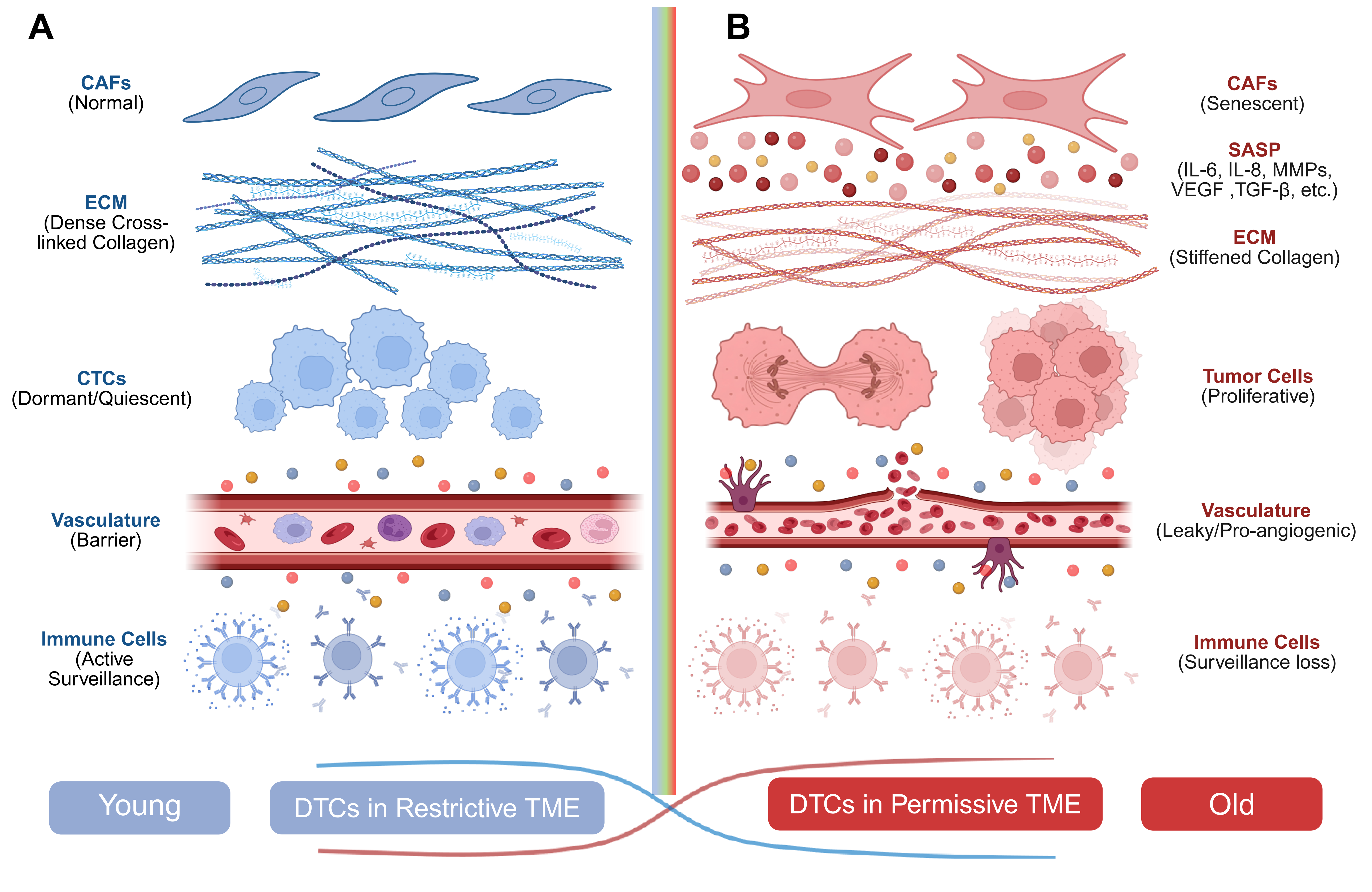
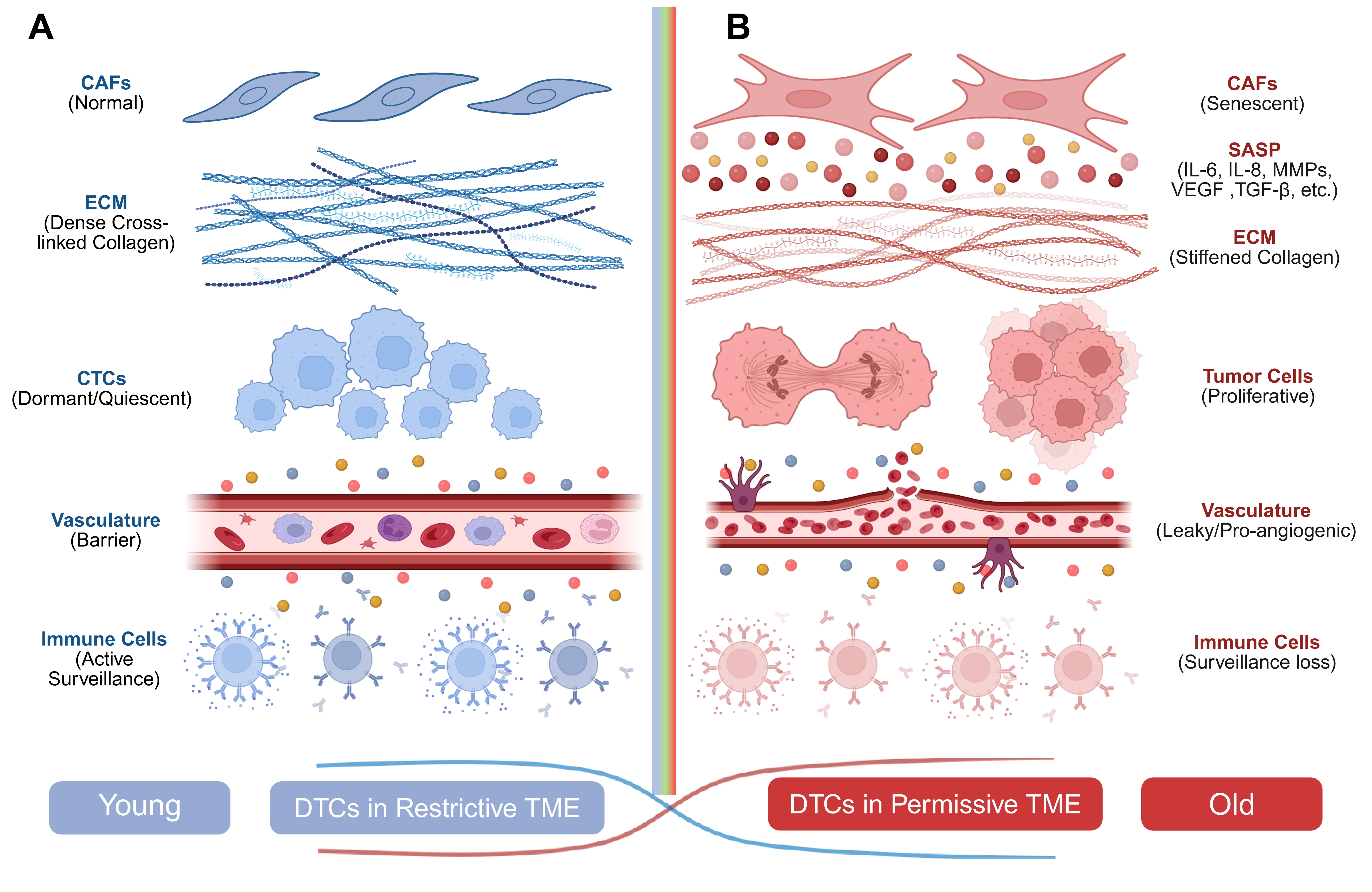
Figure 1. Comparison of the tumor microenvironment in young and aged tissues. (A) Young tissue microenvironment. In young tissues, stromal and immune components cooperate to maintain a restrictive microenvironment that supports DTC dormancy. CAFs preserve a dense and highly cross-linked ECM, intact vasculature maintains endothelial barrier integrity, and effective immune surveillance mediated by NK cells and T lymphocytes constrains tumor cell dissemination and outgrowth. Together, these features establish a dormancy-permissive niche that favors long-term tumor cell quiescence; (B) Aged tissue microenvironment. In aged tissues, the accumulation of senescent stromal cells promotes the secretion of SASP factors, including IL-6, IL-8, MMPs, VEGF, TGF-β, and FGFs. Concurrently, aging is associated with ECM remodeling and altered tissue mechanics, vascular dysfunction and increased angiogenic activity, and progressive impairment of immune surveillance. Collectively, these aging-associated changes transform the tumor microenvironment from a restrictive, dormancy-supportive state into a permissive microenvironment that promotes dormant tumor cell reactivation, metastatic outgrowth, and cancer recurrence. Created in BioRender. Cao, M. (2026) https://BioRender.com/3eby823. DTC: disseminated tumor cell; CAFs: cancer-associated fibroblasts; ECM: extracellular matrix; NK: natural killer; SASP: senescence-associated secretory phenotype; MMPs: matrix metalloproteinases; VEGF: vascular endothelial growth factor; TGF-β: transforming growth factor-β; FGFs: fibroblast growth factors; CTCs: circulating tumor cells; IL-6: interleukin-6; IL-8: interleukin-8.
2. Dormant Tumor Cells and Their Molecular Regulation
Tumor dormancy is a heterogeneous phenomenon that can broadly be categorized into angiogenic dormancy (AD)[14], immune-mediated dormancy (ID)[15], and tumor cell dormancy (TCD). Angiogenic and immune-mediated dormancy primarily reflect population-level equilibrium between tumor cell proliferation and elimination, whereas TCD represents a cell-intrinsic quiescent state that is most closely associated with metastatic latency and late recurrence. Accordingly, this review focuses primarily on cellular dormancy and its relationship with DTCs persistence and metastatic relapse.
Dormant tumor cells are characterized by a reversible growth arrest, typically associated with entry into the G0 or early G1 phase of the cell cycle, and therefore share key features with cellular quiescence. However, quiescence generally represents a transient and readily reversible state of cell-cycle arrest, whereas dormancy can remain quiescent for years or even decades. Consequently, tumor dormancy is recognized as a more deeply enforced and epigenetically stabilized form of growth arrest that enables long-term survival while preserving the capacity for subsequent reactivation[16-18]. A central molecular hallmark of dormancy is an increased ratio of p38 mitogen-activated protein kinase (MAPK) to signal-regulated kinase 1/2 (ERK1/2) signaling activity, which favors stress adaptation and dormancy over proliferation[19]. Environmental stressors, including extracellular matrix (ECM) remodeling, growth factor deprivation, hypoxia, and endoplasmic reticulum (ER) stress, can activate p38-dependent dormancy programs and maintain TCD. Among the transcriptional regulators implicated in dormancy, NR2F1 (COUP-TF1), an orphan nuclear receptor, has emerged as a master regulator in models of head and neck squamous cell carcinoma, breast cancer, and prostate cancer[20]. In proliferating tumor cells, NR2F1 is frequently silenced through promoter methylation. However, under dormancy-supportive microenvironmental conditions, retinoic acid receptor signaling and transforming growth factor-β2 (TGF-β2) can reverse this epigenetic repression and restore NR2F1 expression. NR2F1 induces a SOX9-RARβ-p27 transcriptional program that promotes global chromatin compaction and upregulation of the pluripotency-associated factor NANOG, thereby establishing a deeply quiescent, stem-like, and therapy-resistant cellular state[20]. The clinical relevance of NR2F1 has been validated in DTC-positive breast cancer cohorts, in which patients who relapsed within 12 months exhibited less than 1% NR2F1-high DTCs, whereas higher proportions of NR2F1+ DTCs were strongly associated with prolonged disease-free survival[21]. In addition to transcriptional reprogramming, dormant tumor cells also undergo profound metabolic and phenotypic adaptations that support long-term survival. For example, dormant breast cancer cells utilize sustained autophagic flux to survive during colonization of distant tissues[22]. Similarly, mesenchymal-like dormancy programs driven by factors such as the zinc-finger protein ZFP281 have been identified in early disseminated breast cancer cells within the lung microenvironment[23]. Together, TCD is maintained through coordinated transcriptional, epigenetic, and metabolic adaptations.
3. Dormant Tumor Cells Resides in Specific Tissue Niches
Increasing evidence indicates that tumor dormancy is an actively maintained biological program regulated through dynamic interactions between tumor cells and their surrounding microenvironment. Dormancy-supportive tissue niches provide complex biochemical and biomechanical signals. As a result, disseminated tumor cells will exhibit markedly different behaviors depending on the unique niche in which they reside.
3.1 Bone marrow niche
The bone marrow represents the most extensively characterized reservoir for dormant tumor cells and harbors DTCs in up to 40% of breast cancer patients at diagnosis[24,25]. The dormancy-supportive architecture of this niche has been dissected in considerable detail: CXCL12-CXCR4 signaling specifically directs DTCs homing toward dormancy-supportive perivascular regions where NG2+/Nestin+ perivascular mesenchymal stem cells (pMSCs) secrete TGF-β2 and bone morphogenetic protein 7 (BMP7), which engage transforming growth factor beta receptor type III (TGFBRIII) and bone morphogenetic protein receptor type II (BMPRII) on DTCs and subsequently activate the canonical p38-p27 dormancy pathway[26]. Importantly, dormant tumor cells also actively remodel their surrounding niche to stabilize dormancy. DTCs deposit ECM enriched in type III collagen, which activates discoidin domain receptor 1 (DDR1) signaling and establishes a signal transducer and activator of transcription 1 (STAT1)-dependent positive feedback loop that further reinforces dormancy[27]. In this context, type III collagen structures are characteristic of dormant DTCs, whereas type I collagen fibers are associated with metastatic reactivation. This further self-reinforcing ECM produced by DTCs in the bone marrow niche represents a striking example of how DTCs hijack tissue homeostatic programs to support long-term survival (Figure 2A).
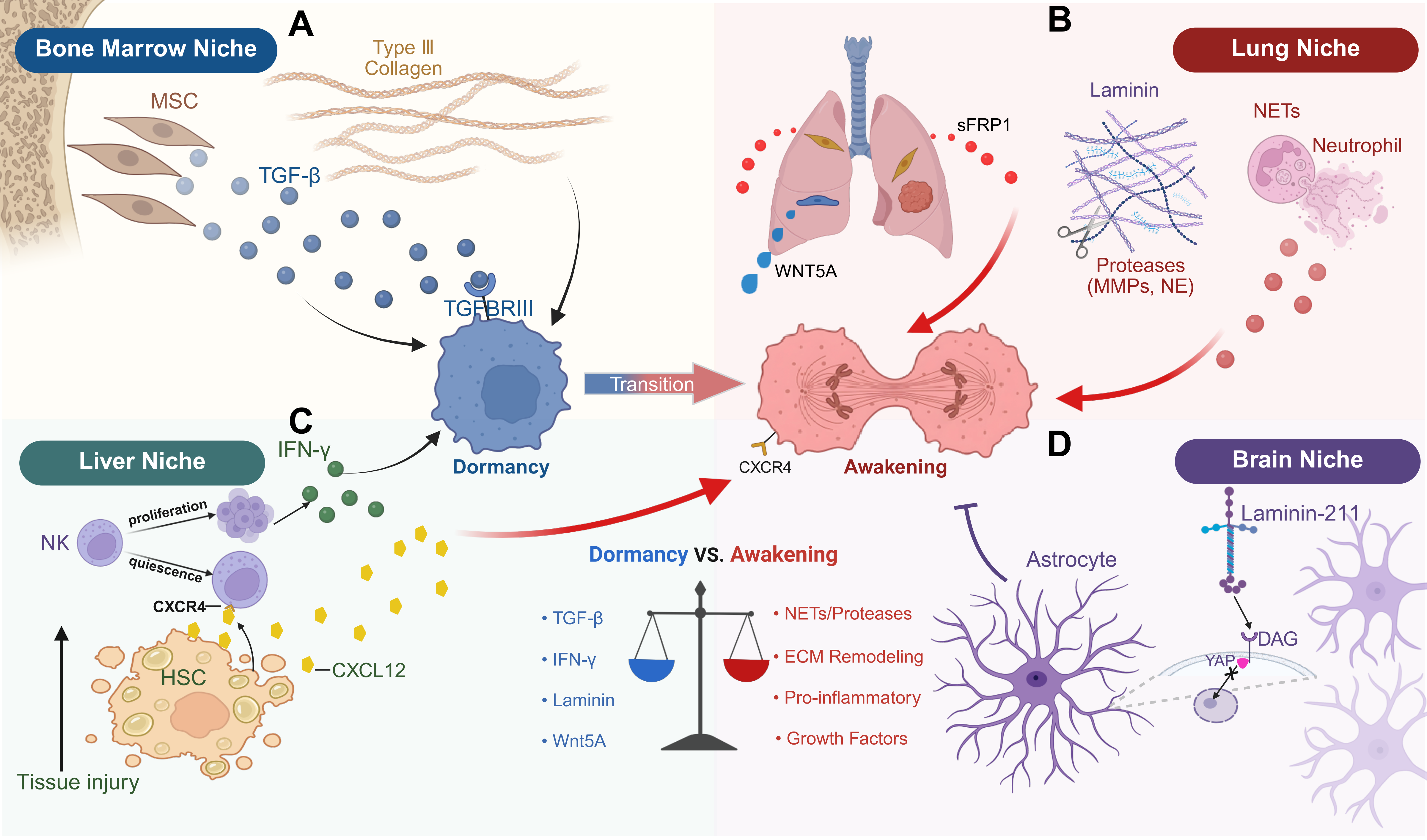
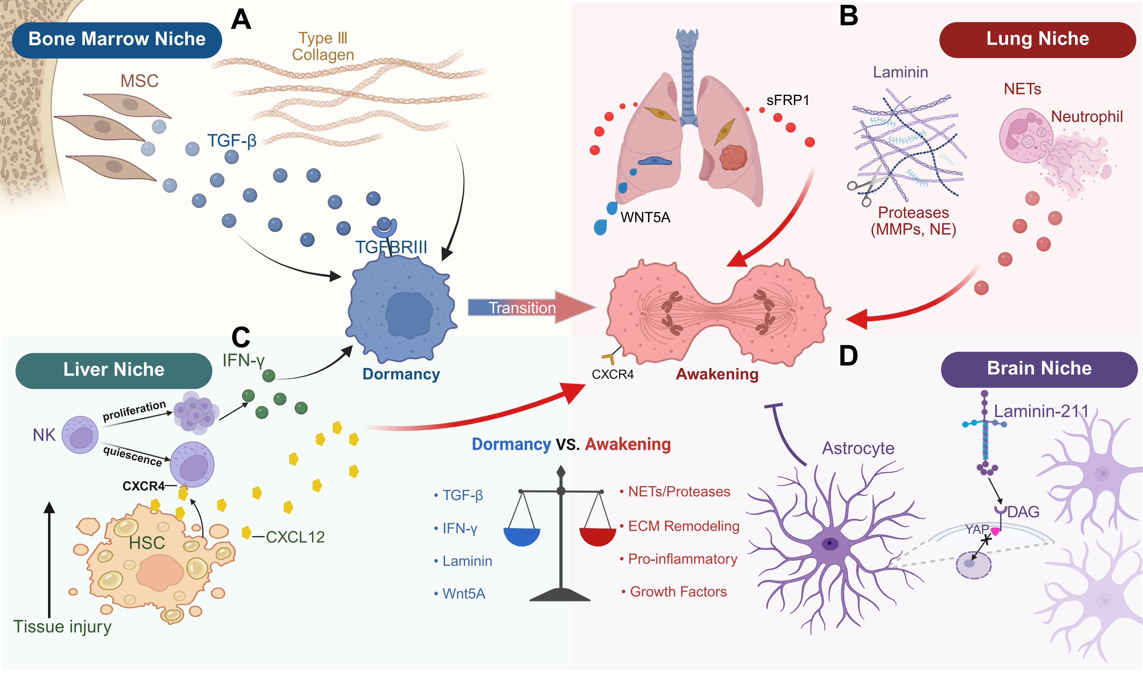
Figure 2. Organ-specific metastatic niches regulate tumor dormancy and awakening. (A) Bone marrow niche. Perivascular MSCs secrete TGF-β2, BMP7, and other dormancy-promoting factors that activate quiescence-associated signaling pathways in DTCs. In parallel, a type III collagen-rich extracellular matrix reinforces dormancy through integrin- and DDR1-dependent signaling; (B) Lung niche. In the pulmonary microenvironment, fibroblast-derived sFRP1 suppresses WNT5A-dependent dormancy programs and promotes metastatic outgrowth. Inflammatory stimuli induce the formation of NETs, whose associated proteases remodel laminin and expose integrin-binding sites that activate pro-proliferative signaling pathways, thereby triggering dormancy escape and metastatic reactivation; (C) Liver niche. Hepatic stellate cells support NK cell-mediated immune surveillance and maintain a dormancy-permissive environment. Sustained production of IFN-γ by liver-resident NK cells contributes to the long-term maintenance of DTCs quiescence, whereas stellate cell activation can disrupt this equilibrium and facilitate metastatic awakening; (D) Brain niche. Astrocyte-derived laminin-211 binds DAG receptors on DTCs, sequestering YAP in the cytoplasm and preventing its nuclear activation, thereby enforcing cellular quiescence. The unique biochemical and biomechanical properties of the brain microenvironment further support dormancy maintenance. Collectively, organ-specific stromal, immune, and extracellular matrix components provide distinct regulatory cues that govern the balance between dormancy maintenance and metastatic reactivation. Created in BioRender. Cao, M. (2026) https://BioRender.com/556e809. MSCs: mesenchymal stem cells; TGF-β2: transforming growth factor-β2; BMP7: bone morphogenetic protein 7; DTCs: disseminated tumor cells; DDR1: discoidin domain receptor 1; NETs: neutrophil extracellular traps; NK: natural killer; IFN-γ: interferon gamma; DAG: dystroglycan; YAP: yes-associated protein; ECM: extracellular matrix; MMPs: matrix metalloproteinases.
3.2 Lung niche
The lung is a major reservoir for late metastatic recurrence in breast cancer, melanoma, and sarcoma, and also provides a comparatively heterogeneous microenvironment for tumor dormancy (Figure 2B). Under physiological conditions, perivascular ECM niches enriched in type IV collagen, laminin, and nidogens support DTCs dormancy[14], as well as tumor cell-intrinsic signaling pathways through which WNT5A–AXL signaling maintains dormancy in melanoma DTCs and ZFP281 programs a mesenchymal-dictated dormancy in early disseminated breast cancer cells[3,23]. Notably, the pulmonary dormancy niche is highly vulnerable to inflammatory disruption. For instance, persistent inflammation stimulates neutrophils to release neutrophil extracellular traps (NETs), whose DNA scaffolds present neutrophil elastase and MMP-9 in close proximity to laminin-111. Sequential proteolytic cleavage of laminin-111 exposes a cryptic integrin α3β1-binding epitope, which subsequently activates the focal adhesion kinase (FAK)–extracellular signal–regulated kinase (ERK)–myosin light chain kinase (MLCK)–yes-associated protein (YAP) signaling cascade and drives dormant tumor cell reactivation[28]. In lung adenocarcinoma DTCs, stimulator of interferon genes (STING) signaling is epigenetically suppressed during dormancy through promoter and enhancer hypermethylation, but becomes reactivated during cell-cycle re-entry. Elevated STING activity subsequently promotes T cell- and natural killer (NK) cell–mediated elimination of cells exiting dormancy, thereby functioning as a tumor cell-intrinsic checkpoint that restricts metastatic outgrowth[29].
3.3 Liver and brain niches
The hepatic dormancy niche is strongly dependent on immune-mediated equilibrium, particularly surveillance by liver-resident NK cells[15]. Correia et al. demonstrated that liver-resident NK cells maintain dormancy of triple-negative breast cancer DTCs through sustained interferon gamma (IFN-γ) secretion: quiescent hepatic stellate cells, characterized by abundant intracellular lipid droplets, preserve the homeostatic microenvironment necessary to support NK cell-mediated surveillance; however, activated stellate cells by chronic liver injury secrete CXCL12, which engages CXCR4 on NK cells and induces NK-cell quiescence, ultimately reducing the hepatic NK-cell population and permitting dormant tumor cells to re-enter the cell cycle and initiate metastatic outgrowth[15] (Figure 2C). In contrast, the brain establishes a highly specialized dormancy-supportive microenvironment characterized by high laminin content, soft biomechanical properties, and the presence of an intact blood–brain barrier. For example, astrocyte-derived laminin-211 provides a specialized dormancy signal by binding to dystroglycan (DAG) receptors on DTCs, thereby sequestering YAP within the cytoplasm and preventing its nuclear translocation, ultimately enforcing cellular quiescence[30] (Figure 2D).
4. Ageing-Educated Microenvironment and Tumor Dormancy
The ageing-associated tumor microenvironment (aTME) undergoes profound and multifactorial remodeling during ageing. These alterations include the accumulation of senescent stromal cells accompanied by persistent secretion of senescence-associated secretory phenotype (SASP) factors[31-33], progressive immunosenescence that compromises NK cell and T cell surveillance[34,35], ECM remodeling that disrupts biomechanical dormancy signals, deterioration of vascular niche architecture[36,37], and inflammageing[38]. Rather than functioning independently, these processes interact dynamically to reshape tissue homeostasis and collectively influence DTCs’ fate.
4.1 Cellular senescence in the ageing-associated tumor microenvironment
Cellular senescence is now formally recognized as one of the twelve hallmarks of ageing[39,40]. Initially, cell senescence evolved as a protective tumor-suppressive mechanism that prevents the proliferation of damaged or stressed cells through stable cell-cycle arrest. With the chronic accumulation of senescent cells, it paradoxically exerts deleterious effects on tissue integrity and tumor control. A defining feature of senescent cells is the secretion of a complex repertoire of bioactive molecules collectively termed the SASP. The SASP includes pro-inflammatory cytokines such as interleukin (IL)-6, IL-8, and IL-1α; chemokines including CCL2, CCL5, and CXCL1; growth factors such as vascular endothelial growth factor (VEGF), hepatocyte growth factor (HGF), and platelet-derived growth factor (PDGF); as well as matrix-remodeling enzymes including matrix metalloproteinase (MMP)-1, MMP-3, and MMP-10[41,42]. During ageing, persistent SASP secretion establishes a state of chronic low-grade inflammation accompanied by ECM remodeling, vascular dysfunction, and the development of immunosuppressive microenvironments, all of which are supposed to affect tumor progression[1,39,43]. Accumulating evidence indicates that senescent stromal cells actively generate microenvironments favorable for tumor progression and metastatic dissemination. For example, senescent fibroblasts enhance melanoma metastasis by promoting angiogenesis[44], senescent osteoblasts facilitate breast cancer bone metastasis through IL-6-driven osteoclastogenesis[45-47], and senescent macrophages accelerate tumor progression through extensive remodeling of the tumor microenvironment[48,49]. Moreover, SASP factors released from non-transformed senescent stromal cells, particularly those accumulating during physiological tissue ageing rather than as a direct consequence of tumor formation, can establish permissive pre-metastatic niches that support DTCs’ survival and eventual reactivation[46,50]. Indeed, this early convergence between senescence and dormancy may critically influence the trajectory of metastatic relapse years or even decades later[51-53]. In addition, the relationship between ageing and dormancy extends beyond the metastatic niche itself. Tumors arising in aged hosts are likely to acquire an enhanced capacity to evade senescence-associated growth arrest during primary tumor evolution, and this phenotype may persist following dissemination, rendering DTCs less responsive to dormancy-inducing signals[54,55].
4.2 Therapy-induced senescence and oncogene-induced senescence
In addition to physiological ageing, therapy-induced senescence (TIS) and oncogene-induced senescence (OIS) represent two major sources of senescent cells. TIS, triggered by chemotherapy, radiotherapy, or targeted therapies, adds an additional layer of complexity to the clinical relevance of tumor dormancy[56]. For instance, the long-term persistence of therapy-induced senescent stromal cells, such as cancer-associated fibroblasts (CAFs), can progressively remodel the tumor microenvironment through sustained SASP secretion. This chronic SASP signaling promotes immune evasion, therapeutic resistance, extracellular matrix remodeling, and ultimately tumor recurrence[57]. Consequently, anti-cancer therapies may generate a pro-recurrence microenvironment despite achieving initial tumor control. In contrast to TIS, OIS functions as an intrinsic barrier against malignant transformation during the earliest stages of tumorigenesis[58,59]. Activation of oncogenes such as RAS, BRAF, or MYC in otherwise normal cells initially induces a burst of proliferation, which is subsequently followed by stable cell-cycle arrest mediated primarily through the p16-INK4a–RB and p19-ARF–p53 tumor suppressor pathways[60,61]. Through this mechanism, OIS restricts pre-neoplastic expansion and prevents the progression of early lesions toward overt malignancy. Interestingly, OIS appears to become progressively compromised during ageing, a phenomenon often referred to as the age-associated bypass of OIS. Multiple ageing-associated alterations weaken OIS-mediated cell growth arrest and permit cells harboring oncogenic lesions to escape senescence and progress toward malignancy. Several studies have unraveled the mechanisms contributing to the age-dependent erosion of OIS. Extrinsically, pro-inflammatory cytokines and chemokines associated with SASP, including IL-6 and IL-8, secreted by neighboring senescent stromal cells, can enhance stem-like properties in adjacent premalignant cells, stimulate cell-cycle re-entry, and recruit immunosuppressive myeloid populations that impair immune-mediated clearance of OIS cells[62]. In parallel, intrinsic ageing-associated alterations further compromise OIS responsiveness. Progressive epigenetic drift impairs activation of the p16-INK4a pathway in response to oncogenic stress through DNA methylation at the CDKN2A locus, dysregulated Polycomb repressive complex activity, and loss of heterochromatin integrity[63]. In addition, ageing-associated mitochondrial dysfunction, chronic oxidative stress, and altered nutrient-sensing pathways, particularly aberrant mTOR activation, disrupt the balance between senescence induction and apoptotic elimination[64-66]. As a consequence, aged cells may enter a fragile “pre-senescent” or “quasi-senescent” state that retains the capacity to resume proliferation upon exposure to additional oncogenic or inflammatory stimuli.
4.3 Ageing-associated extracellular matrix remodeling
ECMs function not only as a structural scaffold, but also as active signaling platforms that provide integrin ligands, store growth factors, and transduce mechanosensory cues that profoundly influence cellular fate decisions. Ageing induces characteristic alterations in ECM composition, organization, and biomechanical properties, and these changes vary substantially across tissues, thereby generating organ-specific microenvironments that influence tumor progression, metastatic dissemination, and dormancy regulation. In the skin, aged fibroblasts generate profoundly altered secretory and ECM-remodeling programs. One of the most prominent alterations is the marked reduction of HAPLN1, a hyaluronan–proteoglycan linker protein that is essential for ECM crosslinking and structural integrity, in the secretome of aged fibroblasts[67,68]. Loss of HAPLN1 produces a more aligned and less crosslinked collagen matrix that simultaneously enhances melanoma cell migration and invasion while impairing T cell infiltration, thereby establishing a microenvironment that is both pro-invasive and immunosuppressive. Subsequent studies further demonstrated that reduced HAPLN1 expression in aged dermal fibroblasts increases endothelial ICAM1 expression, compromises vascular integrity, and facilitates systemic dissemination of tumor cells[69]. Age-associated ECM remodeling also plays a critical role in regulating dormancy escape within metastatic organs. In the lung, aged pulmonary fibroblasts exhibit a reprogrammed secretome enriched in sFRP1, a WNT signaling antagonist that suppresses WNT5A-mediated dormancy signaling in melanoma DTCs and promotes metastatic outgrowth through reactivation of the AXL–MER kinase axis[3,70]. Similarly, CAF-driven deposition of collagen I within metastatic microenvironments can reactivate dormant breast cancer cells through integrin β1–SRC–ERK signaling[49]. Collectively, these findings indicate that ageing-associated ECM remodeling is not merely a passive consequence of tissue degeneration, but rather an active regulator of tumor dormancy and metastatic reactivation.
4.4 Ageing-associated vascular remodeling
The vasculature constitutes a highly specialized niche that critically regulates both tumor dormancy and metastatic reactivation. Stable and quiescent microvasculature actively maintains breast cancer dormancy through secretion of thrombospondin-1 (TSP-1), whereas newly sprouting neovasculature produces periostin and TGF-β1, both of which promote metastatic outgrowth[14]. Thus, the structural and functional state of the vascular niche plays a central role in determining whether disseminated tumor cells remain dormant or resume proliferation. In the ageing bone marrow, reduced blood flow resulting from diminished PDGF signaling and progressive pericyte loss generates a hypoxic microenvironment that disrupts the pro-dormancy perivascular niche and facilitates dormancy escape[49]. In parallel, elevated expression of sFRP2 in aged dermal fibroblasts enhances angiogenesis in melanoma, thereby creating additional vascular routes that support metastatic dissemination[3,70]. More broadly, ageing transforms the vascular niche from a dormancy-maintaining environment into one that increasingly favors tumor progression through several coordinated mechanisms. These include reduced endothelial TSP-1 production associated with age-related neovascular remodeling[14], elevated secretion of pro-angiogenic SASP factors such as VEGF and fibroblast growth factor (FGF)[43,71], progressive loss of pericyte coverage[72], endothelial dysfunction[73], and deterioration of vascular integrity[74]. Such changes not only weaken dormancy-supportive signaling, but also enhance vascular permeability and inflammatory remodeling.
4.5 Ageing-associated stromal metabolic reprogramming
In addition to inflammatory and structural remodeling, ageing also imposes profound metabolic alterations on stromal cells. These changes extend beyond SASP production and include dysregulated lipid metabolism[75-77], mitochondrial dysfunction[78-80], and altered utilization of cellular energy substrates[81,82]. One representative example has been identified in aged dermal fibroblasts, which exhibit enhanced secretion of neutral lipids, particularly ceramides. Melanoma cells can take up these lipids through the fatty acid transporter FATP2, thereby acquiring resistance to BRAF/MEK-targeted therapy-induced cell death[83]. This observation directly links ageing-associated stromal metabolic reprogramming to therapeutic resistance and tumor recurrence in elderly patients. Additionally, senescent cancer-associated fibroblasts undergo characteristic metabolic rewiring marked by enhanced aerobic glycolysis and increased lactate secretion, which supports tumor cell survival, invasion, and metastatic progression[84-86]. Beyond serving as alternative energy substrates, metabolites released by aged stromal cells may also influence redox balance, epigenetic regulation, and stress-adaptation pathways within DTCs[87-90], thereby promoting long-term survival under hostile microenvironmental conditions. These findings suggest that stromal ageing is not merely accompanied by metabolic decline, but rather by active metabolic reprogramming that generates nutrient-rich and stress-adaptive microenvironments favorable for tumor persistence.
5. Ageing and Metastatic Awakening
As a systemic and progressive biological process, ageing introduces chronic stressors that are largely absent in young tissues, including persistent SASP-associated inflammation, fibrotic ECM remodeling, vascular dysfunction, metabolic rewiring, and myeloid-biased hematopoiesis. Consequently, the balance between dormancy maintenance and metastatic reactivation gradually shifts toward tumor outgrowth over time. Understanding how ageing-associated microenvironmental remodeling influences each stage of dissemination, dormancy, and metastatic awakening is therefore essential for developing strategies aimed at preventing dormancy-associated cancer recurrence.
5.1 The effect of ageing on the primary site: Pre-metastatic conditioning
For solid tumors, stromal ageing begins to reshape both the primary tumor microenvironment and future metastatic niches long before DTCs are generated. As mentioned above, aged fibroblasts deposit a profoundly remodeled ECM characterized by reduced HAPLN1 and hyaluronan content, altered collagen organization, and enhanced fibrillar alignment, collectively creating a structurally and biochemically permissive environment for tumor invasion[68]. At the same time, SASP promotes phenotypic switching from proliferative to invasive cellular states for tumor cells, thereby effectively selecting for tumor cells with dissemination competence prior to metastatic escape[91]. At another level, ageing-associated inflammation and stromal senescence also initiate one of the earliest ageing-dependent regulatory axes, namely SASP-educated future metastatic niches, which occurs earlier before DTCs colonize distant organs. Briefly, elevated systemic levels of inflammatory mediators such as IL-6 and IL-8 derived from aged stroma increase chronic inflammatory tone[31,43,92], while myeloid-derived suppressor cells (MDSCs) and M2-polarized macrophages are recruited to pre-metastatic sites[93-95]. Concurrently, chronic rewiring of TGF-β signaling shifts tissue homeostasis away from the dormancy-supportive TGF-β2 signature maintained by healthy mesenchymal stem cells and toward a pro-proliferative TGF-β1-dominant environment[49]. Consequently, tumor cells disseminating in elderly hosts encounter distant organs that have already undergone substantial remodeling in favor of metastatic survival and reactivation (Figure 3A).
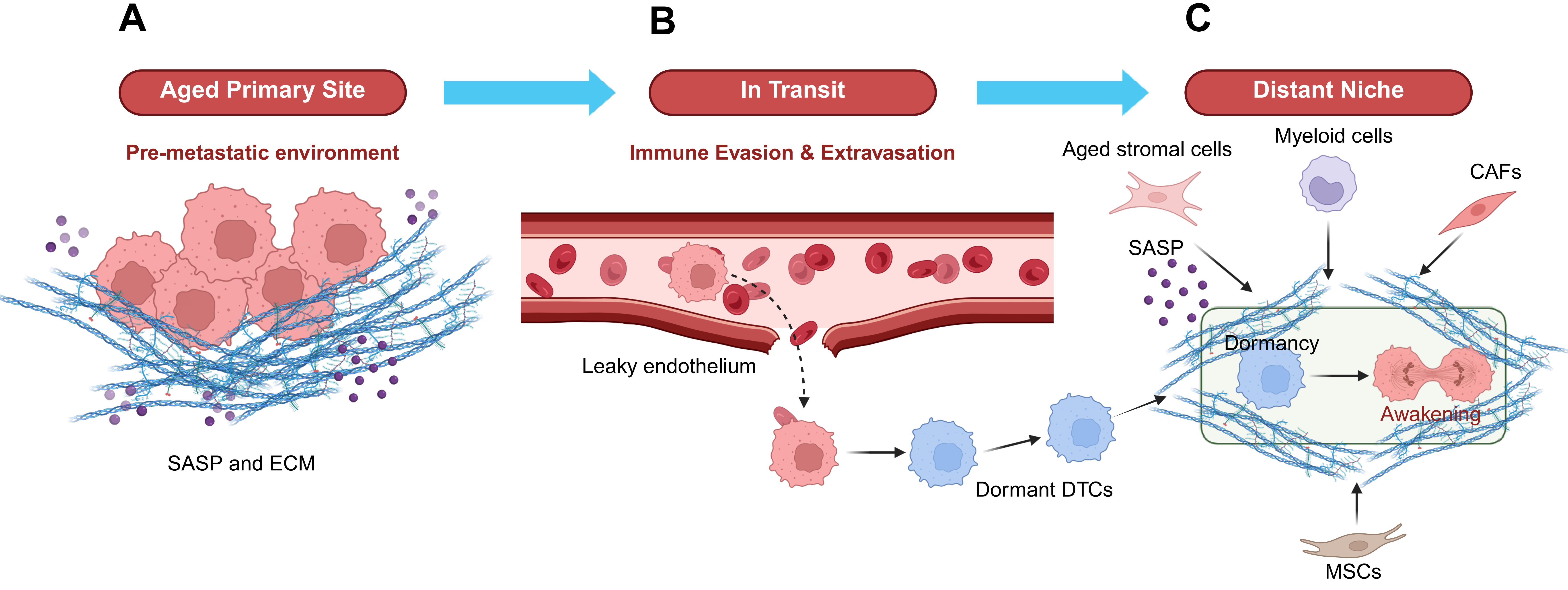
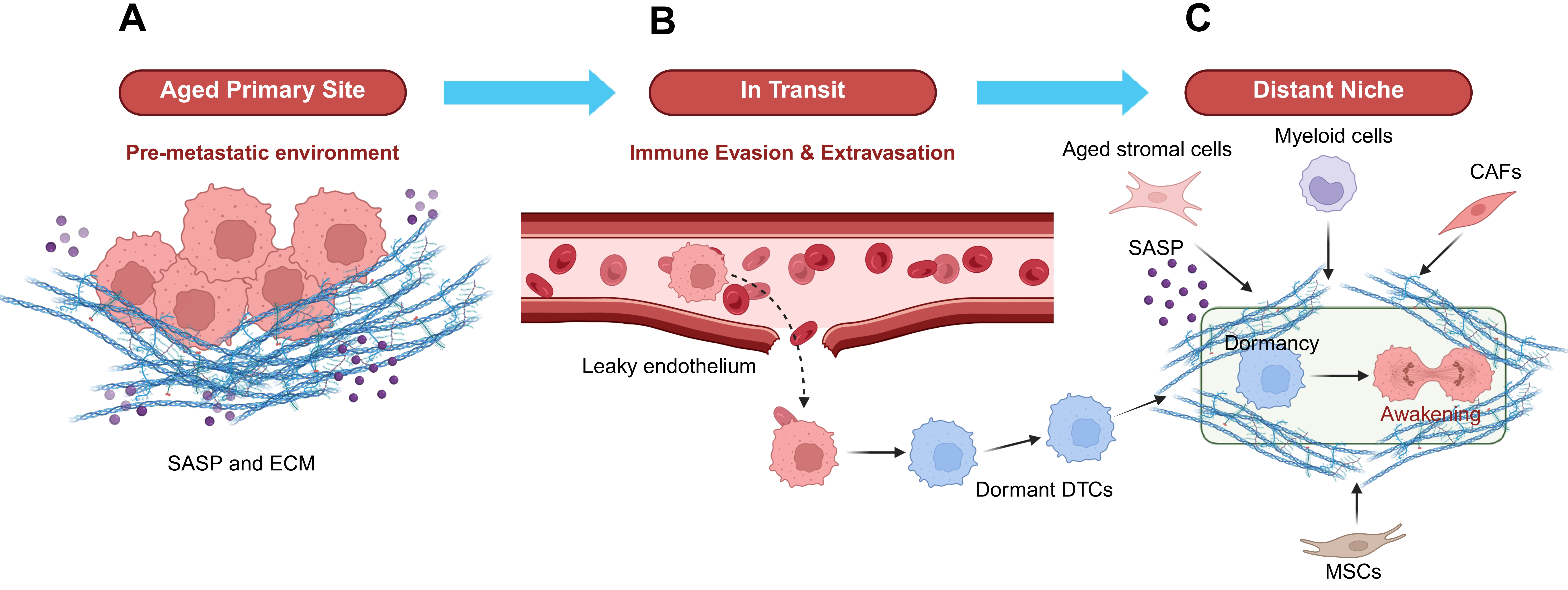
Figure 3. Ageing-driven disruption of tumor dormancy and metastatic awakening. (A) Primary tumor site. Aging-associated accumulation of senescent stromal cells promotes the secretion of SASP factors and drives ECM remodeling, thereby generating a pro-invasive microenvironment that facilitates tumor cell dissemination and pre-metastatic niche conditioning; (B) Systemic transit. During hematogenous dissemination, age-related endothelial dysfunction, increased vascular permeability, and progressive decline in immune surveillance enhance CTC survival, extravasation, and colonization of distant organs; (C) Metastatic niche. Following seeding, DTCs enter a dormant state within tissue-specific microenvironments. As aging progresses, the accumulation of senescent stromal cells, CAFs, myeloid-derived suppressive cells, and SASP-associated inflammatory mediators progressively disrupts dormancy-supportive signaling networks. This aging-driven erosion of dormancy-maintaining mechanisms ultimately shifts the balance from quiescence toward metastatic reactivation, resulting in metastatic outgrowth and late cancer recurrence. Overall, aging influences every stage of the metastatic cascade, from primary tumor evolution and systemic dissemination to dormancy maintenance and metastatic awakening, thereby increasing the likelihood of late cancer recurrence. Created in BioRender. Cao, M (2026) https://BioRender.com/wtxekx5. SASP: senescence-associated secretory phenotype; ECM: extracellular matrix; CTC: circulating tumor cell; DTCs: disseminated tumor cells; CAFs: cancer-associated fibroblasts.
5.2 Ready to go: Circulating tumor cells (CTCs) traverse an aged vascular compartment
Between primary tumor invasion and distant organ colonization lies the transient yet critical stage of CTCs. Survival within the circulation is normally constrained by multiple hostile factors, including shear stress, anoikis, NK cell-mediated cytotoxicity, and complement-dependent lysis. Importantly, many of these constraints become progressively weakened during ageing. NK cell cytotoxic function for clearance of CTCs declines substantially with ageing[96,97]. In parallel, ageing-associated vascular remodeling alters properties of the circulation through which CTCs travel, including endothelial dysfunction, thinning of endothelial barriers, and altered leukocyte adhesion dynamics[98]. Furthermore, HAPLN1-dependent disruption of CDH5-mediated endothelial junctions renders aged vasculature more permeable, while concomitant upregulation of ICAM1 on aged endothelial cells provides additional adhesive substrates that facilitate CTCs attachment and transendothelial migration at sites that would otherwise remain restrictive in younger tissues[69,99]. Importantly, this CTC stage also represents the most clinically accessible diagnostic window throughout the metastatic cascade. Liquid biopsy strategies based on serial CTC enumeration, molecular characterization, and integrated circulating tumor DNA (ctDNA) analysis can be readily implemented in elderly cancer patients[100] (Figure 3B). When combined with ageing-associated biomarker panels, these approaches may enable individualized risk stratification that has not yet been systematically incorporated into current surveillance paradigms[101-103]. Longitudinal CTC monitoring in elderly patients may provide one of the earliest actionable indicators of ongoing metastatic dissemination.
5.3 The effect of ageing on DTCs seeding, dormancy, and awakening
Following extravasation, disseminated tumor cells interact extensively with the microenvironment of the recipient organ, and the nature of this interaction ultimately determines whether DTCs enter long-term dormancy or initiate micrometastatic outgrowth (Figure 3C). In young hosts, intact tissue microenvironments provide coherent and robust dormancy-inducing signals. Examples include TSP-1 secreted by stable perivascular endothelial cells[104], TGF-β2 and BMP7 produced by NG2+/Nestin+ pMSCs in the bone marrow[26], laminin-211 derived from astrocytes within the brain[30], and CXCL12-mediated regulation of NK cell quiescence by activated hepatic stellate cells through CXCR4 signaling in the liver[15]. These niche-derived signals converge on DTCs to establish a coordinated regulatory network that maintains tumor cells in a viable yet non-proliferative state for prolonged periods, potentially spanning decades. With ageing, however, these dormancy-supportive signals become progressively weakened through two major regulatory mechanisms: ECM proteolytic remodeling and organ-specific stromal reprogramming. ECM proteolytic remodeling appears to function as a common downstream executor of dormancy escape across multiple tissues. In the aged lung, collagen I deposited by senescent CAFs engages integrin β1 on DTCs and activates SRC–ERK signaling to override the p38 pathway[36]. In the bone marrow, SASP factors secreted by senescent osteoblasts include RANKL, which stimulates osteoclast activation and subsequently releases matrix-sequestered TGF-β1 and IGF-1, directly promoting DTCs proliferation[105]. Moreover, aged fibroblast-derived sFRP1 suppresses WNT5A-dependent melanoma DTCs dormancy and activates the AXL–MER signaling axis to promote metastatic reactivation[3]. Similarly, aged and fibrotic pulmonary tissues in ERα+ breast cancer accumulate PDGF-C, which activates PDGFRα signaling in DTCs and promotes reactivation[106]. In the liver, ageing-associated activation of hepatic stellate cells disrupts NK cell-mediated dormancy maintenance through CXCL12-driven induction of NK cell quiescence[15]. Notably, ageing does not universally promote metastatic outgrowth. Crist et al. demonstrated that excessive oxidative stress within aged skeletal muscle can instead eliminate DTCs[107], highlighting that certain aTMEs may remain hostile to tumor cell survival.
6. Targeting Tumor Dormancy and Metastatic Awakening
Given the central role of the aTME in regulating DTCs dormancy and reactivation, therapeutic strategies aimed at preventing late-stage recurrence can be broadly categorized into three major directions: biomarker-guided risk stratification, inhibition of dormancy escape signals, and direct targeting of dormant DTCs. At the distant metastatic niche stage, biomarker development becomes increasingly tissue-specific and mechanistically informative. Among currently available candidates, NR2F1 profiling in bone marrow DTCs represents one of the most clinically validated biomarkers of tumor dormancy. Integration of NR2F1 status with ageing-associated biomarkers, including composite SASP signatures, circulating IL-6 and C-reactive protein (CRP) levels, biological ageing clocks, longitudinal ctDNA dynamics, and CTC-based liquid biopsy analyses, may enable multilayered and individualized risk stratification that substantially exceeds the predictive value of any single biomarker alone. Such approaches may be particularly valuable for identifying elderly patients at high risk of late metastatic recurrence long before overt relapse becomes clinically detectable. Therapeutic strategies targeting the distant metastatic niche can be conceptually aligned with the two principal niche-stage regulatory mechanisms described above and are complementary to earlier senolytic interventions. Among the currently proposed candidates, PDGFRα inhibition in ER α+ breast cancer patients at high risk for pulmonary recurrence represents one of the most strongly validated strategies[106]. Additional promising approaches include modulation of the WNT5A–AXL signaling axis in melanoma and CXCR4 blockade aimed at preserving hepatic NK cell surveillance, both of which warrant further clinical investigation.
More directly, by eliminating senescent stromal cells during or after primary tumor treatment, senolytic therapy is considered to reduce the systemic pre-conditioning burden and attenuate the downstream SASP-driven effects that facilitate future DTC reactivation. He et al.[57] demonstrated that the senolytic combination of dasatinib and quercetin prevented DTCs reactivation in preclinical models after chemotherapy, a finding that directly justifies trials of senolytic combinations as adjunctive therapy in elderly patients receiving conventional cytotoxic treatment. In addition, Zhou et al. demonstrated that p21+ senescent CAFs drive CD8+ T cell dysfunction and immune suppression in prostate cancer, whereas senolytic treatment reversed this immunosuppressive phenotype and restored antitumor immune responses[32]. These findings suggest that senolytics can function not only as modulators of SASP-driven dormancy disruption but also as immunological priming agents that improve the immune competence required for immune checkpoint blockade. Consistent with this concept, preclinical studies in aged tumor models showed that senolytic treatment enhanced the efficacy of anti-PD-1 therapy by alleviating tumor-associated immunosenescence, increasing CCR7+ naïve T-cell populations, and reversing immunosenescence-associated transcriptional programs in both T- and B-cell subsets[108]. Together, these findings support a rational sequential therapeutic strategy in which senolytic treatment first clears the immunosuppressive senescent stromal compartment, thereby creating a more permissive immune microenvironment, followed by immune checkpoint blockade to enhance NK- and T-cell-mediated elimination of dormant DTCs and reactivating micrometastases. Although this therapeutic strategy is supported by compelling preclinical evidence, few clinical trials have formally evaluated whether senescence clearance enhances the efficacy of immunotherapy[109]. Addressing this gap should therefore be a priority for future clinical trial design.
A second therapeutic direction aims to actively reinforce dormant states rather than merely preventing their disruption. In this context, pharmacological stabilization of dormancy-associated transcriptional programs has emerged as an attractive strategy. For example, the NR2F1 agonist C26 suppresses metastatic outgrowth by enforcing a stable NR2F1-hi/p27-hi dormancy program in disseminated tumor cells[110]. Similarly, combined treatment with 5-azacytidine and all-trans retinoic acid reprograms DTCs into an NR2F1-dependent quiescent state through restoration of TGF-β–SMAD4 signaling[111]. CDK4/6 inhibitors may further reinforce dormancy-associated cell-cycle arrest through partially overlapping quiescence programs[112,113].
Ultimately, the most ambitious therapeutic objective is the selective eradication of dormant DTCs before metastatic reactivation occurs. Proof-of-concept evidence supporting this strategy has already emerged from CAR-T cell studies demonstrating approximately 98% clearance of dormant pulmonary DTCs[114]. CAR-NK cell therapy then offers a complementary approach with several potential advantages for elderly patients, including a lower risk of cytokine release syndrome and immune effector cell-associated neurotoxicity, as well as the feasibility of off-the-shelf allogeneic products[115,116]. These features may be particularly relevant for older patients, who often have limited tolerance for intensive cellular therapies and may experience reduced manufacturing success with autologous CAR-T cells because of age-associated T-cell dysfunction. Future development of immune cell-based strategies for dormant DTC eradication should therefore incorporate age-specific considerations of safety, efficacy, and manufacturability, while further evaluating the therapeutic potential of allogeneic CAR-NK platforms.
Collectively, therapeutic interventions targeting tumor dormancy will likely require integration with ageing-informed patient stratification and long-term surveillance systems. Combined approaches incorporating senolytic therapies, dormancy-reinforcing agents, immune modulation, and liquid biopsy-based monitoring may ultimately provide a feasible framework for preventing late metastatic recurrence in elderly cancer patients.
7. Discussion and Perspectives
7.1 Limitations of current models on studying ageing and tumor dormancy
Perhaps the most significant methodological limitation hindering progress in tumor dormancy research is the routine use of young adult mice, typically 8-12 weeks of age, despite the fact that cancer recurrence predominantly occurs in patients during the sixth to ninth decades of life[117,118]. As discussed throughout this review, the aged microenvironment regulates dormant DTCs behavior through mechanisms that are largely absent in young hosts, including contraction of MSC quiescence-supportive pools, fibrotic ECM remodeling, impaired NK cell surveillance, vascular dysfunction, and SASP-dominated inflammatory secretomes[1,119]. Consequently, dormancy-modifying interventions validated in young animals are frequently evaluated within biological contexts that poorly reflect the physiology of the patient populations most likely to receive such therapies[120]. The practical limitations associated with aged-cohort studies are substantial and should be explicitly acknowledged. Mortality and attrition rates increase considerably in mice older than 52 weeks, which leads to substantially higher maintenance costs compared with young cohorts. Indeed, beyond 24 months of age, overall experimental costs may approach an order of magnitude above standard catalog pricing[121]. In addition, biological heterogeneity progressively increases with ageing, thereby reducing statistical power and necessitating larger cohort sizes to achieve equivalent effect-size detection[122,123]. Nevertheless, these challenges represent manageable experimental design issues rather than fundamental biological barriers. Importantly, Henry and DeGregori recently proposed a comprehensive framework for aged-cohort dormancy studies, including recommendations regarding strain selection, attrition-adjusted power calculations, and incorporation of genetically diverse mouse populations[118]. Adoption of such age-aware experimental frameworks as field-wide methodological standards represents a critical step toward establishing dormancy models that more accurately reflect the biological context of elderly cancer patients.
7.2 Emerging technologies and future experimental platforms
Organoid-based systems represent another highly promising yet currently underutilized technology for studying ageing-associated tumor dormancy. Patient-derived organoids from colorectal and prostate cancers have already been cultured under dormancy-inducing conditions, and organoid models of castration-resistant prostate cancer dormancy have recently been established[124,125]. In contrast, comparable patient-derived dormancy organoid systems for breast cancer remain underdeveloped, with most studies still relying heavily on established cancer cell lines[126]. Importantly, currently available organoid protocols generally utilize stromal components derived from young or middle-aged donors and have not systematically incorporated aged stromal elements. In parallel, single-cell and spatial profiling technologies have profoundly transformed the resolution at which tumor dormancy and its surrounding microenvironment can be interrogated. Bulk transcriptomic analyses inherently obscure the cellular heterogeneity that likely governs DTCs fate at the single-cell level[127]. Whether an individual DTCs encounters a dormancy-supportive or dormancy-disruptive niche depends critically on its precise spatial localization within tissues, a question inaccessible to population-averaged approaches[128]. Recent advances in spatial transcriptomics, including Visium, MERFISH, and Slide-seq, now permit characterization of the cellular architecture surrounding NR2F1+ dormant DTCs at near-single-cell resolution[129-131]. Integrated single-cell multi-omics approaches combining scRNA-seq, scATAC-seq, and spatial proteomic profiling can simultaneously characterize the transcriptional, epigenetic, and signaling states of dormant DTCs together with their stromal neighbors[132-134]. The major remaining challenge is systematic application of these technologies to paired young-versus-aged metastatic niches in order to define the ageing-specific cellular neighborhoods that progressively destabilize dormancy maintenance.
7.3 A conceptual framework for ageing-associated dormancy erosion
The interplay among SASP-mediated disruption, ECM proteolytic remodeling, vascular destabilization, immunosenescence, and organ-specific stromal reprogramming can be conceptualized as a “dormancy resilience threshold.” In young tissues, multiple redundant dormancy-maintaining mechanisms cooperate to maintain DTCs dormancy above the threshold required for long-term growth arrest. However, with ageing, each regulatory axis progressively shifts toward disruption, and the cumulative weakening of these protective mechanisms eventually reduces the system below the dormancy maintenance threshold, thereby permitting metastatic reactivation. This framework further predicts that recurrence risk in cancers characterized by prolonged dormancy should not follow simple exponential decay kinetics, but instead progressively increase over time as a consequence of cumulative ageing-associated niche deterioration rather than purely stochastic reactivation events. Clinical epidemiological observations strongly support this model. ERα+ breast cancer exhibits persistent recurrence risk extending over 15-20 years of follow-up[135-137], while prostate cancer bone metastases are similarly characterized by exceptionally long latency periods[138]. Importantly, acute perturbations, including chronic pulmonary inflammation induced by tobacco smoke or lipopolysaccharide exposure[28], surgical injury, systemic infection, or even cytotoxic chemotherapy itself, may trigger dormancy escape against this progressively deteriorating microenvironmental background, thereby contributing substantially to the clinical metastatic recurrence.
8. Conclusions
The proposition that ageing plays a mechanistic, rather than merely associative, role in tumor dormancy and cancer recurrence is now supported by a rapidly expanding body of experimental and clinical evidence. Ageing regulates dormancy not through a single linear pathway, but through convergence of multiple interconnected biological processes. Importantly, therapy-induced senescence introduces an additional iatrogenic component that may paradoxically accelerate the very recurrence that anti-cancer treatment seeks to prevent. Together, these findings support the concept that metastatic recurrence is not simply determined by tumor-intrinsic genetics, but also by progressive ageing-dependent deterioration of dormancy-maintaining tissue environments. Currently, the convergence of geroscience and oncology offers a conceptual framework through which late metastatic recurrence may ultimately be approached as a preventable biological process rather than an inevitable consequence of cancer progression. Although substantial challenges remain, the technological and conceptual foundations necessary to achieve this goal are now rapidly emerging.
Acknowledgements
The authors declare that Claude Sonnet 4.6 was used solely for language polishing during the manuscript preparation process. The authors take full responsibility for the integrity, originality, and accuracy of the work.
Authors contribution
Wu F: Writing-original draft.
Meng Y, Cao M, Zhang J: Writing-review & editing.
Wu Y, Tang X: Conceptualization, writing-review & editing.
Conflicts of interest
The authors declare no conflicts of interest.
Ethical approval
Not applicable.
Consent to participate
Not applicable.
Consent for publication
Not applicable.
Availability of data and materials
Not applicable.
Funding
This study was supported by the Fundamental Research Funds for the Central Universities (Grant Nos. 521119200099, 541109030051 and 531119200268).
Copyright
© The Author(s) 2026.
References
-
5. Giancotti FG. Mechanisms governing metastatic dormancy and reactivation. Cell. 2013;155(4):750-764.[DOI]
-
6. Willis RA. The spread of tumours in the human body. 1st ed. & A. Churchil;1934:[DOI]
-
8. Peng P, Qin S, Li L, He Z, Li B, Nice EC, et al. Epigenetic remodeling under oxidative stress: Mechanisms driving tumor metastasis. MedComm–Oncology. 2024;3(4):e70000.[DOI]
-
9. Tao L, Zhou Y, Luo Y, Qiu J, Xiao Y, Zou J, et al. Epigenetic regulation in cancer therapy: From mechanisms to clinical advances. MedComm–Oncology. 2024;3(1):e59.[DOI]
-
17. Saleh T, Tyutyunyk-Massey L, Gewirtz DA. Tumor cell escape from therapy-induced senescence as a model of disease recurrence after dormancy. Cancer Res. 2019;79(6):1044-1046.[DOI]
-
19. Aguirre-Ghiso JA. Models, mechanisms and clinical evidence for cancer dormancy. Nat Rev Cancer. 2007;7(11):834-846.[DOI]
-
24. Braun S, Pantel K, Müller P, Janni W, Hepp F, Kentenich CRM, et al. Cytokeratin-positive cells in the bone marrow and survival of patients with stage I, II, or III breast cancer. N Engl J Med. 2000;342(8):525-533.[DOI]
-
38. Franceschi C, Bonafè M, Valensin S, Olivieri F, De Luca M, Ottaviani E, et al. Inflamm-aging: An evolutionary perspective on immunosenescence. Ann N Y Acad Sci. 2000;908(1):244-254.[DOI]
-
39. López-Otín C, Blasco MA, Partridge L, Serrano M, Kroemer G. Hallmarks of aging: An expanding universe. Cell. 2023;186(2):243-278.[DOI]
-
40. López-Otín C, Pietrocola F, Roiz-Valle D, Galluzzi L, Kroemer G. Meta-hallmarks of aging and cancer. Cell Metab. 2023;35(1):12-35.[DOI]
-
41. Reynolds LE, Maallin S, Haston S, Martinez-Barbera JP, Hodivala-Dilke KM, Pedrosa AR, et al. Effects of senescence on the tumour microenvironment and response to therapy. FEBS J. 2024;291(11):2306-2319.[DOI]
-
42. Dong Z, Luo Y, Yuan Z, Tian Y, Jin T, Xu F, et al. Cellular senescence and SASP in tumor progression and therapeutic opportunities. Mol Cancer. 2024;23(1):181.[DOI]
-
43. Coppé JP, Desprez PY, Krtolica A, Campisi J. The senescence-associated secretory phenotype: The dark side of tumor suppression. Annu Rev Pathol Mech Dis. 2010;5:99-118.[DOI]
-
45. Zhang L, Wu X, Feng Y, Zheng L, Jian J. Selenium donors inhibits osteoclastogenesis through inhibiting IL-6 and plays a pivotal role in bone metastasis from breast cancer. Toxicol Res. 2020;9(4):544-551.[DOI]
-
50. Faget DV, Ren Q, Stewart SA. Unmasking senescence: Context-dependent effects of SASP in cancer. Nat Rev Cancer. 2019;19(8):439-453.[DOI]
-
52. DeLuca VJ, Saleh T. Insights into the role of senescence in tumor dormancy: Mechanisms and applications. Cancer Metastasis Rev. 2023;42(1):19-35.[DOI]
-
53. Kirkland JL. Tumor dormancy and disease recurrence. Cancer Metastasis Rev. 2023;42(1):9-12.[DOI]
-
54. Braig M, Lee S, Loddenkemper C, Rudolph C, Peters AHFM, Schlegelberger B, et al. Oncogene-induced senescence as an initial barrier in lymphoma development. Nature. 2005;436(7051):660-665.[DOI]
-
55. Collado M, Gil J, Efeyan A, Guerra C, Schuhmacher AJ, Barradas M, et al. Senescence in premalignant tumours. Nature. 2005;436(7051):642.[DOI]
-
57. He D, Wu Q, Tian P, Liu Y, Jia Z, Li Z, et al. Chemotherapy awakens dormant cancer cells in lung by inducing neutrophil extracellular traps. Cancer Cell. 2025;43(9):1622-1636.e7.[DOI]
-
58. Zhu H, Blake S, Kusuma FK, Pearson RB, Kang J, Chan KT, et al. Oncogene-induced senescence: From biology to therapy. Mech Ageing Dev. 2020;187:111229.[DOI]
-
60. Sherr CJ. The INK4a/ARF network in tumour suppression. Nat Rev Mol Cell Biol. 2001;2(10):731-737.[DOI]
-
61. Gil J, Peters G. Regulation of the INK4b-ARF-INK4a tumour suppressor locus: All for one or one for all. Nat Rev Mol Cell Biol. 2006;7(9):667-677.[DOI]
-
62. Kuilman T, Michaloglou C, Vredeveld LCW, Douma S, van Doorn R, Desmet CJ, et al. Oncogene-induced senescence relayed by an interleukin-dependent inflammatory network. Cell. 2008;133(6):1019-1031.[DOI]
-
63. Yücel AD, Gladyshev VN. Systemic epigenetic dysregulation as a driver of ageing and a therapeutic target. Nat Rev Mol Cell Biol. 2026;27(7):528-542.[DOI]
-
64. Miwa S, Kashyap S, Chini E, von Zglinicki T. Mitochondrial dysfunction in cell senescence and aging. J Clin Investig. 2022;132(13):e158447.[DOI]
-
65. Tomtheelnganbee E, Sah P, Sharma R. Mitochondrial function and nutrient sensing pathways in ageing: Enhancing longevity through dietary interventions. Biogerontology. 2022;23(6):657-680.[DOI]
-
70. Fane ME, Chhabra Y, Alicea GM, Maranto DA, Douglass SM, Webster MR, et al. Author Correction: Stromal changes in the aged lung induce an emergence from melanoma dormancy. Nature. 2025;638(8051):E31.[DOI]
-
71. Demaria M, O’Leary MN, Chang J, Shao L, Liu S, Alimirah F, et al. Cellular senescence promotes adverse effects of chemotherapy and cancer relapse. Cancer Discov. 2017;7(2):165-176.[DOI]
-
73. Seals DR, Jablonski KL, Donato AJ. Aging and vascular endothelial function in humans. Clin Sci. 2011;120(9):357-375.[DOI]
-
74. Scioli M, Bielli A, Arcuri G, Ferlosio A, Orlandi A. Ageing and microvasculature. Vasc Cell. 2014;6(1):19.[DOI]
-
76. Wu H, Han Y, Rodriguez Sillke Y, Deng H, Siddiqui S, Treese C, et al. Lipid droplet‐dependent fatty acid metabolism controls the immune suppressive phenotype of tumor‐associated macrophages. EMBO Mol Med. 2019;11(11):e10698.[DOI]
-
82. Barilani M, Lovejoy C, Piras R, Abramov AY, Lazzari L, Angelova PR, et al. Age-related changes in the energy of human mesenchymal stem cells. J Cell Physiol. 2022;237(3):1753-1767.[DOI]
-
84. Li Z, Sun C, Qin Z. Metabolic reprogramming of cancer-associated fibroblasts and its effect on cancer cell reprogramming. Theranostics. 2021;11(17):8322-8336.[DOI]
-
86. Zhang F, Guo J, Yu S, University CM, Zheng Y, et al. Cellular senescence and metabolic reprogramming: Unraveling the intricate crosstalk in the immunosuppressive tumor microenvironment. Cancer Commun. 2024;44(9):929-966.[DOI]
-
90. Lyssiotis CA, Kimmelman AC. Metabolic interactions in the tumor microenvironment. Trends Cell Biol. 2017;27(11):863-875.[DOI]
-
91. Tinaburri L, Valente C, Teson M, Minafò YA, Cordisco S, Guerra L, et al. The secretome of aged fibroblasts promotes EMT-like phenotype in primary keratinocytes from elderly donors through BDNF-TrkB axis. J Investig Dermatol. 2021;141(4):1052-1062.e12.[DOI]
-
93. Wang Y, Ding Y, Guo N, Wang S. MDSCs: Key criminals of tumor pre-metastatic niche formation. Front Immunol. 2019;10:172.[DOI]
-
96. Chen ACY, Jaiswal S, Martinez D, Yerinde C, Ji K, Miranda V, et al. The aged tumor microenvironment limits T cell control of cancer. Nat Immunol. 2024;25(6):1033-1045.[DOI]
-
97. Liu Z, Zuo L, Zhou Z, Liu S, Ba Y, Zuo A, et al. Targeting immunosenescence for improved tumor immunotherapy. MedComm. 2024;5(11):e777.[DOI]
-
98. Huynh J, Nishimura N, Rana K, Peloquin JM, Califano JP, Montague CR, et al. Age-related intimal stiffening enhances endothelial permeability and leukocyte transmigration. Sci Transl Med. 2011;3(112):e3002761.[DOI]
-
101. Moqri M, Herzog C, Poganik JR, Justice J, Belsky DW, Higgins-Chen A, et al. Biomarkers of aging for the identification and evaluation of longevity interventions. Cell. 2023;186(18):3758-3775.[DOI]
-
103. Lin D, Shen L, Luo M, Zhang K, Li J, Yang Q, et al. Circulating tumor cells: Biology and clinical significance. Sig Transduct Target Ther. 2021;6:404.[DOI]
-
108. Liu N, Wu J, Deng E, Zhong J, Wei B, Cai T, et al. Reversing immunosenescence with senolytics to enhance tumor immunotherapy. medRxiv [Preprint]. 2024.[DOI]
-
109. Jain SS, Burton Sojo G, Sun H, Friedland BN, McNamara ME, Schmidt MO, et al. The role of aging and senescence in immune checkpoint inhibitor response and toxicity. IJMS. 2024;25(13):7013.[DOI]
-
110. Khalil BD, Sanchez R, Rahman T, Rodriguez-Tirado C, Moritsch S, Martinez AR, et al. An NR2F1-specific agonist suppresses metastasis by inducing cancer cell dormancy. J Exp Med. 2022;219:e20210836.[DOI]
-
114. Goddard ET, Linde MH, Srivastava S, Klug G, Shabaneh TB, Iannone S, et al. Immune evasion of dormant disseminated tumor cells is due to their scarcity and can be overcome by T cell immunotherapies. Cancer Cell. 2024;42(1):119-134.e12.[DOI]
-
115. Zhang P, Zhang G, Wan X. Challenges and new technologies in adoptive cell therapy. J Hematol Oncol. 2023;16(1):97.[DOI]
-
117. Siegel RL, Miller KD, Wagle NS, Jemal A. Cancer statistics, 2023. CA Cancer J Clin. 2023;73(1):17-48.[DOI]
-
120. Anczuków O, Airhart S, Chuang JH, Coussens LM, Kuchel GA, Korstanje R, et al. Challenges and opportunities for modeling aging and cancer. Cancer Cell. 2023;41(4):641-645.[DOI]
-
121. Miller RA, Nadon NL. Principles of animal use for gerontological research. J Gerontol Ser A Biol Sci Med Sci. 2000;55(3):B117-B123.[DOI]
-
123. Kane AE, Howlett SE. Sex differences in frailty: Comparisons between humans and preclinical models. Mech Ageing Dev. 2021;198:111546.[DOI]
-
124. Ohta Y, Fujii M, Takahashi S, Takano A, Nanki K, Matano M, et al. Cell–matrix interface regulates dormancy in human colon cancer stem cells. Nature. 2022;608(7924):784-794.[DOI]
-
125. Lee S, Mendoza TR, Burner DN, Muldong MT, Wu CCN, Arreola-Villanueva C, et al. Novel dormancy mechanism of castration resistance in bone metastatic prostate cancer organoids. Int J Mol Sci. 2022;23(6):3203.[DOI]
-
127. Navin N, Kendall J, Troge J, Andrews P, Rodgers L, McIndoo J, et al. Tumour evolution inferred by single-cell sequencing. Nature. 2011;472(7341):90-94.[DOI]
-
129. Ståhl PL, Salmén F, Vickovic S, Lundmark A, Navarro JF, Magnusson J, et al. Visualization and analysis of gene expression in tissue sections by spatial transcriptomics. Science. 2016;353(6294):78-82.[DOI]
-
130. Chen KH, Boettiger AN, Moffitt JR, Wang S, Zhuang X. Spatially resolved, highly multiplexed RNA profiling in single cells. Science. 2015;348(6233):aaa6090.[DOI]
-
133. Lee J, Hyeon DY, Hwang D. Single-cell multiomics: Technologies and data analysis methods. Exp Mol Med. 2020;52(9):1428-1442.[DOI]
-
136. Colleoni M, Sun Z, Price KN, Karlsson P, Forbes JF, Thürlimann B, et al. Annual hazard rates of recurrence for breast cancer during 24 years of follow-up: Results from the international breast cancer study group trials I to V. J Clin Oncol. 2016;34(9):927-935.[DOI]
-
138. Kim K, Marquez-Palencia M, Malladi S. Metastatic latency, a veiled threat. Front Immunol. 2019;10:1836.[DOI]
Copyright
© The Author(s) 2026. This is an Open Access article licensed under a Creative Commons Attribution 4.0 International License (https://creativecommons.org/licenses/by/4.0/), which permits unrestricted use, sharing, adaptation, distribution and reproduction in any medium or format, for any purpose, even commercially, as long as you give appropriate credit to the original author(s) and the source, provide a link to the Creative Commons license, and indicate if changes were made.
Publisher’s Note
Science Exploration remains a neutral stance on jurisdictional claims in published
maps
and institutional affiliations. The views expressed in this article are solely those
of
the author(s) and do not reflect the opinions of the Editors or the publisher.
Share And Cite


Science Exploration Style
Wu F, Meng Y, Cao M, Zhang J, Wu Y, Tang X. Tumor dormancy and ageing: Understanding cancer recurrence. Ageing Cancer Res Treat. 2026;3:202628. https://doi.org/10.70401/acrt.2026.0030
Tips
Copy completed.
Submit a Manuscript
Author Instructions
Cite this Article
Article Metrics
0
View
0
Download
Cited
Article Updates

- Abstract
- Keywords
- 1. Introduction
- 2. Dormant Tumor Cells and Their Molecular Regulation
- 3. Dormant Tumor Cells Resides in Specific Tissue Niches
- 4. Ageing-Educated Microenvironment and Tumor Dormancy
- 5. Ageing and Metastatic Awakening
- 6. Targeting Tumor Dormancy and Metastatic Awakening
- 7. Discussion and Perspectives
- 8. Conclusions
- Acknowledgements
- Authors contribution
- Conflicts of interest
- Ethical approval
- Consent to participate
- Consent for publication
- Availability of data and materials
- Funding
- References
- Copyright
 Get citation
Science Exploration Style

Wu F, Meng Y, Cao M, Zhang J, Wu Y, Tang X. Tumor dormancy and ageing: Understanding cancer recurrence. Ageing Cancer Res Treat. 2026;3:202628. https://doi.org/10.70401/acrt.2026.0030
copy
Share Link
copy
Subscribe