RAD51 in homologous recombination-mediated DNA damage repair and genome stability: Mechanisms, regulation and implications for disease

Tao Zhou
#
,
Yilin Cui
#
,
Xinxin Liang
#
,
Ming Pang
Yijun He
Weibin Wang
*

*Correspondence to:
Weibin Wang, Department of Radiation Medicine, School of Basic Medical Sciences, Peking University Health Science Center, Beijing 100191, China.
E-mail: weibinwang@bjmu.edu.cn
Ageing Cancer Res Treat. 2027;4:202631. 10.70401/acrt.2026.0035
Received: July 01, 2026Accepted: August 25, 2026Published: August 25, 2026
This article belongs to the Special lssue Replication Stress Responses and Genome Stability: Mechanisms, Regulation, and Disease Implications
Abstract
Homologous recombination (HR) is a high-fidelity DNA repair pathway that preserves genome stability through the accurate repair of DNA double-strand breaks. RAD51 is the central recombinase of HR and catalyzes the key processes of homology search and strand invasion through the formation of nucleoprotein filaments on single-stranded DNA. The assembly, stability, and disassembly of RAD51 filaments are tightly regulated by a complex network of mediators, cofactors, and anti-recombinases, ensuring efficient DNA repair while preventing aberrant recombination. Beyond its canonical role in HR, RAD51 also contributes to replication fork protection and replication stress responses. Dysregulation of RAD51 activity can lead to genome instability, cancer development, hereditary disorders, and cellular senescence. Conversely, elevated RAD51 expression frequently promotes therapeutic resistance in tumors. In this review, we summarize the molecular mechanisms of RAD51-mediated HR repair, discuss the regulatory pathways that control RAD51 activity and dynamics, examine its roles in genome stability and human disease, and highlight recent advances in therapeutic strategies targeting RAD51 and the HR pathway. Understanding the multifaceted functions of RAD51 will facilitate the development of more effective DNA repair-directed cancer therapies.
Keywords
RAD51, homologous recombination, homology search, DNA damage repair, genome stability, cancer therapy, synthetic lethality, precision oncology
1. Introduction
1.1 DNA damage and genome instability
DNA is the fundamental molecule that stores genetic information in all living cells. During every round of cell replication and division, cells must preserve both the integrity of DNA sequences and the stability of chromosomal architecture. However, DNA is far from being a static molecule. Endogenous cellular metabolism continuously generates reactive oxygen species and other byproducts capable of perturbing DNA integrity[1]. In addition, the DNA replication machinery frequently encounters obstacles such as repetitive sequences, secondary DNA structures, and damaged templates, leading to replication stress and fork stalling[2]. Furthermore, exogenous insults, including ultraviolet radiation, ionizing radiation, chemical carcinogens, and anticancer agents, constantly challenge genomic DNA[3]. If these lesions are not repaired in a timely and accurate manner, cells accumulate mutations and chromosomal abnormalities, ultimately resulting in genome instability, tumorigenesis, developmental defects, and cellular senescence[4-7].
DNA damage and genome instability represent central themes in modern cancer biology. Genome instability can be viewed as a double-edged sword. On one hand, it serves as a source of genetic diversity and natural selection, thereby contributing to species evolution. On the other hand, it plays a devastating role in cancer initiation and progression. Therefore, understanding the intricate relationship between DNA damage and genome instability is essential not only for elucidating the mechanisms underlying tumor development but also for identifying novel therapeutic opportunities for cancer treatment.
1.2 HR and NHEJ repair
DNA double-strand breaks (DSBs) are among the most severe forms of DNA damage. To repair DSBs, cells primarily employ two distinct repair pathways[8-10]. One is non-homologous end joining (NHEJ), an error-prone mechanism that directly rejoins broken DNA ends without requiring a homologous template (Figure 1). Although NHEJ operates rapidly and efficiently, it frequently introduces nucleotide insertions, deletions, or chromosomal rearrangements, thereby increasing the risk of mutagenesis. The other pathway is homologous recombination (HR), a high-fidelity repair mechanism that relies on an intact sister chromatid as a template (Figure 1). Predominantly active during the S and G2 phases of the cell cycle, HR enables accurate restoration of damaged DNA and thus serves as a critical safeguard of genome stability. When DSBs are not repaired properly, for example, when HR is defective or when lesions are incorrectly processed by NHEJ, mutations and chromosomal abnormalities accumulate, leading to genome instability. Persistent genome instability, in turn, promotes the accumulation of oncogenic alterations, driving malignant transformation and facilitating tumor progression and metastasis.
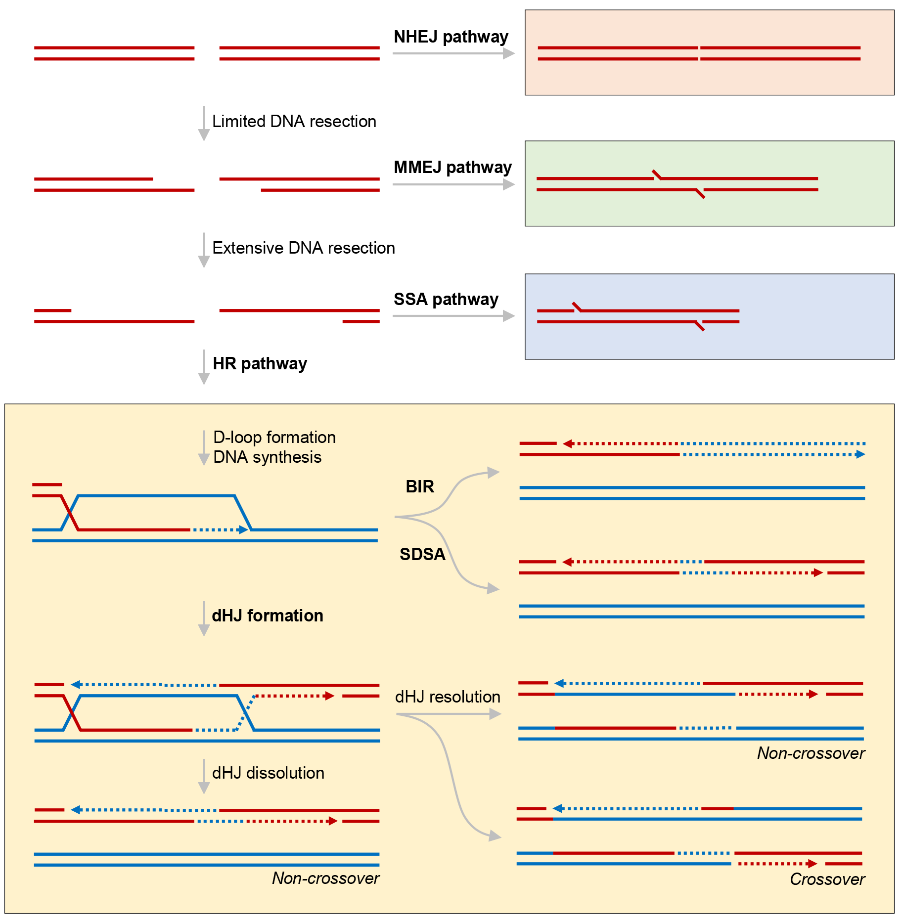
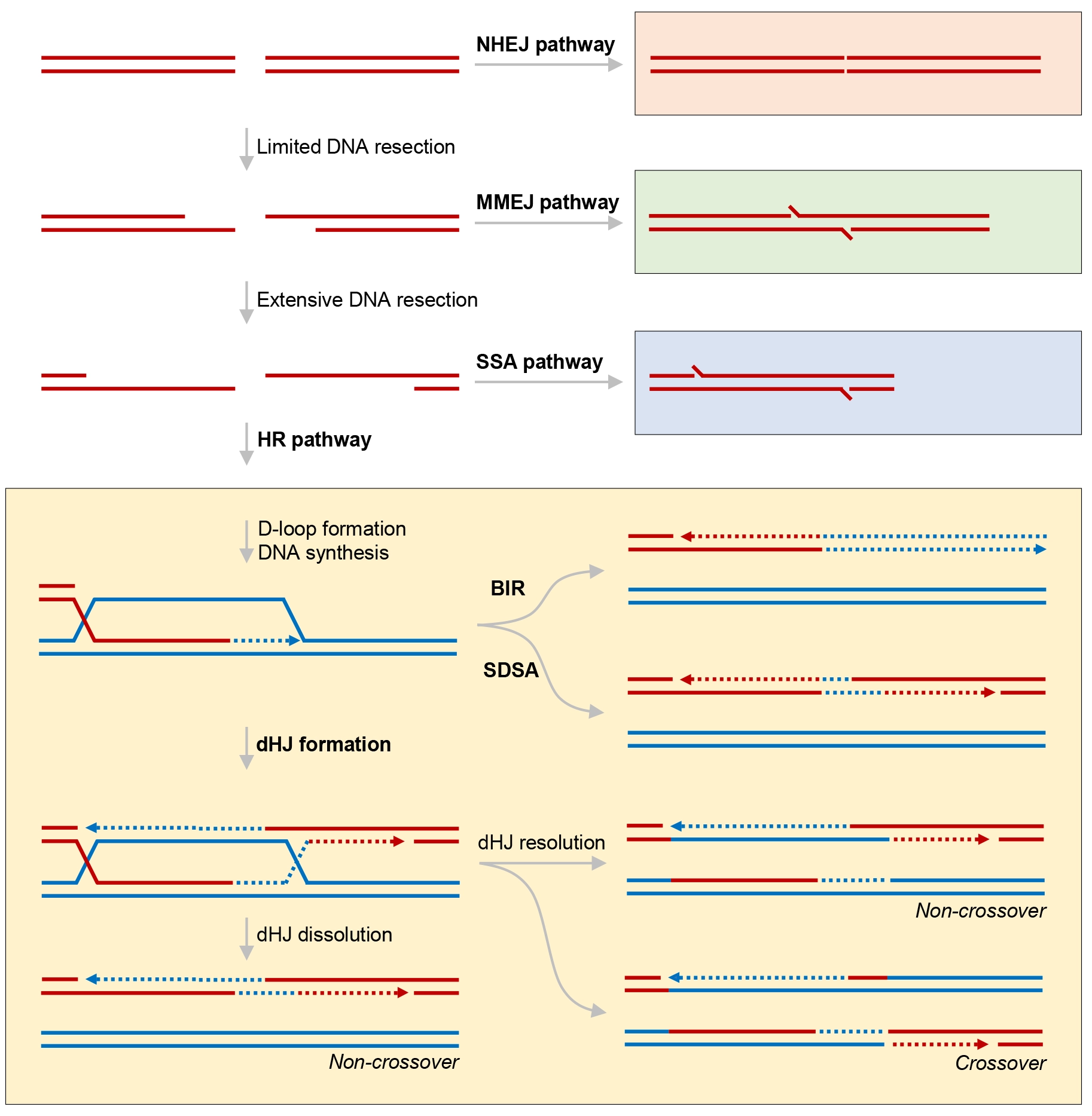
Figure 1. Overview of DSB repair pathways and HR outcomes. DSB: DNA double-strand break; HR: homologous recombination; NHEJ: non-homologous end joining; MMEJ: microhomology-mediated end joining; SSA: single-strand annealing; BIR: break-induced replication; SDSA: synthesis-dependent strand annealing; dHJ: double Holliday junction.
HR and NHEJ are not merely alternative repair pathways but are subject to extensive antagonistic regulation[11]. These pathways differ fundamentally in their cell-cycle dependence, repair fidelity, kinetics, and molecular mechanisms, and together form a complex regulatory network. HR is preferentially utilized during the S and G2 phases and depends on the presence of a sister chromatid template, with recombinases such as RAD51 orchestrating homology-directed repair. In contrast, NHEJ functions throughout the cell cycle and directly ligates DNA ends through factors such as DNA ligase IV. Consequently, the two pathways engage in competition, antagonism, and functional compensation. A comprehensive understanding of these repair mechanisms and their interplay not only provides fundamental insights into the maintenance of genome stability but also establishes the conceptual basis for therapeutic strategies, including radiotherapy and chemotherapy.
DSBs can be repaired through multiple pathways depending on the extent of DNA end resection. In the absence of extensive resection, DSBs are primarily repaired by NHEJ. Limited end resection promotes microhomology-mediated end joining (MMEJ). Extensive DNA end resection generates long single-stranded DNA (ssDNA) overhangs that enable HR or single-strand annealing (SSA). During HR, RAD51-mediated strand invasion leads to displacement loop (D-loop) formation and DNA synthesis. Subsequent repair can proceed through break-induced replication (BIR) or synthesis-dependent strand annealing (SDSA), or through the formation of double Holliday junctions (dHJs), which are resolved either by dissolution to generate non-crossover products or by endonucleolytic resolution to produce crossover or non-crossover outcomes.
1.3 Central role of RAD51 in HR
HR is the principal pathway responsible for the accurate repair of DSBs and is indispensable for the maintenance of genome integrity[12]. At the center of this process lies RAD51, an evolutionarily conserved recombinase that performs the key molecular steps of homology search and strand invasion (Figure 2). As the core catalytic engine of HR, the biological significance of RAD51 extends far beyond its role as a recombinase. The assembly, stabilization, and dynamic remodeling of RAD51 nucleoprotein filaments are tightly regulated by breast cancer gene 2 (BRCA2), the RAD51 paralogs, and numerous accessory factors[13-19]. Together, these regulatory mechanisms form a highly coordinated network that ensures the timely and accurate execution of HR-mediated DNA repair. Beyond its canonical function in DSB repair, RAD51 also plays essential roles in replication-associated genome maintenance, including protection and remodeling of stalled replication forks, replication restart, and the repair of ssDNA gaps generated during DNA replication. These functions highlight RAD51 as a central coordinator that integrates HR with replication stress responses. Consequently, understanding the mechanisms governing RAD51 function has become a central focus in studies of genome stability, cancer biology, and therapeutic intervention.
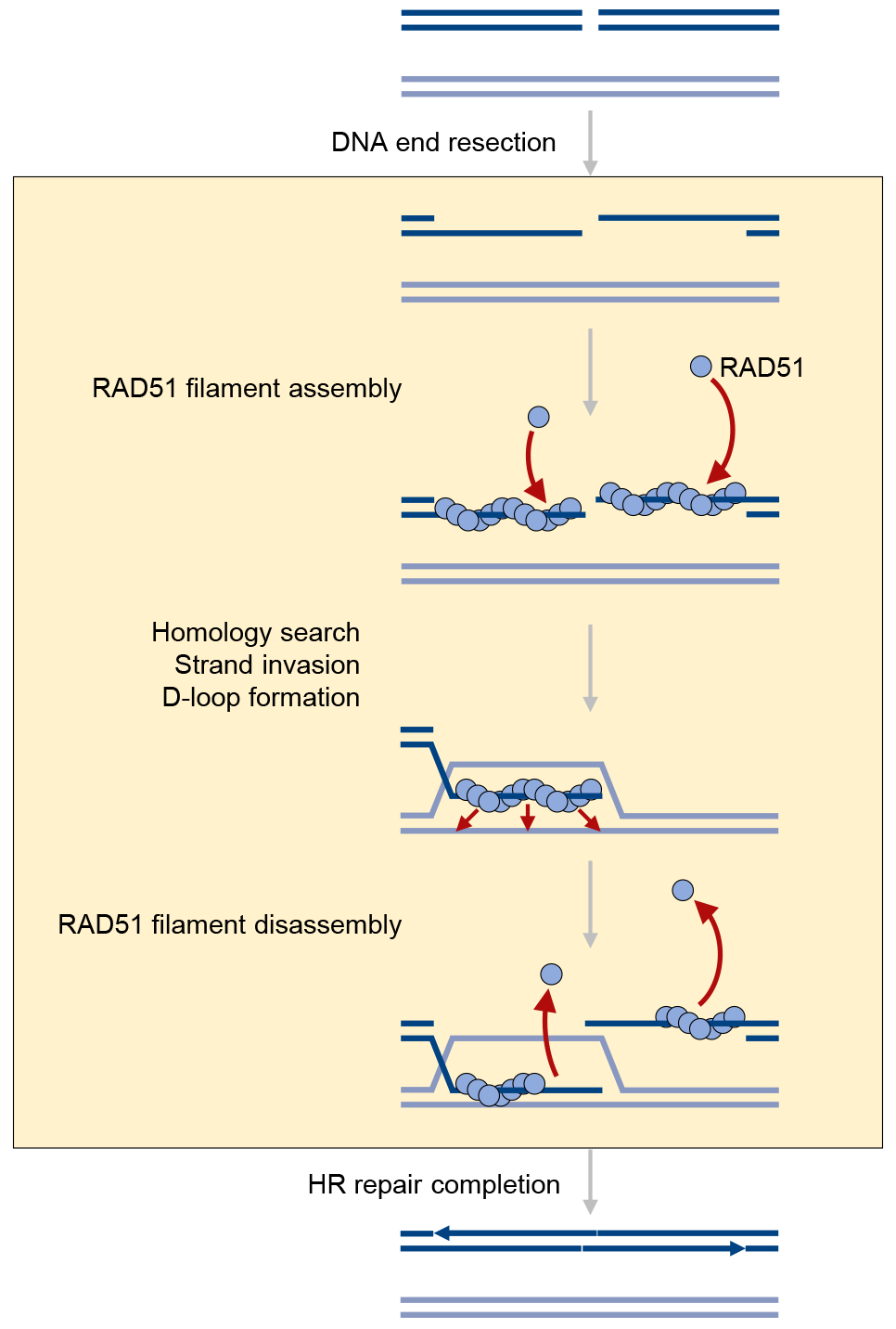
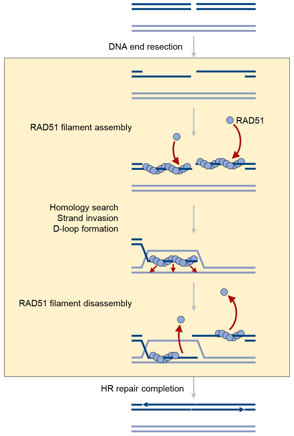
Figure 2. The HR cycle of RAD51: assembly, homology search, strand invasion, and disassembly. HR: homologous recombination.
HR is initiated by DNA end resection, which generates 3’ ssDNA overhangs at DSBs. RAD51 is subsequently assembled onto ssDNA to form a nucleoprotein filament that serves as the catalytic core of HR. The RAD51 filament mediates homology search, strand invasion of the homologous duplex DNA, and D-loop formation, thereby enabling template-directed DNA synthesis. Following completion of repair, RAD51 filaments are actively disassembled by multiple anti-recombinase factors to ensure proper resolution of recombination intermediates and restoration of genome integrity.
1.4 Scope and organization of this review
Although several reviews have summarized the fundamental roles of RAD51, recent studies have significantly expanded our understanding of RAD51 functions and regulatory mechanisms. In this review, we integrate insights from recent advances and provide a more comprehensive perspective, covering the roles of RAD51 from canonical HR repair to replication stress responses, from detailed molecular mechanisms governing RAD51 filament dynamics to its broader implications in genome instability, tumorigenesis, hereditary disorders, aging, and targeted therapeutic strategies. Through this synthesis, we aim to provide a comprehensive framework for understanding the multifaceted roles of RAD51 in genome maintenance and human disease.
2. Major Steps of HR Repair
2.1 DNA end resection
DNA end resection represents a pivotal initiating step of HR[20]. Upon the occurrence of a DSB, the broken DNA ends are first recognized and bound by DNA damage-sensing proteins. Among these, the MRE11-RAD50-NBS1 (MRN) complex plays a central role in the early detection and processing of DSBs. The MRN complex recruits and activates the ataxia telangiectasia mutated (ATM) kinase, which subsequently phosphorylates multiple downstream targets, including H2A histone family member X (H2AX) and mediator of DNA damage checkpoint 1 (MDC1), thereby establishing a DNA damage response signaling platform[21]. This platform not only amplifies damage signaling throughout the nucleus but also contributes to determining whether the break will be repaired through NHEJ or HR.
DNA end resection can generally be divided into three sequential stages[20]. The first two stages involve endonucleolytic cleavage and short-range resection. In cooperation with phosphorylated C-terminal binding protein (CtBP)-interacting protein (CtIP), the MRN complex introduces an initial endonucleolytic incision near the DNA break and performs limited processing of the DNA ends[22-27]. CtIP activity is tightly regulated by the cell cycle. Upon entry into the S and G2 phases, cyclin-dependent kinase (CDK)-mediated phosphorylation enhances the ability of CtIP to promote DNA end resection. Consequently, HR predominantly occurs during S and G2 phases, when sister chromatids have already been generated and are available as accurate repair templates. The third stage involves long-range resection. Following the generation of initial ssDNA intermediates, either the exonuclease 1 (EXO1) pathway or the DNA2-bloom syndrome helicase (BLM) pathway extends the resected region[28-30]. EXO1 functions as a 5’-3’ exonuclease that progressively degrades DNA from the break site. Alternatively, the BLM helicase unwinds double-stranded DNA (dsDNA), allowing DNA2 to selectively cleave the exposed 5’ ssDNA. Through the coordinated activities of these enzymes, extensive 3’ ssDNA overhangs are generated. These long stretches of ssDNA serve as the essential substrates for subsequent HR events, including RAD51 nucleoprotein filament assembly, homology search, and strand invasion. Importantly, the extent of DNA end resection is a major determinant of repair pathway choice. When DNA ends remain unresected, DSBs are preferentially channeled into NHEJ. In contrast, extensive resection suppresses NHEJ and promotes HR as well as other homology-directed repair pathways. Therefore, DNA end resection is not merely a processing step at DNA break ends; rather, it functions as a molecular switch that governs repair pathway selection and ultimately influences genome stability.
2.2 RAD51 filament formation
The 3’ ssDNA generated by DNA end resection is initially coated by replication protein A (RPA). Owing to its high affinity for ssDNA, RPA protects these exposed DNA regions from nucleolytic degradation and prevents the formation of aberrant secondary structures. However, RPA-coated ssDNA is incapable of directly carrying out homology search and strand exchange reactions. To initiate the core steps of HR, RAD51 must replace RPA on ssDNA with the assistance of mediator proteins, most notably BRCA2, and subsequently assemble into a RAD51-ssDNA nucleoprotein filament[13-15,31-34]. This nucleoprotein filament serves as the central platform for homology-directed repair. Once formed, RAD51 filaments are capable of recognizing homologous duplex DNA, promoting strand invasion, and initiating the downstream DNA synthesis events required for repair[35,36].
The integrity and dynamics of RAD51 filament formation are critical determinants of HR efficiency and fidelity. Insufficient RAD51 loading compromises homology search and strand invasion, thereby impairing DSB repair and increasing the likelihood of repair failure. Conversely, excessive stabilization of RAD51 filaments or failure to timely remove RAD51 through the action of anti-recombinases and other negative regulatory factors can impede subsequent repair steps, including DNA synthesis and recombination intermediate resolution. Therefore, RAD51 filaments must achieve a delicate balance between stability and plasticity: they must be sufficiently stable to support efficient homology search and strand exchange, yet remain dynamically remodelable to allow the repair process to progress through its subsequent stages.
2.3 Homology search, strand invasion and RAD51 disassembly
Following the assembly of the RAD51-ssDNA nucleoprotein filament, HR enters the critical stage of homology search[35,36]. During this process, the RAD51 filament scans the genome for duplex DNA sequences that are identical or highly similar to the damaged region. In most cases, the sister chromatid serves as the preferred repair template because it is genetically identical to the broken DNA and is located in close spatial proximity. Although homologous chromosomes can also be used as templates, their utilization in mitotic cells may increase the risk of loss of heterozygosity and other genomic alterations. Therefore, cells generally favor sister chromatids during HR-mediated repair.
RAD51 filaments transiently interact with candidate duplex DNA molecules and assess sequence homology between the ssDNA substrate and one strand of the duplex DNA[37]. Once sufficient homology is identified, the 3’ ssDNA generated by end resection invades the homologous duplex DNA, displacing one strand and pairing with its complementary strand. This process, known as strand invasion, results in the formation of a three-stranded DNA intermediate termed D-loop. D-loop formation positions the invading 3’ hydroxyl end onto the homologous template, thereby providing a substrate for subsequent DNA synthesis. During this process, RAD54 and other factors play key facilitating roles[38-42]. RAD54 interacts with RAD51 filaments and uses adenosine triphosphate (ATP) hydrolysis to drive translocation along duplex DNA, thereby promoting strand invasion and D-loop formation. In addition, its chromatin-remodeling activity alters nucleosome conformation and enhances template accessibility, further supporting HR.
Following strand invasion and D-loop formation, RAD54 also contributes to the removal of RAD51 from heteroduplex DNA and recombination intermediates. This step is essential because persistent RAD51 occupancy can hinder the access of DNA polymerases to the invading 3’ end and thereby impede repair synthesis[43]. Consequently, timely RAD51 disassembly is required for the transition from homology search and strand invasion to DNA synthesis. Thus, RAD51 filaments must be tightly regulated throughout HR: they must remain sufficiently stable to support homology search and strand invasion, yet be efficiently dismantled to expose the 3’ end to DNA polymerases once these processes are completed. This dynamic regulation ensures both the accuracy and efficiency of HR repair.
3. Regulation of RAD51 Activity
3.1 Mediators, loaders, stabilizers and cofactors of RAD51
RAD51 possesses an intrinsic ability to bind ssDNA and assemble into nucleoprotein filaments. However, in cells, this process cannot rely solely on the spontaneous activity of RAD51. Following DNA end resection at DSBs, the resulting 3’ ssDNA is rapidly coated by RPA. Although RPA protects ssDNA, it also prevents the direct loading of RAD51 onto DNA. Therefore, cells require a group of RAD51 mediators and loaders that facilitate the replacement of RPA by RAD51 and promote the assembly of stable, continuous, and recombination-competent RAD51 filaments on ssDNA[44,45]. Among these factors, BRCA2, RAD51 paralogs, RAD51 associated protein 1 (RAD51AP1), and breast cancer 1 (BRCA1) are currently recognized as key regulators of this process (Table 1).
Table 1. Summary of key regulators controlling RAD51 function.
| Protein | HR stage | Major function |
| BRCA2-DSS1 | Presynaptic stage | Replaces RPA with RAD51 and promotes RAD51 filament assembly |
| PALB2 | Presynaptic stage | Recruits BRCA2 to DNA damage sites and facilitates RAD51 loading |
| RAD51B/C/D, XRCC2/XRCC3 | Presynaptic/synaptic stages | Promote RAD51 filament assembly and maturation |
| RAD51AP1 | Synaptic stage | Stimulates homologous DNA pairing and D-loop formation |
| BRCA1-BARD1 | Presynaptic/synaptic stages | Promotes DNA end resection and RAD51-dependent homologous DNA pairing |
| RAD54 | Synaptic stage | Facilitates homology search and filament remodeling |
| Cohesin | Homology search stage | Organizes chromatin environment for homology search |
| RECQ5 | Filament regulation | Dissociates RAD51 filaments |
| FBH1 | Filament regulation | Promotes RAD51 filament disassembly |
| BLM | Post-synaptic stage | Regulates D-loop processing and recombination intermediate resolution |
| RTEL1 | Post-synaptic stage | Disassembles D-loops and controls recombination intermediate processing |
| PARI | Filament regulation | Suppresses replication-associated RAD51 filament extension |
| FIGNL1-FIRRM-MACIR | Filament turnover | Promotes RAD51 filament disassembly and turnover |
PALB2: partner and localizer of BRCA2; HR: homologous recombination; PARI: PCNA-associated recombination inhibitor; FBH1: F-box DNA helicase 1; FIGNL1: fidgetin-like-1; FIRRM: FIGNL1-interacting regulator of recombination and mitosis; MACIR: macrophage immunometabolism regulator; BLM: bloom syndrome helicase;
3.1.1 BRCA2 and partner and localizer of BRCA2 (PALB2)
BRCA2 is the principal RAD51 mediator and loader in mammalian cells[13-15]. It directly interacts with RAD51 and promotes the loading of RAD51 onto resected ssDNA. BRCA2 contains multiple BRC repeats, which serve as binding motifs for RAD51, thereby facilitating RAD51 recruitment and regulating its loading onto DNA. In addition, the C-terminal region of BRCA2 can also bind RAD51 and contributes to the stabilization of RAD51-ssDNA filaments. Through these functional domains, BRCA2 not only delivers RAD51 to ssDNA but also promotes its assembly into an active nucleoprotein filament with the proper orientation and conformation required for HR. DSS1 is an essential cofactor for BRCA2 function[14]. It forms a stable complex with BRCA2 and plays a critical regulatory role during BRCA2-dependent HR repair. Studies have demonstrated that DSS1 not only contributes to the stability and proper conformation of BRCA2 but also exhibits DNA mimicry by emulating the negatively charged characteristics of DNA. This property facilitates BRCA2 recognition of RPA-coated ssDNA, thereby reducing RPA occupancy on ssDNA and creating a favorable environment for RAD51 filament assembly.
The functions of BRCA2 can be broadly summarized into three major roles. First, BRCA2 promotes the replacement of RPA by RAD51, enabling RAD51 binding to ssDNA. Second, BRCA2 suppresses non-productive and non-specific RAD51 nucleation on dsDNA, thereby increasing the efficiency of productive RAD51 filament assembly on ssDNA. Third, BRCA2 stabilizes pre-formed RAD51 filaments and prevents their premature dissociation. Because the RAD51 filament constitutes the central catalytic structure for homology search and strand invasion, BRCA2 deficiency severely compromises HR repair capacity and renders cells highly sensitive to replication stress, poly(ADP-ribose) polymerase (PARP) inhibitors, and DNA crosslinking agents[46,47].
PALB2 serves as a critical molecular bridge linking BRCA1 and BRCA2[48-50]. Its name derives from “Partner and Localizer of BRCA2”. PALB2 interacts with BRCA1 to recruit BRCA2 to sites of DNA damage, while also directly binding BRCA2 to facilitate its RAD51-loading activity at DNA break sites. During HR, the BRCA1-PALB2-BRCA2 axis functions in a highly coordinated and sequential manner. BRCA1 promotes DNA end resection and establishes a repair environment conducive to HR. Subsequently, PALB2 is recruited and facilitates the localization of BRCA2 to damaged chromatin. BRCA2 then loads RAD51 onto RPA-coated ssDNA. This regulatory axis ensures that RAD51 filaments are assembled only at the appropriate stage of the cell cycle, on the correct DNA substrate, and at bona fide sites of DNA damage. When PALB2 is deficient, BRCA2 cannot be efficiently localized to the vicinity of DSBs, resulting in reduced RAD51 focus formation and impaired HR efficiency. Consequently, similar to BRCA2, PALB2 functions as a core tumor suppressor that is indispensable for maintaining HR-mediated DNA repair and genome stability[31,32,50].
3.1.2 RAD51 paralogs
In addition to BRCA2 and PALB2, the RAD51 paralogs serve as essential auxiliary factors for RAD51 filament assembly. Human cells contain five major RAD51 paralogs: RAD51B, RAD51C, RAD51D, XRCC2, and XRCC3. Although these proteins share a certain degree of sequence and structural similarity with RAD51, they generally do not perform the canonical strand-exchange activity mediated by RAD51. Instead, they function primarily through the formation of specialized protein complexes that facilitate RAD51 filament assembly, stabilization, and the subsequent processing of recombination intermediates[16-19].
RAD51 paralogs are organized into two major complexes: the RAD51B-RAD51C-RAD51D-XRCC2 (BCDX2) complex and the RAD51C-XRCC3 (CX3) complex. The BCDX2 complex consists of RAD51B, RAD51C, RAD51D, and XRCC2, whereas the CX3 complex is composed of RAD51C and XRCC3. The BCDX2 complex is generally considered to function during the early stages of HR by promoting RAD51 loading onto ssDNA and stabilizing nascent RAD51 filaments, thereby enhancing the efficiency of RAD51 foci formation[17]. In contrast, the CX3 complex is thought to play a more prominent role in the stabilization and maturation of RAD51 nucleoprotein filaments, as well as in the maintenance and regulation of recombination intermediates[19]. Notably, the CX3 complex has also been closely linked to Holliday junction-associated processes. Because RAD51C is a shared component of both complexes, it is likely to coordinate distinct stages of HR and facilitate the functional transition between early and late recombination events.
The functions of RAD51 paralogs can be broadly categorized into several key aspects. First, they cooperate with the BRCA2-mediated loading machinery to enhance the efficiency of RAD51 assembly onto RPA-coated ssDNA, thereby promoting the formation of RAD51 nucleoprotein filaments. Second, they contribute to the stabilization of preassembled RAD51 filaments and protect them from premature disassembly. Third, RAD51 paralogs participate in the structural quality control of RAD51 filaments, helping to generate recombination-competent filament architectures that are optimal for strand invasion. Fourth, certain RAD51 paralogs also regulate downstream recombination intermediates, indicating that their functions extend beyond the initial RAD51 loading step. Deficiency of RAD51 paralogs commonly results in reduced RAD51 foci formation, impaired HR efficiency, increased chromosomal breakage, and heightened sensitivity to replication stress. Furthermore, germline mutations in several RAD51 paralogs, particularly RAD51C, RAD51D, and XRCC2, have been associated with susceptibility to hereditary breast and ovarian cancers[51-53]. Collectively, these findings underscore the critical importance of the precise regulation of RAD51 filament assembly for the preservation of genome stability.
3.1.3 RAD51AP1
In recent years, RAD51AP1 has emerged as an important accessory regulator of RAD51 filament stability and HR[54,55]. RAD51AP1 directly interacts with RAD51 and preferentially binds branched DNA structures, D-loops, and nucleosome-associated DNA substrates. Unlike BRCA2, which primarily functions in the initial loading of RAD51 onto ssDNA, RAD51AP1 acts predominantly as an accessory factor that enhances both the stability and functional activity of RAD51 filaments. Biochemical studies have demonstrated that RAD51AP1 stimulates RAD51-mediated homologous pairing and D-loop formation, thereby increasing the efficiency of RAD51-dependent recombination within chromatin contexts. Because nucleosomes present substantial physical barriers to strand invasion, RAD51AP1 is thought to facilitate homologous pairing on complex chromatin substrates. Beyond its role in canonical HR, RAD51AP1 has also been implicated in replication stress responses and replication fork protection[56]. Under conditions of replication stress, RAD51AP1 contributes to the maintenance of efficient DNA replication and genome stability[56]. Notably, RAD51AP1 is frequently overexpressed in a variety of human cancers, and elevated RAD51AP1 expression is commonly associated with poor clinical outcomes[57]. Collectively, these findings establish RAD51AP1 as an integral component of the RAD51 regulatory network and highlight its emerging potential as a therapeutic target in cancer.
3.1.4 BRCA1 and BRCA1-associated really interesting new gene (RING) domain 1 (BARD1)
The BRCA1-BARD1 heterodimer not only plays critical roles in DNA end resection and DNA damage checkpoint regulation but also directly promotes RAD51-mediated homologous DNA pairing[58-60]. Studies have shown that BRCA1-BARD1 can interact with both RAD51 and DNA substrates, thereby facilitating RAD51-dependent homologous pairing between ssDNA and homologous dsDNA. Rather than functioning primarily as a RAD51 loader, BRCA1-BARD1 acts more as a recombination cofactor that enhances the functional activity of RAD51 filaments and improves the efficiency of homologous DNA pairing. Through this mechanism, BRCA1-BARD1 directly stimulates the catalytic steps of RAD51-mediated recombination. These observations further suggest that the functions of BRCA1-BARD1 extend beyond their well-established roles in the early stages of HR. In addition to orchestrating DNA end processing and checkpoint signaling, the BRCA1-BARD1 complex can directly participate in the core recombination reactions that drive homology-directed DNA repair, thereby contributing to the efficient and accurate maintenance of genome integrity.
3.2 Regulators of RAD51-mediated homology search dynamics
Compared with NHEJ, HR is generally considered to require more extensive chromatin exploration and DNA movement to locate homologous sequences within the genome. As the central mediator of homology search, RAD51 possesses substantial dynamic potential to facilitate this process. Early studies demonstrated that RAD51 exhibits diffusion behavior in living cells similar to that of BRCA2, providing a kinetic basis for the rapid initiation of HR[61]. Subsequently, it was reported that RAD51-mediated interchromosomal homology search can promote and direct the movement of DSBs at alternative lengthening of telomeres (ALT) telomeres, thereby providing both structural and dynamic support for telomere-telomere HR and telomere elongation[62]. These findings expanded the scope of RAD51 mobility research and offered new perspectives supporting the notion that RAD51 dynamics actively contribute to DSB repair. Using fluorescence-based live-cell tracking approaches, the group of Angela Taddei achieved real-time visualization of Rad51-mediated homology search dynamics in yeast cells[63,64]. Their work revealed that Rad51 does not identify homologous sequences through a purely stochastic scanning process. Instead, homology search is accomplished through a dual strategy that combines local precision searching with efficient global exploration. On one hand, Rad51 nucleoprotein filaments undergo confined local movements around DNA damage sites, preferentially probing neighboring chromatin regions for homologous templates. On the other hand, when local exploration fails to identify suitable homology, Rad51 can engage in larger-scale movements that substantially expand the search territory. This dynamic combination of local scanning and long-range exploration, together with the continuous transitions between compacted and extended filament conformations, is thought to enhance the efficiency of homologous sequence recognition and thereby facilitate the completion of homology search by Rad51. Nevertheless, the molecular mechanisms that drive the three-dimensional movement of RAD51 filaments within the nuclear space, particularly in mammalian cells, remain largely unknown.
The group of Eric Greene demonstrated that yeast Rad54, a key accessory protein in HR[38], utilizes its ATPase activity to hydrolyze ATP and generate the energy required for one-dimensional translocation of Rad51 nucleoprotein filaments along dsDNA. This ATP-driven movement facilitates the search and recognition of homologous duplex DNA by the broken ssDNA substrate, thereby promoting efficient homology matching. In addition, recent studies have revealed a previously unappreciated role for Cohesin in regulating homology search[65-67]. Following DSB formation, Cohesin can extrude chromatin loops anchored at the break site, thereby driving directional one-dimensional scanning of chromatin by RAD51 nucleoprotein filaments while simultaneously restricting the search space within topologically associated domain (TAD) boundaries. Through this mechanism, Cohesin establishes extensive yet spatially constrained homology-search territories spanning hundreds of kilobases of chromatin. Such organization minimizes unproductive exploration and enhances the efficiency of homologous sequence identification. Collectively, these processes enable HR to efficiently locate the correct repair template within a large and highly repetitive genome, ultimately ensuring accurate and high-fidelity DNA repair. Despite these advances, our understanding of the regulatory mechanisms governing RAD51 mobility remains incomplete. Identifying the molecular machinery that coordinates RAD51 filament dynamics across nuclear space, particularly in higher eukaryotes, represents an important challenge for future studies and may provide fundamental insights into how cells achieve rapid and accurate homology-directed DNA repair.
3.3 Anti-recombinases and negative regulators of RAD51
Formation of the RAD51 filament is essential for homology search and strand invasion during HR. However, excessive or overly stable RAD51 filaments can be detrimental rather than beneficial. Aberrant accumulation of RAD51 on DNA may promote inappropriate template usage, persistent D-loop structures, defective replication fork processing, and the generation of toxic recombination intermediates. To prevent these outcomes, cells employ a group of anti-recombinases and negative regulators that restrain RAD51 activity, promote filament disassembly or remodeling, and suppress excessive HR[68]. Prominent members of this regulatory network include RECQ5, F-box DNA helicase 1 (FBH1), BLM, RTEL1, PARI, and the FIGNL1-FIRRM-MACIR complex.
RECQ5 is a member of the RecQ family of helicases and functions as an anti-recombinase that negatively regulates RAD51 activity. RECQ5 can directly interact with RAD51 and promote ATP-dependent disassembly of RAD51 filaments[69]. Loss of the RECQ5-RAD51 interaction markedly impairs the anti-recombinase activity of RECQ5, indicating that direct engagement with RAD51 filaments is central to its function in restricting HR. A key role of RECQ5 is to prevent excessive RAD51 loading onto inappropriate DNA regions. Persistent RAD51 filaments at replication stress sites, transcription-replication conflict regions, or repetitive DNA sequences can trigger aberrant strand invasion and genome rearrangements. By dismantling RAD51-ssDNA filaments, RECQ5 suppresses unnecessary HR activity and helps maintain the balance among DNA replication, transcription, and repair. RECQ5 is also functionally linked to RNA polymerase-associated transcription and transcription-coupled DNA damage responses, suggesting that it may limit aberrant recombination in highly transcribed genomic regions through local control of RAD51 activity.
FBH1 is an F-box-containing DNA helicase that is widely regarded as a mammalian anti-recombinase analogous to the yeast Srs2 protein[70,71]. FBH1 directly binds RAD51 and promotes RAD51 filament disassembly through its ssDNA translocase activity. Biochemical studies have shown that FBH1 can disrupt RAD51 filaments in vitro, and cellular studies support its role as a negative regulator of HR. The functions of FBH1 are multifaceted: it participates in replication fork processing under certain replication stress conditions while simultaneously preventing the accumulation of toxic recombination intermediates by limiting RAD51 filament stability. The presence of an F-box domain further suggests that FBH1 may influence the stability of RAD51 or other repair factors through ubiquitin-dependent mechanisms. Consistent with this idea, studies in fission yeast have shown that Fbh1 negatively regulates Rad51-mediated HR through the combined action of its DNA helicase/translocase activity and ubiquitin ligase-associated functions.
BLM is another member of the RecQ family of helicases and is encoded by the gene mutated in Bloom syndrome. BLM plays dual roles in HR. On one hand, BLM cooperates with DNA2 during long-range DNA end resection, thereby facilitating the initiation of HR[72,73]. On the other hand, BLM also exerts a pronounced inhibitory effect on RAD51 activity that restrains aberrant or excessive RAD51-mediated recombination events and limits the accumulation of recombination intermediates such as D-loops[74,75]. Biochemical studies have demonstrated that BLM can suppress RAD51-mediated D-loop formation and dismantle certain preformed D-loop intermediates. Consistent with this function, BLM-deficient cells exhibit markedly elevated levels of sister chromatid exchange, indicating that BLM plays a critical role in suppressing excessive recombination and correcting inappropriate recombination events. More broadly, BLM regulates both the directionality and quality of HR. It can eliminate aberrant or unstable strand invasion intermediates and, in cooperation with the topoisomerase IIIα (TOPIIIα)-RMI1-RMI2 complex, promote the dissolution of double Holliday junctions[76]. This process suppresses crossover formation and favors non-crossover repair outcomes. Thus, BLM not only supports productive HR but also prevents recombination from progressing toward deleterious outcomes, including excessive sister chromatid exchange and chromosomal rearrangements.
Regulator of telomere length 1 (RTEL1) is another important helicase that negatively regulates RAD51-dependent recombination[77]. Initially identified for its essential role in telomere maintenance, RTEL1 has subsequently emerged as a critical regulator of HR. RTEL1 can dismantle D-loops and other recombination intermediates, thereby restricting excessive or aberrant HR and suppressing crossover formation. Similar to BLM, RTEL1 primarily functions during the later stages of HR by controlling the fate of recombination intermediates. This activity facilitates the timely termination of RAD51-mediated strand invasion events and prevents the persistence of potentially toxic DNA structures. Loss of RTEL1 results in replication fork instability, increased telomere fragility, and genome instability, highlighting its particularly important role in replication-associated HR regulation. In addition, several studies suggest that RTEL1 promotes replication fork recovery and limits the accumulation of aberrant RAD51-dependent recombination events under conditions of replication stress[78].
Proliferating cell nuclear antigen (PCNA)-associated recombination inhibitor (PARI) functions as an important negative regulator of replication-coupled HR[79]. PARI contains both a PCNA-interacting peptide (PIP) box and a small ubiquitin-related modifier (SUMO)-interacting motif (SIM), enabling it to bind SUMOylated PCNA and localize to replication forks. PARI directly interacts with RAD51 and suppresses the stabilization and extension of RAD51 filaments on ssDNA, thereby preventing excessive RAD51 loading and filament growth. The activity of PARI is primarily restricted to the S-phase replication environment, where its principal function is to prevent inappropriate activation of HR at or near replication forks. Loss of PARI generally results in elevated HR activity, whereas PARI overexpression reduces RAD51 foci formation and suppresses HR. Consequently, PARI is widely regarded as a molecular “brake” for replication-associated HR, helping to maintain a balance between replication-associated repair and genome stability.
In recent years, the fidgetin-like-1 (FIGNL1)-FIGNL1-interacting regulator of recombination and mitosis (FIRRM)-macrophage immunometabolism regulator (MACIR) complex has emerged as a major focus in studies of RAD51 filament disassembly[80-84]. FIGNL1 is an AAA+ ATPase, whereas FIRRM, also known as C1orf112, functions as a structural partner that interacts with and stabilizes FIGNL1[82,83]. MACIR (C5orf30) serves as an adaptor that bridges FIGNL1-FIRRM with RAD51-DNA nucleoprotein filaments[84]. Studies from our group and others have demonstrated that the FIGNL1-FIRRM-MACIR complex promotes the dissociation of RAD51 and DMC1 from chromatin, thereby preventing their aberrant accumulation on undamaged chromosomal regions[82-92]. Beyond regulating recombinase turnover, the FIGNL1 complex controls the dynamics of RAD51 and DMC1 during both meiotic recombination and replication fork restart. By controlling RAD51 filament dynamics during replication fork restart, this complex contributes to the resolution of replication-associated recombination intermediates and prevents excessive RAD51 accumulation at stalled forks. Thus, the FIGNL1 complex represents an important link between RAD51 filament turnover and replication stress tolerance. Biochemical analyses further revealed that the complex can remodel RAD51 and DMC1 nucleoprotein filaments and suppress strand invasion activity. Mechanistic studies have shown that FIGNL1 promotes the dissociation of RAD51 from DNA substrates through an ATPase-dependent but non-canonical mechanism, thereby contributing to the maintenance of genome stability. Structural and biochemical investigations have consequently established FIGNL1 as a new key negative regulator of RAD51 recombinase activity. More recent studies have further suggested a complex genetic relationship between BRCA2 and FIGNL1. In BRCA2-deficient cells, additional loss of FIGNL1 can promote the retention of RAD51 at DSBs, thereby altering HR status and influencing tumor responses to DNA-damaging therapies[93]. These findings highlight the dynamic balance between RAD51 assembly factors and anti-recombinases in determining recombination proficiency and therapeutic sensitivity.
4. RAD51 in Genome Stability and Disease
RAD51 is the core recombinase of HR and a key factor in maintaining genome stability. By promoting homology search, strand invasion, replication fork protection, and replication stress responses, RAD51 enables the accurate repair of DSBs and preserves replication continuity. Consequently, dysregulation of RAD51 can lead to two distinct outcomes. On one hand, insufficient RAD51 activity results in HR deficiency, chromosomal breakage, and replication fork collapse. On the other hand, RAD51 overexpression or aberrant activation may drive inappropriate recombination events, therapeutic resistance, and tumor progression[94,95].
4.1 RAD51 dysfunction and genomic instability
RAD51 dysfunction directly compromises the fidelity of HR-mediated DNA repair. During normal HR, DSBs undergo DNA end resection to generate 3’ ssDNA, onto which RAD51 is subsequently loaded with the assistance of BRCA2, PALB2, RAD51 paralogs, and other HR factors, forming a RAD51-ssDNA nucleoprotein filament. This filament serves as the catalytic platform for homology search and strand invasion, allowing the damaged DNA molecule to use the sister chromatid as a template for high-fidelity repair. When RAD51 expression is insufficient, filament assembly is impaired, ATPase activity is disrupted, or filament dynamics become unbalanced, cells are unable to efficiently complete HR. As a result, DSBs may remain unrepaired or be redirected to more error-prone pathways, such as NHEJ or MMEJ, which frequently generate deletions, insertions, and chromosomal rearrangements[96,97].
RAD51 deficiency also exacerbates replication stress-associated genome instability. Following replication fork stalling, RAD51 stabilizes stalled or reversed replication forks and protects nascent DNA strands from degradation by nucleases such as MRE11, DNA2, and EXO1[2,94]. In addition to fork protection, RAD51 participates in the processing and restart of reversed replication forks. RAD51-mediated filament formation facilitates controlled fork remodeling. Moreover, RAD51 contributes to the repair of post-replicative ssDNA gaps and tolerance of replication-associated lesions, allowing cells to bypass DNA damage encountered during genome duplication. When RAD51 cannot be efficiently recruited to or stably maintained at stalled forks, replication forks become more susceptible to collapse and can be converted into single-ended DSBs. Because these lesions lack a corresponding second DNA end, they are generally poor substrates for classical NHEJ and instead rely predominantly on HR-mediated fork restart mechanisms. Consequently, impairment of these repair processes promotes chromosomal breakage, copy number alterations, and complex structural rearrangements. In addition to its canonical role in HR, RAD51 also has several non-canonical functions, including replication fork protection, regulation of fork reversal, and buffering of replication stress. Collectively, these activities restrain the progression of replication-associated DNA lesions into chromosomal instability[94,98].
Notably, excessive RAD51 activity can also compromise genome integrity. Hyperstabilized RAD51 filaments may promote aberrant strand invasion into repetitive sequences or non-allelic homologous regions, thereby triggering non-allelic HR and structural rearrangements such as deletions, duplications, inversions, and translocations. Early studies demonstrated that although RAD51 overexpression can enhance cellular tolerance to DNA damage, it may simultaneously increase the frequency of aberrant recombination events and thereby promote genome instability[95]. Therefore, the contribution of RAD51 to genome maintenance is highly dependent on its dosage, timing, and spatial regulation. While insufficient RAD51 activity leads to repair failure and genome instability, excessive or persistent RAD51 activation likewise poses a substantial risk of pathological recombination and chromosomal rearrangements.
4.2 The role of RAD51 in cancer development
RAD51 is closely linked to both tumor initiation and cancer progression[99]. Genomic instability is a hallmark of cancer, and dysregulation of RAD51 represents one of the major contributors to this instability. Deficiency of RAD51 compromises HR-mediated DNA repair, leading to the accumulation of DSBs, chromosomal breaks, and replication-associated DNA lesions. If these lesions are not efficiently resolved, they can promote the acquisition of driver mutations, copy number alterations, chromosomal translocations, and ultimately tumor evolution. Conversely, RAD51 overexpression is frequently observed in a wide range of human cancers and may facilitate tumor cell survival by enhancing DNA repair capacity, thereby enabling cancer cells to better tolerate replication stress as well as DNA damage induced by radiotherapy and chemotherapy.
From a clinical perspective, elevated RAD51 expression is often associated with poor prognosis[100]. Consistently, pan-cancer analyses have revealed widespread RAD51 overexpression across diverse cancer types and have linked high RAD51 expression to reduced overall survival in several malignancies. These observations can be explained by the biological demands of cancer cells. Tumor cells are typically exposed to high levels of replication stress and endogenous DNA damage; therefore, increased RAD51 expression may enhance their ability to repair therapy-induced DNA lesions, protect stalled replication forks, and sustain proliferation under otherwise unfavorable conditions.
RAD51 is also a critical determinant of therapeutic response in cancer[100]. Radiotherapy, platinum-based agents, topoisomerase inhibitors, and PARP inhibitors all directly or indirectly increase DNA damage and replication stress. Cancer cells with elevated RAD51 activity are often more capable of repairing these lesions and consequently exhibit treatment resistance[95]. In contrast, tumors with BRCA1/2 deficiency or broader HR deficiencies are often more sensitive to PARP inhibitors and platinum compounds. However, acquired resistance can emerge when tumor cells restore RAD51 loading, re-establish replication fork protection, or reactivate HR capacity. Importantly, the role of RAD51 in cancer is not simply tumor-promoting or tumor-suppressive. In normal cells, RAD51 acts as a genome guardian by maintaining HR proficiency and replication fork stability, thereby suppressing tumorigenesis. In established cancers, however, RAD51 overexpression or aberrant activation may enable tumor cells to withstand elevated replication stress and therapeutic pressure, ultimately facilitating tumor progression and treatment resistance. Thus, RAD51 exhibits a context- and stage-dependent role during cancer development. During the early stages of tumorigenesis, RAD51 deficiency may accelerate mutation accumulation and genomic instability, thereby promoting malignant transformation. In contrast, during tumor progression and therapeutic intervention, enhanced RAD51 activity can support cancer cell survival and adaptation, leading to resistance to radiotherapy and chemotherapy.
4.3 RAD51 mutations and human disease phenotypes
RAD51 is an essential component of the HR machinery and is required for cellular viability; consequently, complete loss of RAD51 function is generally incompatible with normal development and survival. Although germline RAD51 variants are relatively rare in humans, they have been linked to several distinct disease phenotypes. One of the best-characterized disorders associated with RAD51 is congenital mirror movements (CMM), a neurodevelopmental condition in which voluntary movement of one limb is accompanied by involuntary mirrored movements of the contralateral limb. Studies have demonstrated that RAD51 haploinsufficiency can cause CMM, suggesting that RAD51 functions not only in DNA repair but also in neural development[101,102]. More recent investigations have further shown that, in addition to classical RAD51 haploinsufficiency, certain non-truncating loss-of-function variants can also give rise to CMM[103,104]. RAD51 variants have also been associated with Fanconi anemia (FA)-like phenotypes. Notably, dominant mutations in RAD51 have been reported to cause a distinct FA subtype[105]. FA is typically characterized by developmental abnormalities, bone marrow failure, and increased cancer susceptibility[106]. The identification of RAD51-associated cases underscores the critical role of RAD51-mediated HR and interstrand crosslink (ICL) repair in development, hematopoietic homeostasis, and tumor suppression. Furthermore, another study demonstrated that dominant RAD51 mutations revealed a specific function of RAD51 in ICL repair[107] that is not entirely equivalent to its canonical role in HR.
In the context of aging, RAD51 is thought to contribute primarily through age-associated declines in DNA repair capacity, progressive accumulation of replication stress, and the consequent activation of cellular senescence pathways[108-112]. DNA damage gradually accumulates in aging cells, and defects in DNA repair are well known to promote premature aging-like phenotypes. As organisms age, or as cells undergo replicative senescence, HR efficiency may progressively decline. Early studies and reviews noted that RAD51 expression is tightly regulated in a cell cycle-dependent manner and becomes markedly reduced or even undetectable in replicatively senescent cells. As a consequence, aged cells may become less capable of repairing replication-associated DSBs through RAD51-dependent HR, thereby exacerbating genomic instability. More recent studies have further strengthened the link between RAD51 and cellular senescence. For example, a 2024 study[111] demonstrated that bleomycin induces suppression of DNA repair and promotes cellular senescence, whereas restoration of RAD51 expression partially alleviated both defective DSB repair and senescence-associated phenotypes. These findings suggest that RAD51 dysfunction may contribute directly to damage-induced cellular senescence. Mechanistically, reduced RAD51 activity results in the persistent accumulation of DSBs and replication-associated lesions, leading to sustained activation of senescence-associated signaling pathways, including the ATM/ataxia telangiectasia and Rad3 related (ATR)-p53-p21 and p16-retinoblastoma (RB) axes, ultimately driving cells into an irreversible state of proliferative arrest[113]. Conversely, excessive RAD51 activity or dysregulated RAD51 filament dynamics may also promote aberrant recombination events, thereby increasing mutation burden and chromosomal structural abnormalities. Taken together, the relationship between RAD51 and aging appears to be governed by a delicate balance. Physiological levels of RAD51 activity are required to preserve genomic integrity and maintain tissue homeostasis throughout life. In contrast, either reduced RAD51 function or aberrant regulation of RAD51 activity can accelerate DNA damage accumulation, promote cellular senescence, and contribute to the development of age-associated disorders.
5. Therapeutic Targeting of RAD51 and HR Pathway
RAD51 and the HR pathway have emerged as important targets in cancer treatment[46]. Many tumor cells exist under conditions of elevated replication stress and persistent DNA damage, making them more dependent than normal cells on HR, replication fork protection, and DNA damage response pathways for survival. As the core recombinase of HR, RAD51 not only mediates DSB repair but also plays critical roles in replication fork protection and the buffering of replication stress. Consequently, targeting RAD51 or the HR pathway can impair the ability of cancer cells to repair DNA damage.
5.1 RAD51 as a cancer therapy target
The rationale for targeting RAD51 in cancer therapy is supported by three major considerations. First, RAD51 is the core effector of HR-mediated DNA repair. When RAD51 filament formation is disrupted, the capacity of tumor cells to repair DSBs and replication-associated lesions is substantially reduced, leading to the accumulation of DNA damage and decreased cellular viability[12]. Second, many cancers exhibit elevated RAD51 expression or enhanced RAD51 activity. This state enables tumor cells to tolerate replication stress and therapy-induced DNA damage, thereby promoting survival and therapeutic resistance[99]. Third, RAD51 activity is closely associated with the response to DNA damage-based therapies, including radiotherapy, platinum compounds, and PARP inhibitors[46]. Consequently, RAD51 serves not only as a therapeutic target but also as a potential biomarker of HR function and treatment response. Collectively, these observations have established RAD51 as a key target in DNA repair-directed cancer therapy, and its roles in HR regulation, therapy resistance, and anticancer drug development have become major areas of both basic and translational research.
Strategies for targeting RAD51 can be broadly categorized into direct and indirect approaches. Direct targeting aims to interfere with critical RAD51 functions through small molecules, peptides, nucleic acid aptamers, or targeted protein degradation technologies. These approaches may disrupt RAD51 DNA binding, ATPase activity, filament assembly, interactions with BRCA2, or D-loop formation. In contrast, indirect targeting strategies suppress RAD51 function by modulating its upstream loading machinery or replication stress response pathways. These include interference with BRCA1-PALB2-BRCA2-mediated RAD51 loading, as well as inhibition of the ATR-CHK1-WEE1 signaling axis and replication fork protection mechanisms. Because RAD51 is also indispensable for the survival of normal proliferating cells, broad or potent inhibition of RAD51 may result in significant toxicities, including bone marrow suppression, gastrointestinal injury, and damage to normal tissues. Therefore, contemporary therapeutic approaches increasingly focus on inducing a state of “functional HR deficiency (functional HRD)” within specific tumor contexts rather than completely blocking RAD51 activity. Such strategies may be particularly effective in tumors characterized by high replication stress, BRCA-proficient cancers that remain highly dependent on RAD51-mediated HR, and tumors that have acquired resistance to platinum-based therapies or PARP inhibitors. A variety of RAD51-targeting small molecules and modulators have been developed to interfere with key steps in the HR process, including RAD51-ssDNA binding, filament assembly, RAD51-BRCA2 interactions, and D-loop formation. Representative tool compounds include B02, RI-1, RI-2, IBR2, and DIDS[114-119] (Table 2). In cell-based and in vitro studies, these compounds reduce RAD51 foci formation, suppress HR activity, increase γH2AX accumulation, and sensitize tumor cells to DNA-damaging agents or PARP inhibitors. Among them, B02 is one of the most extensively characterized RAD51 inhibitors and inhibits RAD51-mediated D-loop formation. RI-1 and its derivative RI-2 suppress RAD51 function through covalent modification of a critical cysteine residue, whereas IBR2 has been reported to impair RAD51 protein stability. DIDS and related analogs inhibit RAD51 binding to ssDNA and block D-loop formation. Nevertheless, most early RAD51 modulators remain research tools, as they often suffer from limited selectivity, suboptimal pharmacokinetic properties, and insufficient drug-like characteristics for clinical development.
Table 2. Representative RAD51-targeting compounds and their therapeutic development status.
| Inhibitor | Mechanism of action | Molecular target/action | Development status | Advantages | Limitations |
| B02 | Inhibits RAD51-DNA interaction and prevents RAD51 filament formation | RAD51 DNA-binding activity | Preclinical | Potent inhibition of HR and widely used tool compound | Limited specificity and pharmacological optimization |
| RI-1 | Covalently modifies RAD51 Cys319 and disrupts RAD51 filament assembly | RAD51 oligomerization/filament formation | Preclinical | Provides mechanistic proof-of-concept for RAD51 targeting | Low potency and stability issues |
| RI-2 | Stabilizes inactive RAD51 conformations and inhibits HR | RAD51 ATPase/filament dynamics | Preclinical | Improved activity compared with RI-1 | Limited in vivo validation |
| IBR2 | Blocks RAD51-mediated strand exchange and promotes RAD51 aggregation | RAD51 filament function | Preclinical | Strong cellular HR inhibition | Mechanism and specificity require further clarification |
| DIDS | Interferes with RAD51 nucleoprotein filament formation | RAD51 DNA-binding function | Preclinical | Early RAD51-targeting strategy | Limited selectivity and toxicity concerns |
| CYT-0851 | Inhibits RAD51-associated DNA repair pathway | RAD51/HR-related repair pathway | Clinical | HR-targeting small molecule entering clinical evaluation | Precise molecular mechanisms remain under investigation |
HR: homologous recombination; DIDS: 4,4'-diisothiocyanostilbene-2,2'-disulfonic acid.
With regard to clinical translation, CYT-0851 has been investigated in clinical studies involving multiple solid tumors, including NCT03997968, both as a monotherapy and in combination with chemotherapeutic agents such as gemcitabine and capecitabine[120]. Preclinical studies have suggested that CYT-0851 inhibits RAD51-associated HR activity and enhances PARP inhibitor sensitivity in breast cancer models[121]. These findings suggest that modulation of the HR/RAD51 axis may provide greater clinical value as part of combination therapies and biomarker-guided patient stratification strategies rather than as a broadly applicable single-agent treatment. Beyond conventional small-molecule inhibitors, several emerging approaches have recently been explored, including strategies targeting protein-protein interaction[122-124]. However, these approaches remain at an early stage of development, and their selectivity, safety, pharmacological properties, and clinical benefits require further validation before therapeutic application.
5.2 Synthetic lethality
Synthetic lethality represents one of the most successful clinical paradigms for targeting the HR pathway in cancer therapy[47,125,126]. The core concept is that inhibition of a single DNA repair pathway is often tolerated because cells can rely on compensatory repair mechanisms; however, when a complementary pathway is already defective, simultaneous disruption of both pathways results in selective cell death. The synthetic lethal interaction between PARP inhibition and BRCA1/2 deficiency is the prototypical example of this strategy. In BRCA1/2-deficient cells, HR repair capacity is compromised, whereas PARP inhibition blocks single-strand break repair, induces PARP1 trapping, and interferes with replication fork progression, ultimately leading to the accumulation of replication-associated DSBs. Because these cells lack efficient HR-mediated repair, they are unable to resolve such lesions and consequently undergo selective cell death. PARP inhibitors have therefore become a cornerstone of precision therapy for HR-deficient (HRD) tumors. Representative approved agents include olaparib, rucaparib, niraparib, and talazoparib[127], which are used across several cancers with BRCA or HR deficiency, including ovarian, breast, prostate, and pancreatic cancers.
RAD51 occupies a pivotal position within PARP inhibitor-mediated synthetic lethality. One of the principal functions of BRCA1, PALB2, and BRCA2 is to promote RAD51 loading and RAD51 filament formation at sites of DNA damage[36]. Failure to form RAD51 foci generally indicates impaired HR capacity and may serve as a potential biomarker associated with increased sensitivity to PARP inhibitors and platinum-based chemotherapy. Conversely, tumors may acquire resistance to PARP inhibitors through restoration of RAD51 loading and HR function. Such restoration can occur through multiple mechanisms, including BRCA1/2 reversion mutations, alterations in the 53BP1 pathway, loss of REV7/RIF1-dependent end protection, or other adaptive changes that re-establish RAD51-mediated repair[128-132]. From a therapeutic perspective, RAD51-targeting strategies offer opportunities to generate new synthetic lethal combinations with PARP inhibitors. In BRCA-wild-type or HR-proficient tumors, PARP inhibitor monotherapy often exhibits limited efficacy. However, simultaneous inhibition of RAD51 or disruption of RAD51 loading may artificially induce a state of HR deficiency, thereby sensitizing tumor cells to PARP inhibition. Consistent with this concept, recent studies have highlighted extensive combinatorial potential among RAD51 inhibitors, ATR/checkpoint kinase 1 (CHK1)/WEE1 pathway inhibitors, and PARP inhibitors[133-135]. Importantly, the therapeutic efficacy of PARP inhibitors is not determined solely by BRCA mutation status. Multiple factors influence treatment response, including the degree of HR deficiency, RAD51 foci formation, replication fork protection capacity, the extent of PARP trapping, tumor microenvironmental conditions, drug exposure, and the emergence of resistant tumor clones. Accordingly, the future of synthetic lethality-based therapy is likely to move beyond patient stratification based on single-gene mutations and toward a more comprehensive framework that integrates functional HRD assessment, replication stress status, and dynamic mechanisms of therapeutic resistance.
5.3 Challenges and future directions
Therapeutic targeting of RAD51 and the HR pathway faces several major challenges. The first challenge is the limited therapeutic window. RAD51 is essential not only for tumor cells but also for normal proliferating cells[136]. Consequently, potent systemic inhibition of RAD51 may result in substantial toxicity and normal tissue damage. Future drug development will therefore require improved tumor selectivity. Potential approaches include exploiting the elevated replication stress characteristic of many cancers, leveraging tumor-specific DNA repair defects, targeting oncogene-driven dependencies, or utilizing advanced drug delivery strategies that render tumor cells more reliant than normal cells on RAD51-mediated HR repair. The second challenge lies in the intrinsic difficulty of drugging RAD51. As a highly conserved DNA-binding ATPase, RAD51 functions through dynamic oligomerization, nucleoprotein filament assembly, and complex protein-DNA interactions. Although numerous early-generation inhibitors have demonstrated activity in vitro, many exhibit limited cellular selectivity, suboptimal pharmacokinetic properties, or unacceptable toxicity profiles. Consequently, future therapeutic strategies may need to move beyond global RAD51 inhibition toward more refined forms of modulation. Examples include disrupting the RAD51-BRCA2 interaction, selectively suppressing aberrantly stabilized RAD51 filaments, targeting tumor-specific states of RAD51 dependency, or developing controllable degraders and allosteric modulators capable of fine-tuning RAD51 activity. The third challenge is the lack of optimal predictive biomarkers. Multiple biomarkers have been proposed, including BRCA1/2 mutations, HRD scores, genomic scars, RAD51 foci formation, replication fork protection status, and resistance-associated mutations, each with distinct strengths and limitations[137]. For example, genomic scar signatures provide evidence of historical HR deficiency but do not necessarily reflect the current functional status of HR within a tumor. In contrast, RAD51 foci analysis offers a more direct assessment of real-time HR competency, yet its implementation is influenced by specimen handling, timing of sample acquisition, and the extent of DNA damage induction. RAD51 foci are commonly evaluated by immunofluorescence-based assays following DNA damage induction, often combined with S/G2-phase markers to specifically assess HR activation capacity. However, differences in antibody performance, DNA damage induction protocols, sample processing procedures, foci scoring criteria, and cutoff definitions remain major challenges for assay standardization and clinical implementation. Compared with commercially available genomic HRD assays, which measure accumulated genomic scars, RAD51 foci provide a functional readout of current HR proficiency. Increasing evidence suggests that tumors with impaired RAD51 foci formation exhibit increased sensitivity to PARP inhibitors, whereas restoration of RAD51 loading may contribute to acquired PARP inhibitor resistance. Nevertheless, prospective clinical validation is required to establish RAD51 foci as a standardized functional HRD biomarker. Future patient stratification strategies will likely require integrated assessment of HR gene alterations, RAD51 foci formation, replication stress biomarkers, circulating tumor DNA (ctDNA) dynamics, and adaptive changes that emerge during treatment. The fourth challenge is the remarkable complexity of resistance mechanisms. Resistance to PARP inhibitors can arise through diverse pathways, including BRCA1/2 reversion mutations, restoration of RAD51 loading, recovery of replication fork protection, alterations in PARP1 itself, enhanced drug efflux, and perturbations of the 53BP1-RIF1-Shieldin axis[138]. As a result, future therapeutic approaches should not focus solely on individual targets but instead employ mechanism-informed combination strategies tailored to specific resistance states and evolutionary trajectories.
Although substantial advances have been made in understanding and therapeutically exploiting RAD51 and the HR pathway, many important questions remain unresolved. One promising direction is the development of more selective and controllable RAD51 modulators rather than broadly acting inhibitors that completely suppress RAD51 activity. In parallel, there is a pressing need to establish comprehensive and clinically translatable frameworks for dynamic functional HRD assessment. Such frameworks should incorporate multiple functional parameters, including RAD51 foci formation, replication fork protection capacity, and other measures of ongoing DNA repair activity, thereby enabling more precise patient stratification and treatment selection. Future studies should also focus on designing personalized combination therapies based on the molecular mechanisms underlying PARP inhibitor resistance. Moreover, increasing attention is being directed toward the potential interplay between RAD51-targeted therapies and cancer immunotherapy. Inhibition of HR or impairment of RAD51 function can promote the accumulation of DNA damage, micronucleus formation, and replication stress, all of which may activate the cGAS-STING innate immune signaling pathway[139]. Overall, the long-term promise of targeting RAD51 and the HR pathway does not lie in replacing existing DNA damage-based therapies. Rather, its greatest potential resides in expanding the population of patients who benefit from HRD-directed treatment through precise molecular stratification, dynamic functional assessment of DNA repair capacity, and rationally designed combination regimens. Such strategies may ultimately overcome acquired resistance to PARP inhibitors and platinum-based therapies while broadening the clinical impact of DNA repair-targeted cancer treatment.
Authors contribution
Wang W: Conceptualization, supervision, writing-review & editing, formal analysis, investigation, writing-original draft.
Zhou T, Cui Y, Pang M, He Y: Writing-review & editing, formal analysis.
Liang X: Investigation, writing-original draft.
Conflicts of interest
The authors declare no conflicts of interest.
Ethical approval
Not applicable.
Consent to participate
Not applicable.
Consent for publication
Not applicable.
Availability of data and materials
Not applicable.
Funding
This work was supported by grants from the Beijing Natural Science Foundation (Grant No. 7262071), the National Natural Science Foundation of China (Grant No. 82372612), and Peking University (Grant No. PKU2023LCXQ011).
Copyright
© The Author(s) 2026.
References
-
10. Pannunzio NR, Watanabe G, Lieber MR. Nonhomologous DNA end-joining for repair of DNA double-strand breaks. J Biol Chem. 2018;293(27):10512-10523.[DOI]
-
14. Zhao W, Vaithiyalingam S, San Filippo J, Maranon DG, Jimenez-Sainz J, Fontenay GV, et al. Promotion of BRCA2-dependent homologous recombination by DSS1 via RPA targeting and DNA mimicry. Mol Cell. 2015;59(2):176-187.[DOI]
-
18. Greenhough LA, Galanti L, Liang CC, Boulton SJ, West SC. Cryo-electron microscopic visualization of RAD51 filament assembly and end-capping by XRCC3-RAD51C-RAD51D-XRCC2. Science. 2026;391(6788):eaea1546.[DOI]
-
19. Chun J, Buechelmaier ES, Powell SN. Rad51 paralog complexes BCDX2 and CX3 act at different stages in the BRCA1-BRCA2-dependent homologous recombination pathway. Mol Cell Biol. 2013;33(2):387-395.[DOI]
-
26. Reginato G, Cannavo E, Cejka P. Physiological protein blocks direct the Mre11-Rad50-Xrs2 and Sae2 nuclease complex to initiate DNA end resection. Genes Dev. 2017;31(23-24):2325-2330.[DOI]
-
29. Niu H, Chung WH, Zhu Z, Kwon Y, Zhao W, Chi P, et al. Mechanism of the ATP-dependent DNA end-resection machinery from Saccharomyces cerevisiae. Nature. 2010;467(7311):108-111.[DOI]
-
45. Prakash R, Zhang Y, Feng W, Jasin M. Homologous recombination and human health: The roles of BRCA1, BRCA2, and associated proteins. Cold Spring Harb Perspect Biol. 2015;7(4):a016600.[DOI]
-
50. Xia B, Sheng Q, Nakanishi K, Ohashi A, Wu J, Christ N, et al. Control of BRCA2 cellular and clinical functions by a nuclear partner, PALB2. Mol Cell. 2006;22(6):719-729.[DOI]
-
55. Kuhlen L, Argunhan B, Liang P, Zhong J, Masino L, Zhang X. RAD51AP1 is a versatile RAD51 modulator. Proc Natl Acad Sci U S A. 2025;122(49):e2514728122.[DOI]
-
61. Reuter M, Zelensky A, Smal I, Meijering E, van Cappellen WA, de Gruiter HM, et al. BRCA2 diffuses as oligomeric clusters with RAD51 and changes mobility after DNA damage in live cells. J Cell Biol. 2014;207(5):599-613.[DOI]
-
64. Piazza A, Taddei A. Spatial controls of homology search. Curr Opin Genet Dev. 2026;98:102450.[DOI]
-
65. Marin-Gonzalez A, Rybczynski AT, Nilavar NM, Nguyen D, Li AG, Karwacki-Neisius V, et al. Cohesin drives chromatin scanning during the RAD51-mediated homology search. Science. 2025;390(6777):eadw1928.[DOI]
-
66. Teloni F, Takacs Z, Mitter M, Langer CCH, Prlesi I, Steinacker TL, et al. Cohesin guides homology search during DNA repair using loops and sister chromatid linkages. Science. 2025;390(6777):eadw0566.[DOI]
-
74. Patel DS, Misenko SM, Her J, Bunting SF. BLM helicase regulates DNA repair by counteracting RAD51 loading at DNA double-strand break sites. J Cell Biol. 2017;216(11):3521-3534.[DOI]
-
77. Vannier JB, Sarek G, Boulton SJ. RTEL1: Functions of a disease-associated helicase. Trends Cell Biol. 2014;24(7):416-425.[DOI]
-
78. Dixit S, Nagraj T, Bhattacharya D, Saxena S, Sahoo S, Chittela RK, et al. RTEL1 helicase counteracts RAD51-mediated homologous recombination and fork reversal to safeguard replicating genomes. Cell Rep. 2024;43(8):114594.[DOI]
-
80. Kumar R. FIGNL1 hexamer dissociates RAD51-filament: A new mechanism. Trends Biochem Sci. 2025;50(4):287-289.[DOI]
-
81. Tsaridou S, van Vugt MATM. FIRRM and FIGNL1: Partners in the regulation of homologous recombination. Trends Genet. 2024;40(6):467-470.[DOI]
-
85. Carver A, Yu TY, Yates LA, White T, Wang R, Lister K, et al. Molecular basis of FIGNL1 in dissociating RAD51 from DNA and chromatin. Science. 2025;387(6732):426-431.[DOI]
-
86. Zainu A, Dupaigne P, Bouchouika S, Cau J, Clément JAJ, Auffret P, et al. FIGNL1-FIRRM is essential for meiotic recombination and prevents DNA damage-independent RAD51 and DMC1 loading. Nat Commun. 2024;15:7015.[DOI]
-
87. Tischler JD, Tsuchida H, Bosire R, Oda TT, Park A, Adeyemi RO. FLIP(C1orf112)-FIGNL1 complex regulates RAD51 chromatin association to promote viability after replication stress. Nat Commun. 2024;15:866.[DOI]
-
90. Stok C, Tsaridou S, van den Tempel N, Everts M, Wierenga E, Bakker FJ, et al. FIRRM/C1orf112 is synthetic lethal with PICH and mediates RAD51 dynamics. Cell Rep. 2023;42(7):112668.[DOI]
-
93. Kuthethur R, Safa Nasrin VZ, Sengodan SK, Fonseca C, Braunshier S, Nagar N, et al. FIGNL1 inhibits homologous recombination in BRCA2 deficient cells by dissociating RAD51 filaments. Science. 2025;390(6772):eadt1210.[DOI]
-
94. Bhat KP, Cortez D. RPA and RAD51: Fork reversal, fork protection, and genome stability. Nat Struct Mol Biol. 2018;25(6):446-453.[DOI]
-
102. Gallea C, Popa T, Hubsch C, Valabregue R, Brochard V, Kundu P, et al. RAD51 deficiency disrupts the corticospinal lateralization of motor control. Brain. 2013;136(11):3333-3346.[DOI]
-
107. Wang AT, Kim T, Wagner JE, Conti BA, Lach FP, Huang AL, et al. A dominant mutation in human RAD51 reveals its function in DNA interstrand crosslink repair independent of homologous recombination. Mol Cell. 2015;59(3):478-490.[DOI]
-
112. Lombard DB, Chua KF, Mostoslavsky R, Franco S, Gostissa M, Alt FW. DNA repair, genome stability, and aging. Cell. 2005;120(4):497-512.[DOI]
-
118. Zhu J, Zhou L, Wu G, Konig H, Lin X, Li G, et al. A novel small molecule RAD51 inactivator overcomes imatinib-resistance in chronic myeloid leukaemia. EMBO Mol Med. 2013;5(3):353-365.[DOI]
-
122. Scott DE, Francis-Newton NJ, Marsh ME, Coyne AG, Fischer G, Moschetti T, et al. A small-molecule inhibitor of the BRCA2-RAD51 interaction modulates RAD51 assembly and potentiates DNA damage-induced cell death. Cell Chem Biol. 2021;28(6):835-847.e5.[DOI]
-
130. Xu G, Chapman JR, Brandsma I, Yuan J, Mistrik M, Bouwman P, et al. REV7 counteracts DNA double-strand break resection and affects PARP inhibition. Nature. 2015;521(7553):541-544.[DOI]
-
135. Lee SG, Kim J, Jeong E, Myung K. DNA damage response inhibitors in cancer therapy: Mechanisms, clinical development, and combination strategies. DNA Repair. 2025;153:103887.[DOI]
-
137. Hoppe MM, Sundar R, Tan DSP, Jeyasekharan AD. Biomarkers for homologous recombination deficiency in cancer. J Natl Cancer Inst. 2018;110(7):704-713.[DOI]
-
138. Li H, Liu ZY, Wu N, Chen YC, Cheng Q, Wang J. PARP inhibitor resistance: The underlying mechanisms and clinical implications. Mol Cancer. 2020;19(1):107.[DOI]
Copyright
© The Author(s) 2027. This is an Open Access article licensed under a Creative Commons Attribution 4.0 International License (https://creativecommons.org/licenses/by/4.0/), which permits unrestricted use, sharing, adaptation, distribution and reproduction in any medium or format, for any purpose, even commercially, as long as you give appropriate credit to the original author(s) and the source, provide a link to the Creative Commons license, and indicate if changes were made.
Publisher’s Note
Science Exploration remains a neutral stance on jurisdictional claims in published
maps
and institutional affiliations. The views expressed in this article are solely those
of
the author(s) and do not reflect the opinions of the Editors or the publisher.
Share And Cite


Science Exploration Style
Zhou T, Cui Y, Liang X, Pang M, He Y, Wang W. RAD51 in homologous recombination-mediated DNA damage repair and genome stability: Mechanisms, regulation and implications for disease. Ageing Cancer Res Treat. 2027;4:202631. https://doi.org/10.70401/acrt.2026.0035
Tips
Copy completed.
Submit a Manuscript
Author Instructions
Cite this Article
Article Metrics
0
View
0
Download
Cited
Article Updates

- Abstract
- Keywords
- 1. Introduction
- 2. Major Steps of HR Repair
- 3. Regulation of RAD51 Activity
- 4. RAD51 in Genome Stability and Disease
- 5. Therapeutic Targeting of RAD51 and HR Pathway
- Authors contribution
- Conflicts of interest
- Ethical approval
- Consent to participate
- Consent for publication
- Availability of data and materials
- Funding
- References
- Copyright
 Get citation
Science Exploration Style

Zhou T, Cui Y, Liang X, Pang M, He Y, Wang W. RAD51 in homologous recombination-mediated DNA damage repair and genome stability: Mechanisms, regulation and implications for disease. Ageing Cancer Res Treat. 2027;4:202631. https://doi.org/10.70401/acrt.2026.0035
copy
Share Link
copy