O-GlcNAcylation in aging and age-related diseases: From molecular mechanisms to therapeutic potential

Dongjie Liu
1,2

,
Xinyi Niu
1
,
Ying Zou
1
,
Jing Li
2,*
,
Xiaoqian Liu
1,*
*Correspondence to:
Jing Li, Beijing Key Laboratory of DNA Damage Response and College of Life Sciences, Capital Normal University, Beijing 100048, China.
E-mail: jing_li@mail.cnu.edu.cn
Xiaoqian Liu, State Key Laboratory of Microbial Technology, Shandong University, Qingdao 266237, Shandong, China. E-mail: liuxiaoqian@sdu.edu.cn
Xiaoqian Liu, State Key Laboratory of Microbial Technology, Shandong University, Qingdao 266237, Shandong, China. E-mail: liuxiaoqian@sdu.edu.cn
Ageing Cancer Res Treat. 2027;4:202623. 10.70401/acrt.2026.0036
Received: May 15, 2026Accepted: August 26, 2026Published: August 27, 2026
Abstract
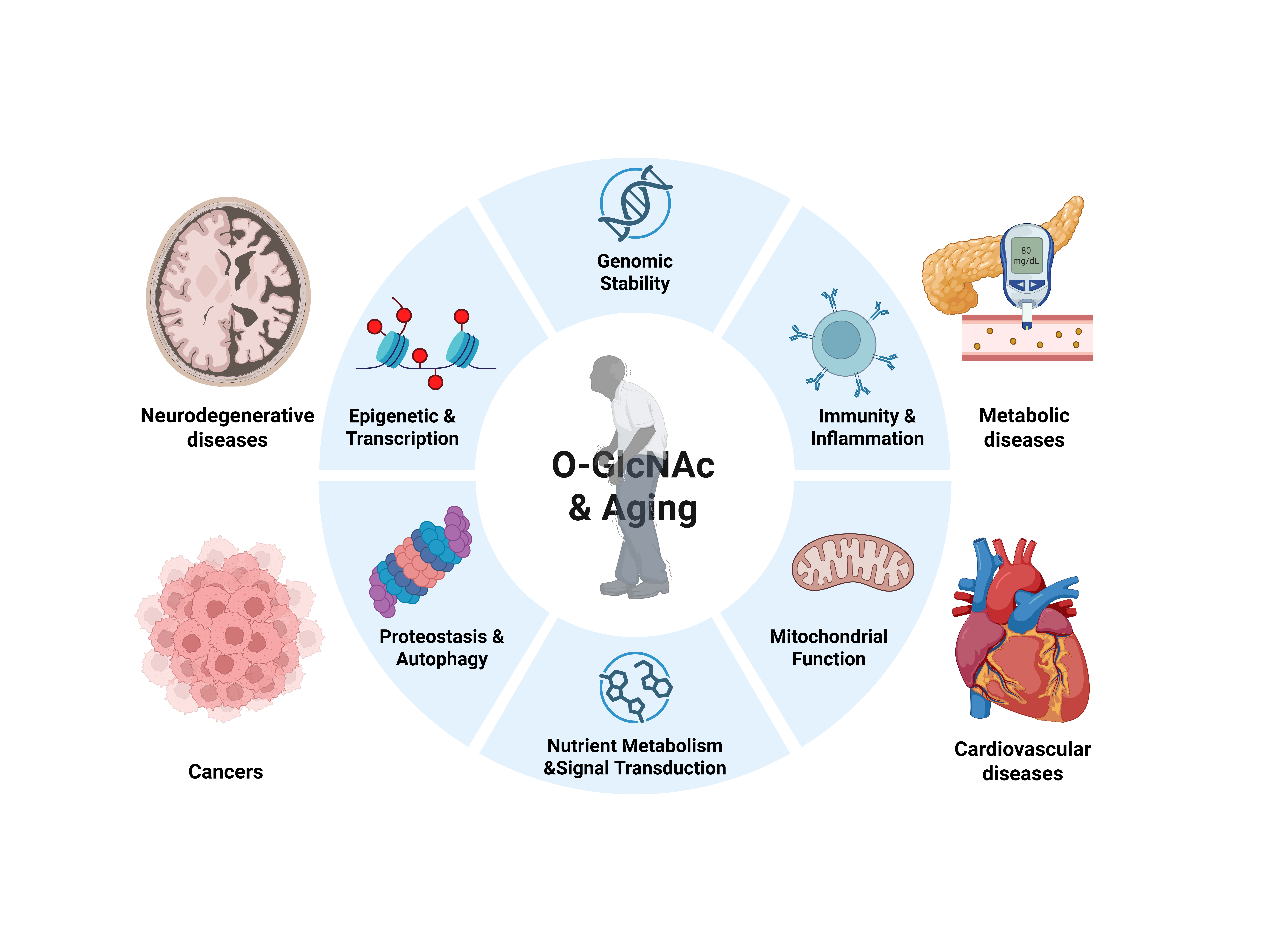
O-GlcNAcylation, a dynamic and nutrient-sensitive post-translational modification, has emerged as a key regulator of aging processes. In this review, we summarize the biological basis of O-GlcNAcylation and explore its emerging roles in aging and age-related diseases. Then we focus on the molecular mechanisms of O-GlcNAcylation in the context of key hallmarks of aging, including genomic stability, epigenetic regulation, proteostasis, autophagy, nutrient metabolism, mitochondrial function, and immunity. Finally, we discuss the therapeutic potential of targeting O-GlcNAcylation to alleviate aging-related decline and treat age-associated diseases.
Graphical Abstract
Keywords
O-GlcNAcylation, aging, age-related diseases, aging hallmarks
1. Introduction
The accelerating pace of aging of the global population poses a major public health challenge, with aging recognized as a key risk factor for numerous diseases[1]. In 2025, a comprehensive framework published in Cell updates 14 integrated hallmarks of aging, including genomic instability, telomere attrition, epigenetic alterations, loss of proteostasis, disabled autophagy, deregulated nutrient sensing, mitochondrial dysfunction, cellular senescence, stem cell exhaustion, altered intercellular communication, chronic inflammation, dysbiosis, extracellular matrix (ECM) changes, and psychosocial isolation[2]. As a nutrient sensor, O-linked β-N-acetylglucosamine (O-GlcNAc), first discovered by Torres and Hart in 1984, is a unique and dynamic post-translational modification (PTM) that integrates metabolic signaling with protein function[3]. The role of O-GlcNAcylation in aging has gained increasing attention, and growing evidence has highlighted that aging is accompanied by alterations in global O-GlcNAc levels[4]. Furthermore, O-GlcNAcylation has been implicated in regulating several of the core hallmarks of aging[5,6]. Consequently, aberrant O-GlcNAc signaling is associated not only with the aging process itself but also with a range of age-related diseases, such as neurodegenerative diseases, metabolic disorders, cancers, and cardiovascular diseases[7]. These findings suggest that targeting O-GlcNAcylation holds promise as a novel therapeutic strategy for alleviating aging-related decline and treating age-associated diseases. In this review, we first provide a brief overview of the biological basis of O-GlcNAcylation, and then focus on its emerging roles in aging and age-related diseases, address the underlying molecular mechanisms related to the core hallmarks of aging, and discuss the targeted therapeutic potential.
2. Fundamental Biology of O-GlcNAcylation
O-GlcNAcylation is a monosaccharide modification that occurs on serine (Ser) and threonine (Thr) residues of intracellular proteins in the cytoplasm, nucleus, and mitochondria[8]. This modification differs from classical O-glycosylation, which is abundant on extracellular and secreted glycoproteins and can form complex and elongated glycan structures, as well as from N-glycosylation, which features asparagine (Asn)-linked glycoconjugates on both intracellular and extracellular proteins[9]. It utilizes UDP-GlcNAc as the donor substrate, which is produced by the hexosamine biosynthetic pathway (HBP) through integrating metabolism of glucose, amino acids, fatty acids, and nucleotides (Figure 1a)[8,10]. O-GlcNAc cycling is controlled by two enzymes; O-GlcNAc transferase (OGT) is responsible for adding O-GlcNAc moieties to target proteins, while O-GlcNAcase (OGA) is required for its removal (Figure 1a)[11]. The opposing yet balanced actions of OGT and OGA establish a dynamic O-GlcNAc equilibrium that converts metabolic cues and stress signals into fine-tuned regulation of cellular processes.
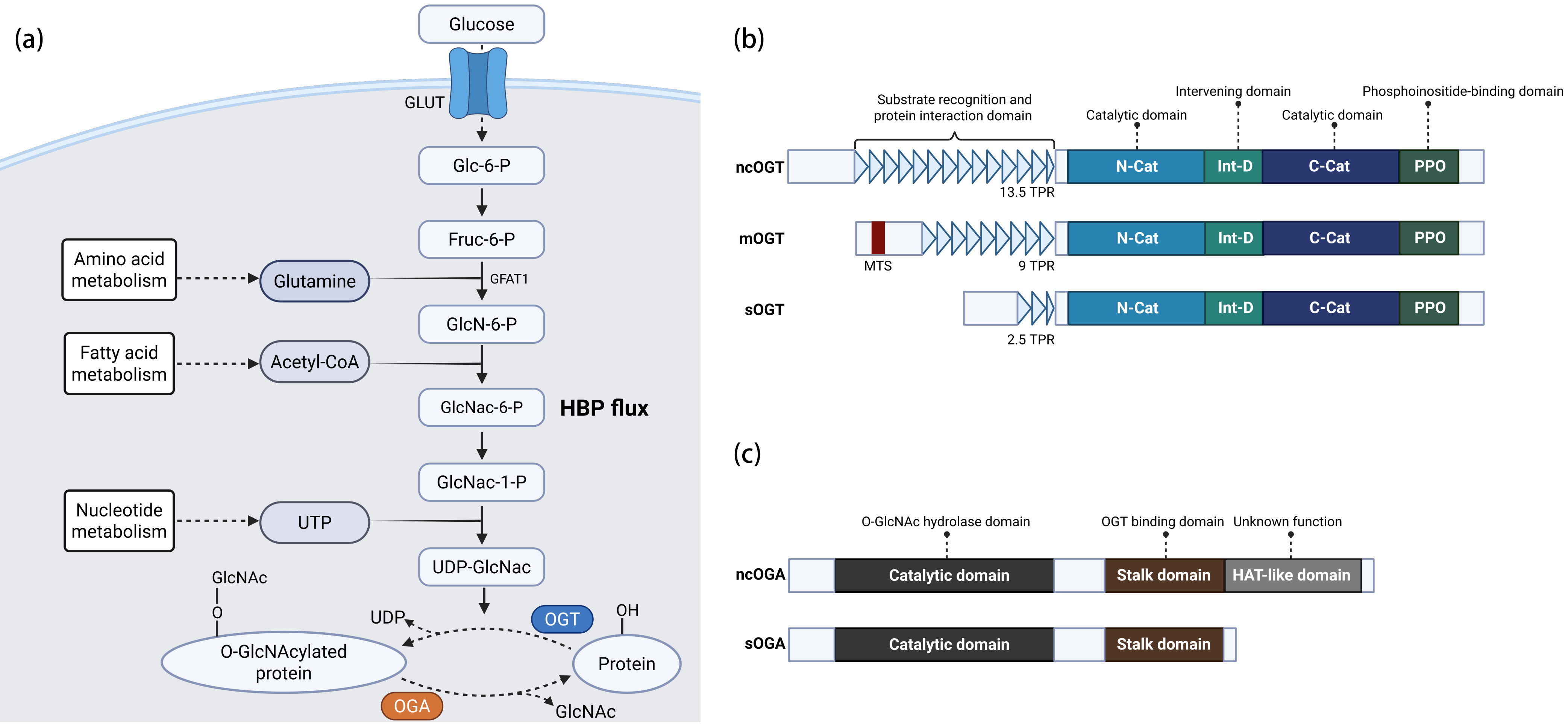
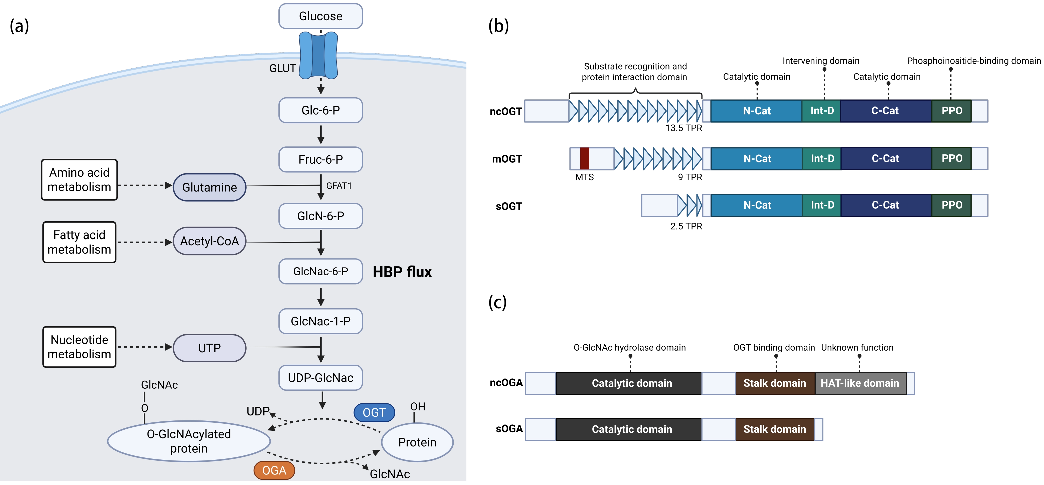
Figure 1. O-GlcNAcylation in HBP and structure of OGT and OGA. (a) The HBP and O-GlcNAc cycling; (b) Human OGT domain architecture and three OGT isoforms; (c) Human OGA domain architecture and two OGA isoforms. Created in BioRender.com. Zou, Y. (2026) https://app.biorender.com/illustrations/69fc7fb9aefcc9b85b2c912c?slideId=9e1d17be-9eaa-4f05-9a50-3183453f9505. OGT: O-GlcNAc transferase; OGA: O-GlcNAcase; TPRs: Tetratricopeptide repeats; N-Cat: N-terminal catalytic domain; Int-D: Intervening domain; C-Cat: C-terminal catalytic domain; MTS: Mitochondrial targeting sequence; ncOGT: Nucleocytoplasmic OGT; mOGT: Mitochondrial OGT; sOGT: Short OGT; ncOGA: Nucleocytoplasmic OGA; sOGA: Short OGA; HBP: hexosamine biosynthetic pathway; GLUT1: Glucose transporter type 1; GFAT1: Glutamine-fructose-6-phosphate amidotransferase 1.
2.1 Structure, substrate recognition and catalytic kinetics of OGT
The human OGT gene is located on the X chromosome at Xq13.1 and comprises 22 exons according to current genome annotations (https://www.ncbi.nlm.nih.gov/gene/8473#genomic-context). It encodes a protein characterized by multiple tandem tetratricopeptide repeats (TPRs) at the N-terminus (Figure 1b)[12]. The two catalytic domains, designated as N-catalytic (N-Cat) and C-catalytic (C-Cat), adopt a glycosyltransferase-B (GT-B) fold that is conserved among glycosyltransferases (Figure 1b)[12]. These two domains are separated by an intervening domain (Int-D) of unknown function (Figure 1b)[12]. Additionally, a phosphoinositide-binding (PPO) domain has been identified downstream of the N-Cat (Figure 1b)[12]. OGT is highly conserved across evolution; for instance, the nematode ortholog shares approximately 80% amino acid sequence identity with human OGT[13]. To date, nine characterized mutations along with numerous others under investigation are associated with OGT Congenital Disorder of Glycosylation (OGT-CDG), a disorder primarily presenting with global developmental delay, intellectual disability, variable neurological features, and subtle facial dysmorphisms[14].
Three major isoforms of OGT are produced due to alternative splicing, which differ primarily in the number of N-terminal TPR domains[12]. The longest isoform, known as nucleocytoplasmic OGT (ncOGT), contains 13.5 TPRs and a complete N-terminal TPR and a C-terminal catalytic domain, and is primarily distributed in the nucleus and cytoplasm, where it is responsible for O-GlcNAcylating the majority of substrate proteins (Figure 1b). A shorter isoform, containing 9 TPRs and a mitochondrial targeting sequence (MTS), localizes to the inner mitochondrial membrane as mitochondrial OGT (mOGT), where it is reported to maintain the mitochondrial proteome and function, although with extremely low levels (Figure 1b)[15]. The smallest isoform, short OGT (sOGT), which contains only 2.5 TPRs, remains the least characterized but is speculated to be involved in apoptosis (Figure 1b)[16].
Studies of the crystal structure of OGT in a complex with substrate peptides demonstrate that the N-terminal TPR domain functions as a dynamic gatekeeper, where substrate binding to specific TPR pockets induces a conformational change, thereby exposing the active site[17,18]. This process is facilitated by conserved Asn residues within the TPR motifs, which are crucial for substrate recognition. Although the function of Int-D is poorly understood, Blankenship et al. reported that it acts as a specific regulator of OGT interactions and substrate O-GlcNAcylation[19]. Although OGT is reported to catalyze a broad range of substrates without a strict consensus sequence, a semi-consensus sequence, including P-P-(V/T)-g(S)-(S/T)-A and (P/T)-P-(V/T)-g(T)-(S/T)-(A/T), has been identified[18]. OGT follows a random ordered Bi-Bi mechanism: UDP-GlcNAc binds first, followed by the peptide substrate, which binds above the sugar donor primarily through backbone rather than side-chain interactions[20]. Nevertheless, the scarcity of solved OGT-substrate crystal structures warrants further studies to elucidate its substrate recognition and catalytic kinetics.
2.2 Structure and substrate recognition of OGA
The OGA-encoding gene, meningioma expressed antigen 5 (MGEA5), is located on human chromosome 10q24.32 and comprises 18 exons, which undergo alternative splicing to generate two major isoforms[21]. The nucleocytoplasmic isoform (ncOGA) contains an N-terminal O-GlcNAc hydrolase domain, a C-terminal pseudo-histone acetyltransferase (HAT)-like domain of unknown function, and a central stalk region that is responsible for OGT binding (Figure 1c)[22]. In contrast, the short isoform (sOGA) lacks the HAT-like domain and localizes to the endoplasmic reticulum and lipid droplets (Figure 1c)[22]. OGA forms a stable dimer in solution, with the substrate-binding cleft formed between the catalytic domain of one monomer and the stalk region of the sister monomer[23]. Structural comparisons of the human OGA complex reveal that the GlcNAc moieties of glycopeptide substrates align perfectly within the active site, establishing a conserved interaction network that underscores the essential role of O-GlcNAc binding in substrate recognition by OGA[23].
2.3 Regulation of O-GlcNAcylation homeostasis
The regulation of OGT and OGA activity is governed by several mechanisms in response to nutrient availability and stress stimuli. A group of OGT-interacting proteins, known as adaptors, modulate enzyme activity or substrate recruitment. For instance, glucose deprivation triggers mitogen-activated protein kinase (MAPK) to recruit OGT to neurofilament-H (NFH) for O-GlcNAcylation, while fasting promotes host cell factor (HCF)-mediated targeting of OGT to peroxisome proliferator-activated receptor γ coactivator 1α (PGC-1α), enhancing its stability[24,25]. Recently, Griffin et al. established the networking of O-GlcNAc transferase interactors and substrates (NOTISE) by systematically integrating interactome and O-GlcNAcome datasets, demonstrating that the interactor BRCA1 associated protein-1 (BAP1) can site-specifically enhance or inhibit O-GlcNAcylation[26]. Additionally, biophysical and biochemical screenings have identified readers that directly recognize and bind to O-GlcNAc moieties, such as 14-3-3, which expands the known regulatory landscape of O-GlcNAcylation[27]. Furthermore, other PTMs also modulate the activities of OGT and OGA. For instance, phosphorylation of OGT at T985 by phosphoinositide 3-kinase β (PI3Kβ) and unc-51-like autophagy activating kinase 1 (ULK1) enhances its catalytic activity, thereby promoting tumor growth[28,29]. Zhang et al. demonstrated that F-Box Protein 31 (FBXO31)-mediated ubiquitination of OGT downregulated its activity to suppress endometrial malignancy[30]. In addition, OGA SUMOylation facilitates its interaction with heat shock cognate protein 70 (HSC70) and regulates autophagy[31]. OGA protein stability can also be enhanced by O-GlcNAcylation of RNA binding motif protein 14 (RBM14)[32]. Moreover, cryo-EM structures of the OGT-OGA complex reveal that their interaction leads to reciprocal inhibition of their enzymatic activities[33]. Overall, O-GlcNAc homeostasis is mutually regulated by OGT and OGA to maintain an optimal range of O-GlcNAcylation.
3. Dynamic Changes of O-GlcNAc during Aging
The dynamic changes of O-GlcNAc during cellular senescence and organismal aging have been illustrated (Table 1). In aged C. elegans, the gradual reduction in global O-GlcNAcylation and OGT expression is driven by the age-associated decline of ELT-2, a transcription factor homologous to human GATA4[34]. ELT-2 loss decreases O-GlcNAcylation and shortens the lifespan of C. elegans[34]. An elevation of the UDP-GlcNAc levels via gain-of-function mutations of glutamine fructose-6-phosphate amidotransferase 1 (GFAT1), the rate-limiting enzyme in the HBP, reduces protein aggregation and extends lifespan of C. elegans[35]. O-GlcNAcylation of another transcription factor skinhead-1 (SKN-1) at Ser470/Thr493, the ortholog of human NRF2, extends lifespan and oxidative stress resistance in C. elegans[36]. Wheatley et al. demonstrated that OGT and O-GlcNAc levels were decreased in the hippocampus of aged mice[37]. The knockout of neuronal OGT in adult mice recapitulated features of neurodegeneration, whereas increasing OGT expression in the hippocampus of aged mice rescued age-associated cognitive deficits[37]. In addition, age-related loss of O-GlcNAc levels in neural stem cells (NSCs) leads to decreased neurogenesis and increased gliogenesis in the hippocampus via loss of O-GlcNAcylation of STAT3 at Thr717[38]. Oxidative stress sensor glutathione peroxidase 7 (GPx7) interacts with OGA via disulfide bonds to restrict its activity, thereby elevating O-GlcNAcylation to counteract age-dependent oxidative stress in spinal motor neurons[39]. Failure of this protective mechanism leads to amyotrophic lateral sclerosis (ALS)-like phenotypes in aged mice, while pharmacological OGA inhibition rescues neuron loss[39]. Moreover, senescent human dental pulp stem cells exhibit elevated OGA expression and reduced O-GlcNAcylation of Krüppel-like factor 2 (KLF2) at Ser177, which suppresses mitophagy[40]. OGT-mediated O-GlcNAcylation of leptin promotes its degradation and inactivates the nuclear factor-κB (NF-κB) pathway, thereby inhibiting mouse mesenchymal stem cell (MSC) senescence[41]. Contradictorily, increased O-GlcNAc levels are observed in other aged organs, such as liver, along with highly O-GlcNAcylated carbamoyl phosphate synthetase 1 (CPS1)[42]. In aged mouse testis, elevated O-GlcNAcylation leads to meiotic defects, thereby causing a decline in spermatogenesis[43]. Enhancing O-GlcNAc levels via OGA inhibition impairs spermatogenesis in young mice, whereas OGT inhibition rescues age-related spermatogenic impairment in aged testis[43]. Moreover, oga mutant Drosophila with increased O-GlcNAc levels exhibited a shorter lifespan[44]. In addition, elevated O-GlcNAcylation stabilizes GATA4 via O-GlcNAc modification at Ser406, thereby driving the senescence-associated secretory phenotype (SASP) in osteoarthritic cartilage, while inhibition of OGT attenuates osteoarthritis[45]. Taken together, the dynamics of O-GlcNAcylation during aging vary across cell types and tissues, underscoring the critical importance of balanced OGT and OGA activities. It is probably governed by differential metabolic states or upstream regulatory mechanisms for maintaining O-GlcNAc homeostasis in different cell types and tissues. Disruption of this balance during aging leads to either abnormally high or low O-GlcNAc levels, both of which contribute to aging-related defects. Therefore, systematic analysis of tissue-specific O-GlcNAc dynamics and the underlying mechanisms during aging is necessary.
Table 1. Targets of O-GlcNAc during organismal aging and cellular senescence.
| Target/Site | O-GlcNAc change | Model | Aging-related processes | Main effect | Reference |
| N/A | ↓ | C. elegans | Organismal aging | Age-associated decline of ELT-2 reduces OGT expression | [34] |
| N/A | ↑ | C. elegans | Organismal aging | Gain-of-function of GFAT1 reduces protein aggregation and extends lifespan | [35] |
| SKN-1(S470 / T493) | ↑ | C. elegans | Organismal aging | Extends lifespan and increases oxidative stress resistance | [36] |
| N/A | ↓ | Mouse | Brain aging | Recapitulates features of neurodegeneration | [37] |
| STAT3 (T717) | ↓ | Mouse | NSC aging | Decreases neurogenesis and increases gliogenesis | [38] |
| N/A | ↑ | Mouse; human ALS samples | Spinal motor neurons aging | GPx7 suppresses OGA activity and elevates O-GlcNAcylation to counteract age-dependent oxidative stress | [39] |
| KLF2 (S177) | ↓ | Human dental pulp stem cells | Stem cell senescence | Inhibits mitophagy and promotes cell senescence | [40] |
| Leptin (S50) | ↑ | Mouse | MSC senescence | Suppresses NF-κB signaling and inhibits MSC senescence | [41] |
| CPS1 (S537) | ↑ | Mouse | Liver aging | Suppresses CPS1 activity and promotes liver aging | [42] |
| N/A | ↑ | Mouse | Reproductive aging | Disrupts meiotic integrity and causes a decline in spermatogenesis | [43] |
| N/A | ↑ | Drosophila | Organismal aging | Oga mutant exhibits a shorter lifespan | [44] |
| GATA4 (S406) | ↑ | Mouse; human osteoarthritic samples | Osteoarthritic cartilage aging | Drives SASP | [45] |
OGT: O-GlcNAc transferase; GFAT1: glutamine fructose-6-phosphate amidotransferase 1; SKN-1: skinhead-1; NSC: neural stem cell; ALS: amyotrophic lateral sclerosis; GPx7: glutathione peroxidase 7; OGA: O-GlcNAcase; KLF2: Krüppel-like factor 2; MSC: mesenchymal stem cell; NF-κB: nuclear factor-κB; CPS1: carbamoyl phosphate synthetase 1; SASP: senescence-associated secretory phenotype.
4. Molecular Mechanisms Linking O-GlcNAcylation to Aging Hallmarks
Recent advances in O-GlcNAc detection technologies and multi-omics approaches have enabled the identification of over 8,000 O-GlcNAcylated proteins and tens of thousands of modification sites[46]. Accordingly, specialized glycosylation databases, such as O-GlcNAcAtlas 4.0 (https://oglcnac.org/atlas/) and The O-GlcNAc Database (https://www.oglcnac.org/), have been established[47,48]. Notably, O-GlcNAcylation frequently interacts with other PTMs, including phosphorylation, acetylation, ubiquitination, and poly(ADP-ribosyl)ation, either by modulating "writers" and "erasers", or by directly competing for the same amino acid residues on substrate proteins[49]. These modified substrates participate broadly in diverse biological processes. In the following sections, we will focus on summarizing the regulatory roles of O-GlcNAcylation in the context of aging-related hallmarks (Figure 2 and Table 2).
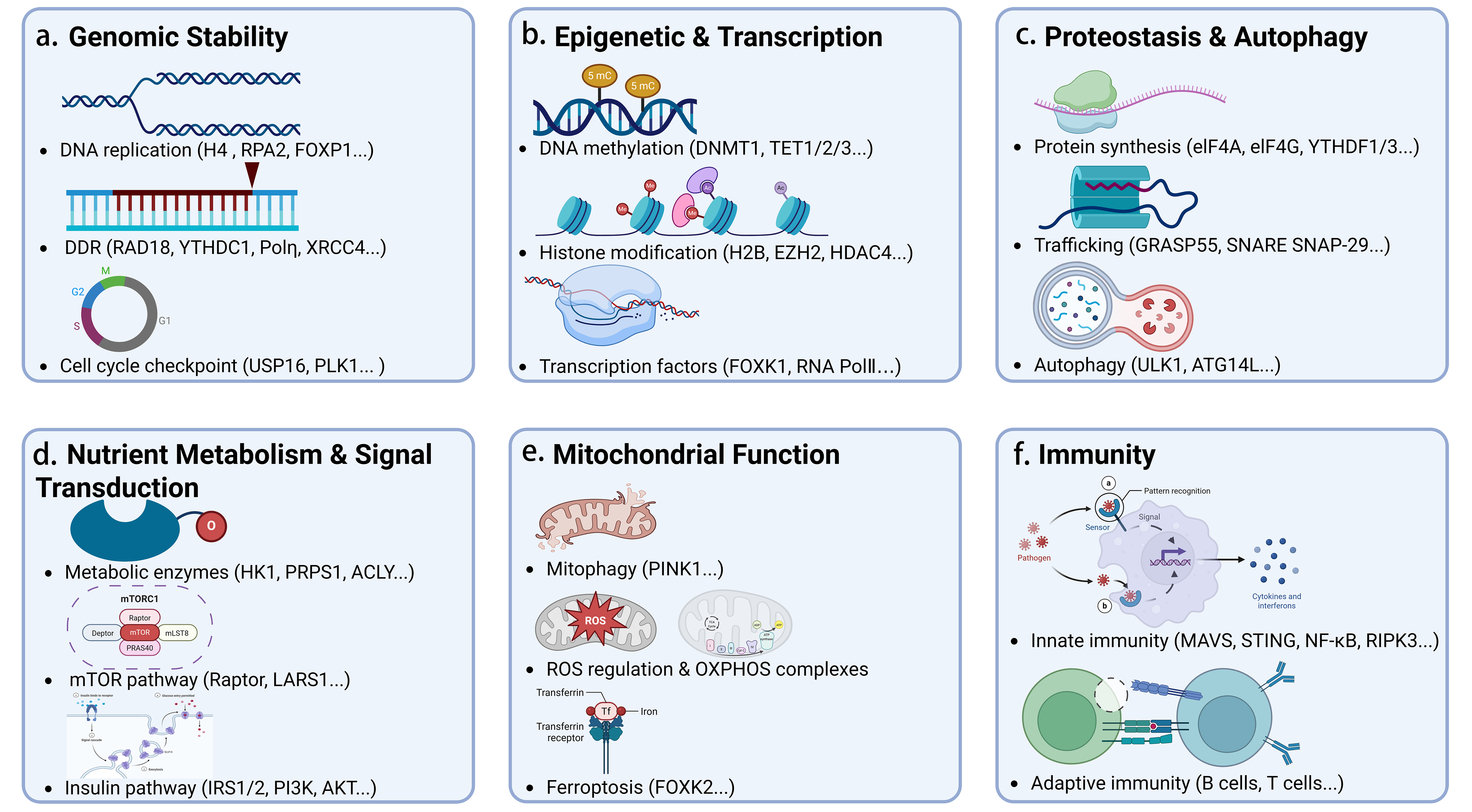
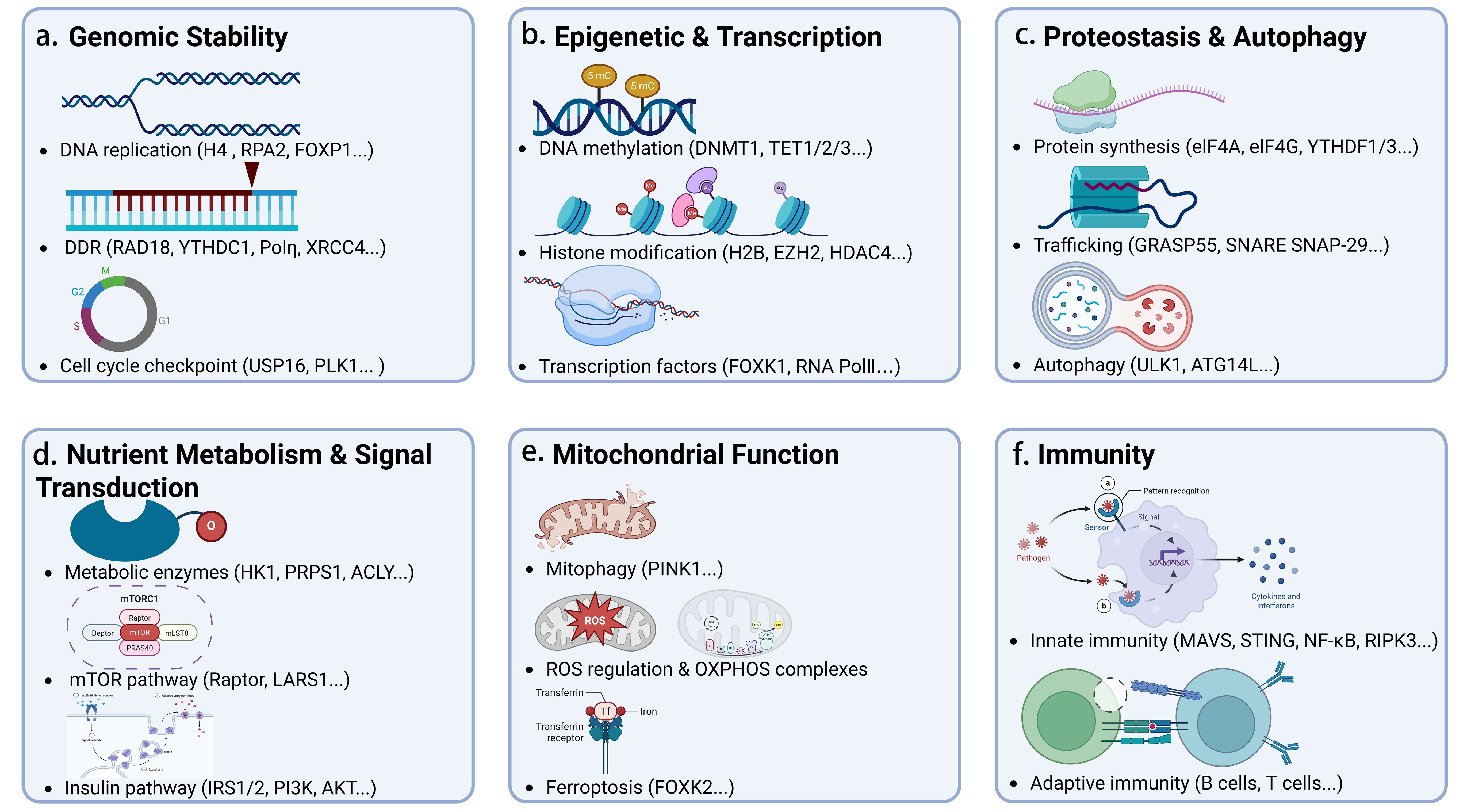
Figure 2. The regulatory roles of O-GlcNAcylation in the key hallmarks of aging. (a) Maintaining genomic stability; (b) Coordinating epigenetic landscapes and transcription; (c) Regulating proteostasis and autophagy; (d) Modulating nutrient metabolism and signal transduction; (e) Governing mitochondrial function; (f) Shaping immunity and inflammation. Created in BioRender.com. Zou, Y. (2026). https://app.biorender.com/illustrations/6a04129a8ca63ef18f87dc53?slideId=9d5c25b2-7e3f-4010-b43e-8a70c7f29156. DDR: DNA damage response; DNMT1: DNA methyltransferase 1; TET1/2/3: ten-eleven translocation methylcytosine dioxygenase 1/2/3; H2B: histone H2B; EZH2: enhancer of zeste homolog 2; HDAC4: histone deacetylase 4; FOXO1: forkhead box O1; eIF4A / eIF4G: eukaryotic translation initiation factor 4A / 4G; YTHDF1/3: YTH N6-methyladenosine RNA-binding protein 1/3; GRASP55: Golgi reassembly-stacking protein 55; SNARE: soluble N-ethylmaleimide-sensitive factor attachment protein receptor; SNAP-29: synaptosomal-associated protein 29; ULK1: unc-51-like autophagy activating kinase 1; ATG14L: autophagy-related 14-like protein; HK1: hexokinase 1; PRPS1: phosphoribosyl pyrophosphate synthetase 1; ACLY: ATP citrate lyase; mTOR: mammalian target of rapamycin; LARS1: leucyl-tRNA synthetase 1; IRS1/2: insulin receptor substrate 1/2; PI3K: phosphoinositide 3-kinase; AKT: protein kinase B; PINK1: PTEN-induced kinase 1; ROS: reactive oxygen species; OXPHOS: oxidative phosphorylation; FOXK2: forkhead box K2; MAVS: mitochondrial antiviral-signaling protein; STING: stimulator of interferon genes; NF-κB: nuclear factor-κB; RIPK3: receptor-interacting serine/threonine-protein kinase 3.
Table 2. Targets of O-GlcNAc in the context of aging-related hallmarks.
| Target/Site | Model | Aging-related hallmarks | Main effect | Reference |
| H4 (S47) | Cell lines | Genome stability | Recruits DDK and promotes DNA replication | [52] |
| RPA2 (S4/ S8) | Cell lines | Genome stability | Impairs Chk1 activation and induces replication stress | [53] |
| FOXP1 (S396) | Cell lines | Genome stability | Inhibits its interaction with ATR and response to replication stress | [54] |
| YTHDC1 (S396) | Cell lines | Genome stability | Enhances its binding with m6A RNA and HR | [55] |
| Polη (T457) | Cell lines | Genome stability | Promotes its ubiquitination and ensures TLS fidelity | [56] |
| RAD18 (S130/ S164/ T468) | Cell lines | Genome stability | Promotes its E3 ligase activity in HR and TLS repair | [57] |
| NONO (S147) | Cell lines | Genome stability | Stabilizes its interaction with SFPQ and regulates NHEJ | [58] |
| XRCC4 (T308) | Cell lines | Genome stability | Enhances its stability and regulates NHEJ | [60] |
| Ogg1 | Genome stability | Impairs its activity and BER | [61] | |
| USP16 (T203) | Cell lines | Genome stability | Promotes cell-cycle progression | [64] |
| DNMT1 (S878) | Cell lines | Epigenetic regulation | Suppresses its enzymatic activity and DNA methylation | [71] |
| TET2 | Cell lines | Epigenetic regulation | Recruits OGT to chromatin and promotes histone O-GlcNAcylation during transcription | [72] |
| H2B (S112) | Cell lines | Epigenetic regulation | Promotes transcription and DNA repair | [74] |
| H4 (T71) | Cell lines | Epigenetic regulation | Maintains heterochromatin | [75] |
| EZH2 (S76/ S729) | Cell lines | Epigenetic regulation | Promotes H3K27me3 deposition | [76] |
| FOXK1 | Cell lines; Mouse | Epigenetic regulation | Reprograms transcription | [77] |
| HIRA (S231) | Primary human fibroblasts | Epigenetic regulation | Regulates nucleosome assembly and accelerates cellular senescence | [82] |
| SWI/SNF | Primary human fibroblasts | Epigenetic regulation | Regulates chromatin remodeling and activates SASP genes | [83] |
| eIF4A (S322/323), eIF4G (S61) | Cell lines | Proteostasis | Regulates translation initiation and fine-tunes protein synthesis | [86] |
| YTHDF1 (S157/196-198), YTHDF3 (T205/S229) | Cell lines | Proteostasis | Attenuates translation and regulates stress granule dynamics | [87] |
| Gigaxonin (S272/T277) | Cell lines | Proteostasis | Regulates intermediate filament turnover | [90] |
| OTX2 (S135/S136/T137) | Cell lines | Proteostasis | Enhances its solubility and limits protein aggregation | [91] |
| GRASP55 (S389/S390/T403/ T404/T413) | Cell lines | Autophagy | Regulates protein trafficking and secretion, and autophagosome-lysosome fusion | [93] |
| SNAP29 (S2/S61/T130/S153) | Cell lines; C. elegans | Autophagy | Blocks autophagosome-lysosome fusion | [94,95] |
| ULK1 (T754) | Cell lines | Autophagy | Enables it to bind and phosphorylate ATG14L, and initiates autophagy | [96] |
| ULK1 (S409/ S410) | Cell lines | Autophagy | Stabilizes ULK1 and promotes autophagosome-lysosome fusion | [97] |
| Raptor (T700) | Cell lines; Mouse | Nutrient sensing | Regulates glucose-induced mTORC1 activation | [99] |
| LARS1 (S1042) | Cell lines | Nutrient sensing | Couples leucine metabolic fate to mTORC1 | [100] |
| HK1 (T259) | Cell lines | Nutrient sensing | Organizes glycolytic metabolon | [102] |
| PRPS1 (S83/T166) | Cell lines | Nutrient sensing | Promotes nucleotide synthesis and tumorigenesis | [103] |
| VDAC2 | Cell lines | Mitochondrial function | Regulates mitochondrial function | [107] |
| FOXK2 (S424) | Cell lines | Mitochondrial function | Suppresses ferroptosis | [109] |
| Drp1 | Cell lines | Mitochondrial function | Causes dramatic changes in mitochondrial morphology and mass | [110] |
| Lyn (S19) | Mouse | Immune regulation and inflammation | Impairs BCR signaling, enhances mature B cell apoptosis, and reduces antibody production | [114] |
| FOXP3 (T38/S57/S58/S270/S273) | Mouse | Immune regulation and inflammation | Regulates T cell proliferation, development, transformation, and differentiation | [115] |
| MAVS (S324/T328/S329/S300/S338/T342/S347/S366) | Mouse and BMDMs | Immune regulation and inflammation | Promotes its ubiquitination and IRF3 activation to drive IFN-β production | [116,117] |
| STING (T229) | Mouse | Immune regulation and inflammation | Facilitates its ubiquitination and antiviral function | [118] |
| NF-κB (T305) | Cell lines | Immune regulation and inflammation | Activates its transcription activity and downstream cytokine production | [119] |
| TAB1 (S395) | Cell lines | Immune regulation and inflammation | Activates TAK1 signaling and cytokine production | [120] |
| RIPK3 (T467) | BMDMs | Immune regulation and inflammation | Limits septic inflammation | [121] |
| STAT1 | Mouse | Dysbiosis | Regulates gut inflammation | [122] |
| FOXO1 | Mouse | Dysbiosis | Regulates L cell differentiation | [123] |
| Dumpy | Drosophila | Extracellular matrix | Regulates cell-matrix interactions | [124] |
| E-cadherin, FAK, Zyxin, and Integrin | Cell lines | Cel communication | Regulates cell adhesion | [125,126] |
DDK: DBF4-dependent kinase; HR: homologous recombination; TLS: translesion synthesis; NHEJ: non-homologous end joining; BER: base excision repair; OGT: O-GlcNAc transferase; SWI/SNF: switch/sucrose non-fermentable; SASP: senescence-associated secretory phenotype; mTORC1: mammalian target of rapamycin complex 1; BCR: B-cell receptor; IRF3: interferon regulatory factor 3; NF-κB: nuclear factor-κB; ATR: ataxia telangiectasia and Rad3-related protein; m6A: N6-methyladenosine; Chk1: checkpoint kinase 1; BMDMs: bone marrow-derived macrophages.
4.1 Maintaining genomic stability
Genomic instability, one of the critical hallmarks of aging, is characterized by elevated DNA damage and impaired DNA repair capacity, leading to cell cycle arrest and thereby cellular senescence[50]. The roles of O-GlcNAcylation in safeguarding genomic integrity have been intensively explored. A comprehensive review by Wu et al. elaborated on the functions of OGT and OGA in DNA replication and various DNA damage response (DDR) pathways, including homologous recombination (HR), non-homologous end joining (NHEJ), base excision repair (BER), nucleotide excision repair (NER), and translesion synthesis (TLS) repair (Figure 2a)[51]. For example, OGT-mediated O-GlcNAcylation of histone H4 at Ser47 facilitates the recruitment of the DBF4-dependent kinase (DDK) to chromatin, leading to MCM helicase phosphorylation and activation of replication origins[52]. O-GlcNAcylation of replication protein RPA2 at Ser4/Ser8 antagonizes its phosphorylation, thereby impairing Chk1 activation and inducing replication stress[53]. Conversely, forkhead box P1 (FOXP1) O-GlcNAcylation inhibits its interaction with ATR, while replication stress-induced, Chk1-dependent FOXP1 phosphorylation at Ser396 relieves this inhibition to promote ATR activation[54]. Li et al. revealed that O-GlcNAcylation of YTHDC1 at Ser396, an m6A reader, enhanced its binding with m6A RNA and ionizing radiation-induced foci (IRIF) formation to promote HR repair[55]. O-GlcNAcylation of DNA polymerase η (Polη) at Thr457 promotes its CRL4CDT2-dependent ubiquitination and timely removal from replication forks, ensuring TLS fidelity[56]. Additionally, RAD18 O-GlcNAcylation at Ser130/Ser164/Thr468 promotes its E3 ligase activity, facilitating PCNA monoubiquitination and Polη IRIF formation to coordinate both HR and TLS repair[57]. In NHEJ repair, O-GlcNAcylation of NONO at Ser147 stabilizes its interaction with splicing factor proline and glutamine rich (SFPQ) to regulate SET and mariner transposase domain methyltransferase (SETMAR) alternative splicing, while OGA-mediated deglycosylation is required for timely removal of NONO and Ku70/80 from damage sites[58,59]. XRCC4 O-GlcNAcylation at Thr308 inhibits TRIM21-mediated ubiquitin-proteasomal degradation, enhancing its stability and conferring DSB repair resistance in cancer cells[60]. Moreover, O-GlcNAcylation of BER factor Ogg1 induced by hyperglycemia impairs its activity, leading to accumulation of oxidative mtDNA lesions in diabetic hearts[61].
Cell cycle checkpoints are surveillance mechanisms activated by the DDR signaling network to halt cell cycle progression, thereby allowing time for DNA repair and preserving genomic integrity[62]. Quantitative chemoproteomic profiling has revealed dynamic O-GlcNAcylation changes during cell cycle progression[63]. Moreover, ubiquitin specific peptidase 16 (USP16) O-GlcNAcylation at Thr203 promotes deubiquitination of H2AK119ub to regulate gene expression and also facilitates polo-like kinase 1 (PLK1) deubiquitination, ensuring proper chromosome segregation and cytokinesis[64]. Nutrient-driven hyper-O-GlcNAcylation activates DDR and promotes hyperproliferation[65]. In contrast, suppression of O-GlcNAcylation has been reported to promote senescence-like phenotypes in cancer cells by impairing DNA damage repair and reducing proliferative capacity[66]. Together, these findings highlight O-GlcNAcylation coordinates replication origin licensing, DNA repair pathway choice, and cell cycle checkpoint activity to sustain genome stability and cell cycle progression. Nevertheless, the specific O-GlcNAc targets governing genome stability during cellular senescence and organ aging per se remain to be elucidated.
4.2 Coordinating epigenetic landscapes and transcription
Aging is marked by multilayered epigenetic alterations, including global DNA hypomethylation, altered histone modifications (e.g., reduced H3K9me3), dysregulated chromatin remodeling, and heterochromatin loss[67]. These disturbances collectively drive aging-related gene dysregulation and genomic instability[68]. Extensive studies have demonstrated the involvement of O-GlcNAcylation in epigenetic regulation, as comprehensively reviewed elsewhere[69,70]. O-GlcNAcylation contributes to epigenetic control at multiple levels (Figure 2b). Firstly, O-GlcNAcylation modulates DNA methylation by targeting key enzymes: O-GlcNAcylation of DNA methyltransferase 1 (DNMT1) at Ser878 suppresses its enzymatic activity[71]; OGT forms stable complexes with ten-eleven translocation (TET) proteins, particularly TET2, which facilitates OGT recruitment to chromatin and regulates histone O-GlcNAcylation during gene transcription[72]. Secondly, histones H2A, H2B, H3, and H4 are all direct substrates of O-GlcNAcylation[73]. For instance, O-GlcNAcylation of H2B at Ser112 promotes its Lys120 monoubiquitylation, thereby regulating transcription and DNA repair[74]. Additionally, a novel O-GlcNAcylation at Thr71 of H4 is a component of heterochromatin[75]. Beyond direct histone modification, O-GlcNAcylation also influences histone modifications by modulating the activity of their writers and erasers. O-GlcNAcylation of the histone methyltransferase EZH2 at Ser76 and Ser729 regulates its stability and enzymatic activity, thereby promoting H3K27me3 deposition[76]. Furthermore, O-GlcNAcylation impacts the epigenetic landscape and gene expression by modifying transcription factors, such as FOXK1 and FOXA1, as well as that of RNA polymerase II (PolII)[77-79]. Moreover, OGT is required for Polycomb-mediated gene repression, highlighting a functional role of O-GlcNAcylation in Polycomb regulation[80]. In a separate mechanism, O-GlcNAcylation of the Polycomb group repressor polyhomeotic prevents aberrant protein aggregation and contributes to its functional maintenance[81].
Although extensive studies have elucidated the roles of O-GlcNAcylation in epigenetic regulation, how it contributes to epigenetic alterations during cellular senescence and organismal aging remains largely unexplored. It is reported that OGT overexpression accelerates, whereas OGT depletion delays senescence in primary human fibroblasts, via O-GlcNAcylation of the histone cell cycle regulator HIRA at Ser231 and regulation of nucleosome assembly[82]. Recently, Zhang et al. identified a dynamic chromatin O-GlcNAcylation profile during oncogene-induced senescence in primary human fibroblasts, revealing that O-GlcNAc facilitates the formation of dual-function complexes: the chromatin remodeling complex switch/sucrose non-fermentable (SWI/SNF) to activate SASP genes, whereas repressor complex NuRD to repress cell cycle regulators[83].
4.3 Regulating proteostasis and autophagy
Aging-associated loss of proteostasis disrupts the dynamic balance of protein synthesis, folding, transport, and degradation[84]. Moreover, disabled macroautophagy leads to accumulation of aberrant protein aggregates, resulting in cellular damage[85]. As a key PTM, O-GlcNAcylation coordinates multiple facets of proteostasis and autophagic flux (Figure 2c). O-GlcNAcylation of the translation initiation factors eIF4A at Ser322/323 and eIF4G at Ser61 fine-tunes protein synthesis[86]. Additionally, O-GlcNAcylation of RNA m6A readers YTHDF1 at Ser157 and Ser196-198, and YTHDF3 at Thr205 and Ser229, attenuates their translation-promoting function and regulates stress granule dynamics[87]. Importantly, O-GlcNAcylation stabilizes nascent polypeptide chains by counteracting cotranslational ubiquitination and controls ubiquitin-mediated proteasomal degradation[88,89]. O-GlcNAcylation of the E3 ligase adaptor gigaxonin at Ser272 and Thr277 regulates intermediate filament turnover[90], while O-GlcNAcylation of maternal transcription factor OTX2 at Ser135, Ser136, and Thr137 enhances its solubility and limits protein aggregation[91]. Furthermore, O-GlcNAcylation regulates protein trafficking and secretion by modifying coat protein complexes COPI/COPII, Golgi protein GRASP55, and membrane fusion protein complex SNARE[92]. Upon glucose starvation, reduced O-GlcNAcylation of GRASP55 promotes its puncta formation and facilitates autophagosome-lysosome fusion by bridging LC3-II and LAMP2[93]. The SNARE complex component, SNAP-29, is O-GlcNAcylated in a nutrient-dependent manner; nutrient deprivation reduces SNAP-29 O-GlcNAcylation, thereby promoting SNARE complex formation and autophagosome maturation, whereas arsenic-induced hyper-O-GlcNAcylation of SNAP-29 conversely blocks autophagosome-lysosome fusion, leading to autophagic dysfunction[94,95]. The key autophagy initiation factor ULK1 is O-GlcNAcylated at Thr754, enabling it to bind and phosphorylate ATG14L, thereby initiating autophagy[96]. Additionally, during HPV infection, ULK1 O-GlcNAcylation at Ser409/Ser410 stabilizes the kinase by antagonizing chaperone-mediated autophagy, thereby promoting autophagosome-lysosome fusion[97]. Of note, O-GlcNAcylation plays a critical role in protein aggregate formation, especially during neuronal aging and neurodegenerative diseases, which will be discussed in the next section. Collectively, these findings reveal O-GlcNAcylation as a master regulator that integrates nutrient and stress signals to orchestrate proteostasis and autophagy across diverse physiological and pathological contexts.
4.4 Modulating nutrient metabolism and signal transduction
Nutrient availability regulates O-GlcNAcylation not only through UDP-GlcNAc levels but also by modulating OGT/OGA expression and location, adaptor proteins, and substrate quality[98]. Reciprocally, O-GlcNAcylation functions as a stress and nutrition sensor, responding to environmental and physiological signals to regulate metabolic signal transduction pathways, thereby impacting glucose, lipid, and nucleotide metabolism (Figure 2d)[98]. Mechanistically, O-GlcNAcylation directly modulates key nodes of major nutrient-sensing pathways. In the mammalian target of rapamycin complex 1 (mTORC1) pathway, O-GlcNAcylation of Raptor at Thr700 is associated with glucose-induced mTORC1 activation[99], and O-GlcNAcylation of leucyl-tRNA synthetase 1 (LARS1) at Ser1042 integrates leucine and glucose availability to regulate mTORC1 and leucine metabolism[100]. Crosstalk between O-GlcNAc and AMPK systems suggests cooperative regulation of nutrient-sensitive cellular processes governing metabolism, growth, proliferation, and tissue function[101]. Moreover, O-GlcNAcylation further extends to metabolic enzymes and energy sensors. For example, modification of hexokinase-1 (HK1) at Thr259 promotes glycolytic metabolon assembly on the mitochondrial outer membrane, coordinating glycolytic and mitochondrial adenosine triphosphate (ATP) production[102], while O-GlcNAcylation directly regulates de novo nucleotide synthesis via modification of the rate-limiting enzyme phosphoribosyl pyrophosphate synthetase 1 (PRPS1) at Ser83 and Thr166[103]. Thus, these studies position O-GlcNAcylation as a master hub orchestrating crosstalk among major nutrient signaling pathways to maintain metabolic homeostasis, whereas its disruption underlies aging-associated metabolic disorders and cancer progression, a topic we will discuss in detail in the next section.
4.5 Governing mitochondrial function
Aging is associated with mitochondrial dysfunction, leading to an imbalance between energy supply and demand, thereby contributing to age-related diseases such as neurodegenerative and cardiovascular diseases[104]. Emerging evidence indicates that O-GlcNAcylation broadly targets mitochondrial proteins and modulates multiple mitochondrial functions, including biogenesis, electron transport chain activity, oxidative stress, mitophagy, and ferroptosis (Figure 2e)[105]. Proteomic profiling has revealed widespread O-GlcNAcylation of mitochondrial proteins, particularly within the oxidative phosphorylation (OXPHOS) system, directly impacting mitochondrial respiration, ATP production, and reactive oxygen species (ROS) release[106]. For instance, the mitochondrial voltage-dependent anion channel 2 (VDAC2) serves as an O-GlcNAc substrate[107]. Additionally, OGT regulates mitochondrial function by maintaining PTEN-induced kinase 1 (PINK1)-dependent mitophagy[108], and ROS-induced O-GlcNAcylation of the transcription factor FOXK2 inhibits ferroptosis[109]. Consistent with these findings, increased O-GlcNAcylation of dynamin-related protein 1 (Drp1) via OGA deletion causes dramatic changes in mitochondrial morphology and mass[110]. O-GlcNAcylation couples glucose metabolism to mitochondrial bioenergetics, sustaining energy homeostasis and mitochondrial plasticity in neurons of the brain[111]. As will be discussed below, dysregulation of mitochondrial O-GlcNAcylation drives pathological consequences in aging-related diseases.
4.6 Shaping immunity and inflammation
Inflammaging, defined as the long-term consequence of chronic low-grade inflammation during aging, is a major driver of aging and age-related diseases[112]. O-GlcNAcylation plays a crucial regulatory role in modulating immune cell homeostasis, activation, and inflammatory responses (Figure 2f)[113]. In the adaptive immune system, O-GlcNAcylation is indispensable for B cell development and function; its deficiency impairs B-cell receptor (BCR) signaling, enhances mature B cell apoptosis, and reduces antibody production, partly through O-GlcNAcylation of tyrosine protein kinase Lyn at Ser19[114]. Similarly, defects in protein O-GlcNAcylation of FOXP3 at Thr38, Ser57, Ser58, Ser270, and Ser273 impair T cell proliferation, development, transformation, and differentiation[115]. In the innate immune system, O-GlcNAcylation is critical for antiviral immunity via modifying key signaling adaptors including mitochondrial antiviral signaling protein (MAVS), where O-GlcNAcylation promotes K63-linked ubiquitination and interferon regulatory factor 3 (IRF3) activation to drive IFN-β production[116,117]. Moreover, stimulator of interferon genes 1 (STING) is O-GlcNAcylated at Thr229 upon HSV-1 infection, which facilitates its K63-linked ubiquitination and antiviral function[118]. Deficient O-GlcNAcylation of NF-κB at Thr305 impaired p300-mediated acetylation of NF-κB on Lys310, thereby reducing its transcriptional activity and downstream cytokine production[119]. Additionally, O-GlcNAcylation of TGF-β activated kinase 1 (TAK1) binding partner TAB1 at Ser395 is required for full TAK1 activation and downstream NF-κB-mediated cytokine production[120]. Conversely, OGT also exerts anti-inflammatory effects by O-GlcNAcylating receptor interacting serine/threonine kinase 3 (RIPK3) at Thr467, which blocks RIPK3-RIPK1 interaction and necroptosis, thereby limiting septic inflammation, indicating that O-GlcNAcylation functions as a bidirectional regulator of inflammation[121]. Collectively, these findings demonstrate that O-GlcNAcylation is a key regulator of immune cell homeostasis and inflammatory signaling. However, whether O-GlcNAcylation contributes to inflammaging requires further study.
4.7 Other hallmarks
Although not extensively studied, O-GlcNAcylation has also been implicated in other hallmarks of aging. For instance, reduced OGT expression in human inflammatory bowel diseases (IBD) leads to defective O-GlcNAcylation of STAT1, which compromises epithelial barrier integrity, induces Paneth cell dysfunction, and promotes microbial dysbiosis, thereby predisposing to intestinal inflammation[122]. Similarly, deletion of Ogt in intestinal L cells disrupts FOXO1 O-GlcNAcylation, which drives L cell hyperplasia, elevates GLP-1 secretion, and perturbs gut microbial composition[123]. O-GlcNAcylation of Dumpy, a membrane-anchored extracellular protein, is essential for epithelial cell-matrix interaction[124]. Moreover, key regulators of cell adhesion and focal adhesion turnover, including E-cadherin, focal adhesion kinase (FAK), Zyxin, and Integrin, have been identified as O-GlcNAcylated targets and function in cell communication[125,126]. While the roles of O-GlcNAcylation in cellular senescence and stem cell exhaustion have been discussed in Section 3, its involvement in telomere attrition and physiological isolation remains largely unexplored, with only limited studies available to date.
5. O-GlcNAc and Age-Related Diseases
Aberrant protein O-GlcNAcylation plays a critical role in the onset and progression of various age-related diseases, including neurodegenerative diseases, metabolic disorders, cancers, and cardiovascular diseases (Table 3).
Table 3. Targets of O-GlcNAc in age-related diseases.
| Target/Site | O-GlcNAc change | Model | Aging-related diseases | Main effect | Reference |
| Tau (T123/S208/S400/S409/S412) | ↓ | Human AD samples, mouse | AD | Promotes abnormal Tau aggregation and neurofibrillary tangle formation | [128,129] |
| a-synuclein (T72) | ↑ | Cell lines, mouse | PD | Reduces α-synuclein aggregation | [131,132] |
| TDP-43 (T199/T233) | ↑ | Cell lines, Drosophila | ALS | Reduces TDP-43 pathology | [134] |
| ATP5A (T432) | ↓ | Human samples; Mouse; Cell lines | AD | Impairs mitochondrial function and ATP production | [136] |
| RIPK3 (T467) | ↑ | Human samples; Mouse | AD | Suppresses necroptosis | [137] |
| IRS1/2, PI3K, and AKT | ↑ | Cell lines | T2DM | Regulates insulin signaling | [139] |
| mTORC1 | ↓ | Mouse | T2DM | Drives diabetes progression | [140] |
| Bmal1 | ↑ | Mouse | T2DM | Impairs diabetes-associated cognitive function | [141] |
| ChREBP | ↑ | Mouse | Obesity | Enhances hepatic lipogenesis | [143] |
| PLIN1 | ↑ | Mouse | Obesity | Inhibits lipolysis | [145] |
| TopIIα (S1469) | ↑ | Human; Cell | Breast cancer | Enhances its chromatin binding activity and drives chemoresistance | [152] |
| MITF (S49) | ↑ | Mouse | Breast cancer | Promotes its nuclear translocation and suppresses CDK4/6 inhibitor-induced senescence | [153] |
| FOXA1 (T432 / S441/ S443) | ↑ | Human; Mouse; Cell | Breast cancer | Enhances its stability and chromatin assembly, and promotes cancer metastasis | [78] |
| PGK1 (T255) | ↑ | Human; Mouse; Cell | Colorectal cancer | Coordinates glycolysis and TCA cycle to drive tumor growth | [154] |
| MDH1 (S189) | ↑ | Human; Mouse; Cell | Pancreatic cancer | Enhances its activity to sustain glutamine catabolism | [156] |
| ACLY (S979) | ↑ | Cell; Mouse | Cancer metabolism | Promotes lipogenesis and tumor cell proliferation | [157] |
| eEF1A | ↑ | Mouse | Liver cancer | Links microbiota to HCC progression | [158] |
| HGS | ↑ | Cell; Mouse | Tumor immunity | Promotes immune evasion by inhibiting PD-L1 lysosomal degradation | [159] |
| ENO1 (T19), PD-L1 (S249) | ↑ | Human; Mouse; Cell | Colorectal cancer | Enhances immune evasion | [160] |
| β-catenin | ↑ | Mouse | Tumor immunity | Maintains stem-like T-cell function | [162] |
| CaMKII (S280) | ↑ | Mouse | Diabetic cardiomyopathy | Promotes arrhythmogenesis | [169] |
| p53 | ↑ | Mouse; Cell | Diabetic cardiomyopathy | Aggravates diabetic microvascular injury | [171] |
| AKT | ↑ | Mouse; Cell | Diabetic cardiomyopathy | Promotes cardiomyocyte hypertrophy | [172] |
AD: Alzheimer’s disease; ALS: amyotrophic lateral sclerosis; T2DM: type 2 diabetes mellitus; ATP: adenosine triphosphate; TCA: tricarboxylic acid; HCC: hepatocellular carcinoma; RIPK3: receptor-interacting serine/threonine-protein kinase 3; IRS1/2: insulin receptor substrate 1/2; PI3K: phosphoinositide 3-kinase; AKT: protein kinase B; mTORC1: mammalian target of rapamycin complex 1; PLIN1: perilipin 1; MITF: melanocyte inducing transcription factor; CDK4/6: cyclin-dependent kinase 4/6; PGK1: phosphoglycerate kinase 1; MDH1: malate dehydrogenase 1; ACLY: ATP citrate lyase; PD-L1: programmed death-ligand 1; PD: Parkinson’s disease; FOXA1: forkhead box A1; eEF1A: eukaryotic elongation factor 1 alpha; HGS: hepatocyte growth factor-regulated tyrosine kinase substrate; CaMKII: calcium/calmodulin-dependent protein kinase II.
5.1 Neurodegenerative diseases
In the brain, O-GlcNAcylation is a key post-translational modification involved in proteostasis, thereby influencing plaque and neurofibrillary tangle formations in neurodegenerative diseases[91,127,128]. In Alzheimer's disease (AD), regional downregulation of OGT expression leads to reduced O-GlcNAcylation and subsequent Tau hyperphosphorylation, promoting abnormal Tau aggregation and neurofibrillary tangle formation[128,129]. Impaired cerebral glucose metabolism, a common feature in AD, reduces UDP-GlcNAc production via the HBP, thereby weakening O-GlcNAcylation capacity and further exacerbating AD pathology[130]. O-GlcNAcylation of a-synuclein reduces pathological aggregate formation and protects against 6-OHDA-induced Parkinson's disease (PD) pathology[131,132]. Reduced O-GlcNAcylation leads to dopaminergic neuronal dysfunction and degeneration, whereas increased O-GlcNAcylation protects against a-synuclein-induced pathological damage, preserving neuronal integrity and function[133]. In ALS, abnormal aggregation of TAR-DNA-binding protein-43 (TDP-43) is a key pathological feature, and O-GlcNAcylation can regulate TDP-43 splicing function, thereby disrupting its pathogenic mechanism[134]. In Huntington's disease (HD), inhibition of O-GlcNAcylation enhances basal autophagic flux and facilitates clearance of toxic mutant huntingtin aggregates[135].
Beyond its role in proteostasis, O-GlcNAcylation also contributes to neurodegeneration through other aging-related hallmarks. For instance, reduced O-GlcNAcylation of ATP synthase subunit ATP5A at Thr432 impairs mitochondrial function and ATP production in AD, and OGA inhibition can rescue this defect[136]. Moreover, O-GlcNAcylation regulates the mitochondrial integrated stress response by modulating ATF4 expression and its downstream target GRP75, although this response is blunted in AD models[111]. Additionally, O-GlcNAcylation of RIPK3 ameliorates AD pathology by inhibiting necroptosis[137]. In summary, aberrant O-GlcNAcylation exerts dual roles in neurodegeneration, and targeting its OGT/OGA balance holds therapeutic promise against neurodegenerative pathologies.
5.2 Metabolic disorders
As a nutrient sensor, aberrant O-GlcNAcylation is closely associated with metabolic disorders, particularly type 2 diabetes mellitus (T2DM) and obesity. T2DM, characterized by chronic hyperglycemia, increases metabolic flux through the HBP, thereby promoting UDP-GlcNAc production and subsequently elevating protein O-GlcNAcylation levels[138]. Sustained elevation of O-GlcNAcylation leads to excessive inhibition of insulin receptor substrate 1 (IRS1), PI3K, and protein kinase B (AKT), resulting in impaired insulin signaling and the development of insulin resistance, which constitutes a key molecular basis for the onset and progression of T2DM[139]. In pancreatic β cells, loss of O-GlcNAcylation modulates mTORC1 and autophagy, driving diabetes progression[140]. In diabetic mice, O-GlcNAcylation of the circadian clock protein Bmal1 impairs cognitive function[141], while O-GlcNAcylation coupled with Hippo signaling promotes vascular dysfunction in diabetic retinopathy[142].
Obesity, driven by nutrient excess and chronic low-grade inflammation, is critically linked to O-GlcNAcylation. O-GlcNAcylation enhances the transcriptional activity of carbohydrate response element-binding protein (ChREBP), promoting the expression of lipogenic genes and thereby exacerbating lipid accumulation[143]. Adipocyte OGT is essential for high-fat diet (HFD)-induced hyperphagia by driving lipid desaturation and accumulation of the appetite-inducing endocannabinoid N-arachidonyl ethanolamine (AEA)[144]. Moreover, adipocyte OGT promotes diet-induced obesity by inhibiting lipolysis via O-GlcNAcylation of perilipin 1 (PLIN1), which reduces its phosphorylation and prevents fat mobilization[145]. In addition, in human white adipose tissue, obesity-associated glutamine depletion reduces UDP-GlcNAc levels and attenuates O-GlcNAcylation, thereby driving a pro-inflammatory transcriptional response[146]. Beyond adipocytes, OGT in AgRP neurons in the dorsomedial hypothalamus suppresses the browning of white fat by maintaining neuronal excitability, thereby conserving energy during fasting and promoting weight gain[147]. Conversely, OGT in the ventromedial hypothalamus drives sympathetic-mediated lipolysis and curbs obesity, as its deletion leads to rapid weight gain, reduced energy expenditure, and impaired lipid mobilization[148]. Additionally, OGT in CaMKII-positive neurons of paraventricular nucleus of the hypothalamus protects against overeating [149]. Overall, in T2DM and obesity, O-GlcNAcylation serves as a key integrative node in nutrient sensing and signal transduction, coupling metabolic status with cellular function to exert bidirectional effects that either maintain homeostasis or promote disease progression.
5.3 Cancers
Tumor cells undergo metabolic reprogramming characterized by enhanced glucose uptake (the Warburg effect) and increased glutamine metabolism, which drive HBP flux, elevate UDP-GlcNAc production, and consequently increase protein O-GlcNAcylation levels[150]. O-GlcNAcylation is broadly implicated in cancer pathogenesis, spanning its critical functions in genomic stability, transcription, metabolism, and immunity[151]. Multiple proteins involved in DDR, such as Ku, XRCC4, and Polη, are O-GlcNAcylated and function in tumorigenesis, a topic we have already covered in the above section[56,59,60]. In breast cancer, O-GlcNAcylation of topoisomerase IIα (TopIIα) at Ser1469 strengthens its chromatin binding and catalytic activity, thereby driving chemoresistance[152]. O-GlcNAcylation also regulates drug resistance by activating melanocyte inducing transcription factor (MITF) via O-GlcNAcylation at Ser49, promoting its nuclear translocation and suppressing cyclin-dependent kinase 4/6 (CDK4/6) inhibitor-induced senescence in breast cancer[153]. Additionally, O-GlcNAcylation of the transcription factor FOXA1 at Thr432/Ser441/Ser443 enhances its stability and chromatin assembly, recruiting repressive complexes to adhesion-related gene loci, thereby promoting breast cancer metastasis[78].
Regarding tumor metabolism, O-GlcNAcylation of phosphoglycerate kinase 1 (PGK1) at Thr255 coordinates glycolysis and tricarboxylic acid (TCA) cycle to drive tumor growth[154]. Increased OGT in cancers decreases a-ketoglutarate, which stabilizes hypoxia inducible factor 1α (HIF-1α) by suppressing its hydroxylation and degradation, thereby promoting glycolysis and survival stress signaling[155]. O-GlcNAcylation of malate dehydrogenase 1 (MDH1) at Ser189 also enhances its activity to sustain glutamine catabolism in pancreatic cancer[156]. ATP-citrate lyase (ACLY), a key rate-limiting enzyme in de novo fatty acid synthesis, is O-GlcNAcylated at Ser979 to promote lipogenesis and tumor cell proliferation[157]. High dietary fructose promotes hepatocellular carcinoma (HCC) progression via microbiota-derived acetate, which elevates UDP-GlcNAc and O-GlcNAcylation of translation elongation factor eEF1A[158].
In tumor immunity, O-GlcNAcylation of hepatocyte growth factor-regulated tyrosine kinase substrate (HGS) promotes immune evasion by inhibiting programmed death-ligand 1 (PD-L1) lysosomal degradation[159]. O-GlcNAcylation of glycolytic enzyme enolase-1 (ENO1) at Thr19 drives glycolysis, while O-GlcNAcylation at Ser249 stabilizes PD-L1, thereby coupling aerobic glycolysis with immune evasion to promote colorectal tumor growth[160]. Additionally, OGT inhibition activates the cGAS-STING pathway by inducing genomic instability, thereby promoting type I interferon-dependent antitumor immunity[161]. Mannose metabolism enhances T cell anti-tumor activity by increasing β-catenin O-GlcNAcylation, preserving transcription factor 7 (Tcf7) expression and stemness[162]. Together, these findings suggest that targeting O-GlcNAcylation represents a promising therapeutic target for cancer treatment.
5.4 Cardiovascular disease
O-GlcNAcylation plays a critical role in the pathogenesis of cardiovascular diseases, particularly through its function in mitochondrial regulation[163]. Excessive O-GlcNAcylation in the heart impairs complex I activity, leading to dilated cardiomyopathy and premature death, whereas reducing O-GlcNAc levels via OGA overexpression is protective[164]. In diabetes, hyperglycemia elevates mitochondrial O-GlcNAcylation, mislocalizes OGT within mitochondria, and impairs complexes I, III, and IV activity, thereby contributing to mitochondrial dysfunction and diabetic cardiomyopathy[164]. In myocardial ischemia/reperfusion injury, transient O-GlcNAcylation induced by ischemic preconditioning or OGA inhibition (e.g., O-(2-acetamido-2-deoxy-D-glucopyranosylidene)amino-N-phenylcarbamate (PUGNAc)) reduces infarct size and improves functional recovery[165]. Mechanistically, O-GlcNAcylation protects the heart by inhibiting mitochondrial permeability transition pore opening, maintaining mitochondrial membrane potential, regulating calcium homeostasis, and activating SIRT3 to clear ROS[166]. It also suppresses calpain-mediated proteolysis during reperfusion to preserve myocardial integrity[167]. However, sustained O-GlcNAcylation under chronic conditions such as diabetes drives diabetic cardiomyopathy[164]. Hyperglycemia increases HBP flux and UDP-GlcNAc production, leading to persistent O-GlcNAcylation that aberrantly activates CaMKII, disrupts calcium homeostasis, and increases arrhythmia susceptibility[168,169]. Diabetes-induced O-GlcNAcylation activates CaMKII, which sustains endothelial metabolic memory and continuous release of miR-15-16-enriched small extracellular vesicles, thereby causing long-term cardiac damage even after blood glucose normalization[170]. Additionally, excess O-GlcNAcylation causes high levels of p53, leading to coronary microvascular disease in T2DM[171]. O-GlcNAcylation of AKT promotes cardiomyocyte hypertrophy, while AMPK activation counteracts cardiac hypertrophy by reducing O-GlcNAcylation[172,173]. Moreover, O-GlcNAcylation can also promote angiogenic transdifferentiation to reverse vascular ischemia[174]. Collectively, O-GlcNAcylation exerts dual roles in cardiovascular health, protective during acute stress but detrimental under chronic pathological conditions, highlighting the importance of its dynamic balance in cardiac disease pathogenesis.
6. Therapeutic Strategies for Targeting O-GlcNAcylation
A range of therapeutic strategies targeting O-GlcNAcylation have been developed, spanning from conventional global modulation of O-GlcNAc cycling, including OGT inhibitors, OGA inhibitors, and HBP-targeting approaches, to emerging precision approaches that allow substrate- or tissue-selective strategies (Table 4).
Table 4. Therapeutic strategies for targeting O-GlcNAcylation.
| Strategy | Compounds/Tools | Stage | Advantages | Limitations | Reference |
| OGT inhibitors | Alloxan | Preclinical | Cell-permeable | Off-target effects and cellular toxicity | [177] |
| Ac-5S-GlcNAc | Preclinical | Cell-permeable and anti-tumor effects | Affect N-glycosylation and specificity concerns | [178] | |
| ST045849 | Preclinical | Cell-permeable and relatively low toxicity | Off-target effects | [179] | |
| OSMI-1 to 4 | Preclinical | Cell-permeable and relatively low toxicity | Lack in vivo applications and clinical trials | [180,181,203] | |
| Goblin1 and 2 | Preclinical | Enhanced inhibition | Poor cellular permeability | [182] | |
| OGA inhibitors | PUGNAc and streptozotocin | Preclinical | Cell-permeable | Poor selectivity and off-target effects | [185] |
| NButGT and Thiamet-G | Preclinical | High stability and brain-penetrant; neuroprotective in multiple models | Lack clinical trials and long term validation | [187,188] | |
| MK-8719 | Phase I | Highly optimized and next-generation drugs and brain-penetrant | Lack long term validation | [189] | |
| LY3372689 | Phase I/II for AD | [190,191] | |||
| ASN90 | Phase I for AD | [190,191] | |||
| BIIB113 | Phase I for AD | [190,191] | |||
| HBP-targeting approaches | DON and Azaserine, | Preclinical | GFAT1 inhibitor; antitumor efficacy | Poor specificity and substantial toxicity | [192] |
| FR054 | Preclinical | PGM3 inhibitor; enhanced selectivity | Unknown durability, specificity, and safety | [193] | |
| GAL-012 | Preclinical | UAP1 inhibitor; enhanced selectivity | [194] | ||
| JHU-083 and DRP-104 | Preclinical | Improved tolerability and efficacy of DON | [195,196] | ||
| Substrate- or tissue-selective strategies | Nanobody-OGT, Nanobody-splitOGA | Cell-based validation only | High specificity; no global O-GlcNAc perturbation | Require ectopic fusion/expression; lack in vivo validation | [197,198] |
| Dual-specificity RNA aptamer (OGT + target protein) | Cell-based validation only | Targets endogenous proteins; inducible expression possible | Delivery and stability challenges; in vivo data lacking | [199] | |
| OGTACs | Cell-based validation only | Small molecule-based; potentially druggable | Require ectopic OGT/tag systems; in vivo validation needed | [200] | |
| Glycoconjugate drugs, glycan-based nanotherapies | Preclinical | Disease-specific targeting; reduces systemic toxicity | Do not directly modulate O-GlcNAc cycling; Require identification of reliable glycan biomarkers | [201,202] |
OGT: O-GlcNAc transferase; OGA: O-GlcNAcase; OSMI: OGT small-molecule inhibitor; PUGNAc: O-(2-acetamido-2-deoxy-D-glucopyranosylidene)amino-N-phenylcarbamate; NButGT: N-butyl-GlcNAc-thiazoline; AD: Alzheimer’s disease; HBP: hexosamine biosynthetic pathway; GFAT1: glutamine:fructose-6-phosphate amidotransferase 1; DON: 6-diazo-5-oxo-L-norleucine; PGM3: phosphoacetylglucosamine mutase 3; UAP1: UDP-N-acetylhexosamine pyrophosphorylase 1; splitOGA: split O-GlcNAcase.
6.1 OGT Inhibitors
Pharmacological OGT inhibitors have been implicated in reducing global protein O-GlcNAcylation especially in cancers, and are broadly classified as three types, including substrate analogs, high-throughput screening (HTS)-derived small molecules, and bisubstrate inhibitors[175,176]. The early substrate analog, like alloxan, occupies the UDP-GlcNAc pocket and inhibits OGT in a dose-dependent manner, but it also generates ROS, causing off-target effects and cellular toxicity[177]. While UDP-S-GlcNAc and related compounds lack cell permeability, the prodrug Ac-5S-GlcNAc has been widely used as a cell-permeable OGT inhibitor, effectively reducing global O-GlcNAcylation levels[178]. However, concerns remain regarding its selectivity, as Ac-5S-GlcNAc requires intracellular metabolic conversion to generate the active OGT inhibitor and may influence related UDP-GlcNAc-dependent processes[178]. HTS has identified inhibitors such as ST045849 and the OGT small-molecule inhibitor (OSMI) series (OSMI-1 to 4), which are widely used due to their favorable pharmacokinetics, cellular permeability, and relatively low toxicity[179,180,203]. Among them, OSMI-1 has been reported to enhance TRAIL-induced apoptosis in colon cancer cells[181]. More advanced bisubstrate inhibitors, including Goblin1 and 2, covalently bridge donor and acceptor moieties to block catalysis, but their poor cellular permeability restricts their use to in vivo applications[182]. Despite these advances, no OGT inhibitor has yet entered clinical trials, underscoring the urgent need for compounds with improved specificity, potency, and cell permeability for both laboratory research and clinical therapy.
6.2 OGA Inhibitors
Inhibition of OGA has emerged as a promising strategy to elevate O-GlcNAc levels, particularly in the context of neurodegenerative diseases[183,184]. Early inhibitors such as PUGNAc and streptozotocin provided proof-of-concept but were limited by poor selectivity and off-target effects[185,186]. These shortcomings prompted the rational design of more selective OGA inhibitors, including thiazoline-based compounds such as N-butyl-GlcNAc-thiazoline (NButGT) and the highly brain-penetrant Thiamet-G, which act as transition state analogs and enable O-GlcNAc modulation within the central nervous system (CNS)[187,188]. In multiple mouse models of tauopathy, amyloid pathology, and synucleinopathy, Thiamet-G consistently demonstrated protective effects, including reduced protein aggregation, attenuated neuroinflammation, and improved cognitive and motor function[133,137]. Structure-guided medicinal chemistry efforts by Merck-Alectos led to the identification of MK-8719, the first-in-class clinical candidate, which advanced to Phase I trials[189]. Subsequent medicinal chemistry efforts yielded next-generation OGA inhibitors, including ASN90 (Asceneuron), LY3372689 (Eli Lilly), and BIIB113 (Biogen). ASN90 demonstrated favorable CNS exposure and therapeutic potential in preclinical models of tau and α-synuclein proteinopathies[190]. BIIB113 showed favorable pharmacokinetic properties, high OGA target engagement, and acceptable tolerability in a first-in-human Phase I study[191]. LY3372689 (ceperognastat) subsequently advanced into Phase II clinical evaluation for early-stage Alzheimer’s disease (https://clinicaltrials.gov/study/NCT05063539). Collectively, early-phase clinical studies indicate that OGA inhibition can achieve brain target engagement with acceptable short-term tolerability; however, the long-term safety, therapeutic efficacy, and potential risks associated with sustained O-GlcNAc elevation remain to be fully determined.
6.3 HBP-targeting approaches
Beyond direct O-GlcNAc enzyme targeting, modulation of the HBP offers an alternative approach to influence UDP-GlcNAc availability. Early HBP-related interventions employed broad glutamine antagonists, such as 6-diazo-5-oxo-L-norleucine (DON) and Azaserine, which inhibit multiple glutamine-dependent amidotransferases, including GFAT1, thereby indirectly reducing HBP flux and UDP-GlcNAc production[192]. Although these compounds demonstrated antitumor efficacy in vivo, their lack of pathway selectivity and substantial toxicity limited further development[192]. To overcome these limitations, more selective inhibitors have been developed, including the phosphoacetylglucosamine mutase 3 (PGM3) inhibitor FR054 and the UDP-N-acetylglucosamine pyrophosphorylase 1 (UAP1) inhibitor GAL-012, both of which suppress UDP-GlcNAc synthesis in preclinical models of cancer cells[193,194]. Prodrugs such as JHU-083 and DRP-104, designed to improve tolerability and efficacy of DON, are under investigation[195,196]. Despite these advances, the durability, specificity, and safety of HBP-targeting strategies remain to be fully established, and further preclinical and clinical evaluation is needed before their therapeutic translation.
6.4 Substrate- or tissue-selective strategies
To circumvent the off-target effects of global O-GlcNAc modulation, several innovative technologies enabling substrate or tissue-specific O-GlcNAc editing have emerged. Nanobody-OGT fusion systems and nanobody-split O-GlcNAcase (splitOGA) fusions allow selective addition or removal of O-GlcNAc on target proteins in living cells[197,198]. Dual-specificity RNA aptamers that bridge endogenous OGT to specific protein targets have been developed, enabling inducible and controllable O-GlcNAcylation of individual proteins without affecting global modification levels[199]. O-GlcNAcylation targeting chimeras (OGTACs) have been reported as bifunctional small molecules that recruit ectopic OGT to specific proteins and enable protein-specific O-GlcNAcylation in living cells[200]. Beyond direct O-GlcNAc editing approaches, glycan-based targeting platforms have been explored as complementary delivery strategies to improve tissue selectivity and reduce systemic toxicity[201,202]. Of note, they do not directly modulate O-GlcNAc cycling and remain largely dependent on disease-associated glycan biomarkers[201,202]. Taken together, while these approaches have demonstrated proof-of-concept efficacy and specificity in cell-based assays, they remain experimental tools requiring further in vivo validation and delivery optimization before clinical translation.
7. Conclusion and Perspective
In conclusion, O-GlcNAcylation serves as a master nutrient-sensing hub that integrates metabolic cues with key aging hallmarks. Despite substantial progress, it should be noted that the majority of studies on the roles of O-GlcNAcylation in aging hallmarks and the development of O-GlcNAc-targeted therapeutic strategies have been conducted in cell lines, with some employing animal models of age-related diseases, such as cancers and neurodegenerative diseases. However, limited investigation has been performed in the context of normal organismal aging or cellular senescence, underscoring the need for further studies in these areas. Additionally, the opposing yet balanced actions of OGT and OGA fine-tune this dynamic modification in response to nutrient flux, yielding context-dependent outcomes shaped by tissue type, substrate identity, site occupancy, disease stage, stress duration, and the nature of OGT/OGA perturbation. Consequently, several fundamental questions remain unanswered. How do cell type- and substrate-specific O-GlcNAcylation dynamics differentially influence aging trajectories and disease susceptibility? Can tissue-selective or substrate-targeted OGT/OGA modulators be developed to restore O-GlcNAc homeostasis without eliciting systemic toxicity, while achieving sustained therapeutic benefits? Furthermore, whether targeting O-GlcNAc homeostasis can influence organismal aging remains to be determined. Addressing these challenges will require integrated approaches that combine chemical biology, multi-omics profiling, advanced imaging, and conditional animal genetics. Future research should focus on mapping the O-GlcNAc regulatory network at single-cell and spatiotemporal resolution across tissues during aging, and translating these mechanistic insights into targeted interventions for age-associated disorders and evaluating their potential impact on healthspan.
Acknowledgements
The authors declare that ChatGPT 5.6 Luna was used solely for language polishing during the manuscript preparation process. The authors are responsible for the accuracy and scientific content of the article.
Authors contribution
Liu D, Niu X: Writing-original draft, writing-review & editing.
Liu X: Writing-original draft, writing-review & editing, supervision.
Zou Y: Visualization, writing-review & editing.
Li J: Writing-review & editing, supervision.
Conflicts of interest
The authors declare no competing interests.
Ethical approval
Not applicable.
Consent to participate
Not applicable.
Consent for publication
Not applicable.
Availability of data and materials
Not applicable.
Funding
This study is funded by the Natural Science Foundation of Shandong Province Youth Program, Category B (ZR2025QB67), Youth Projects of Shandong Taishan Scholars (tsqn202408017), and Shandong University Qilu Young Scholar Grant (62450082463091) .
Copyright
© The Author(s) 2026.
References
-
4. Banerjee PS, Lagerlöf O, Hart GW. Roles of O-GlcNAc in chronic diseases of aging. Mol Aspects Med. 2016;51:1-15.[DOI]
-
9. Reily C, Stewart TJ, Renfrow MB, Novak J. Glycosylation in health and disease. Nat Rev Nephrol. 2019;15(6):346-366.[DOI]
-
10. Xu N, Zhao Y, Chi W, Yuan Y, Li J. Understanding O-GlcNAc transferase (OGT): Every amino acid matters. J Biol Chem. 2025;301(11):110760.[DOI]
-
14. Mayfield JM, Hitefield NL, Czajewski I, Vanhye L, Holden L, Morava E, et al. O-GlcNAc transferase congenital disorder of glycosylation (OGT-CDG): Potential mechanistic targets revealed by evaluating the OGT interactome. J Biol Chem. 2024;300(9):107599.[DOI]
-
21. Alonso J, Schimpl M, van Aalten DMF. O-GlcNAcase: Promiscuous hexosaminidase or key regulator of O-GlcNAc signaling? J Biol Chem. 2014;289(50):34433-34439.[DOI]
-
27. Toleman CA, Schumacher MA, Yu SH, Wenjie Z, Cox NJ, Smith TJ, et al. Structural basis of O-GlcNAc recognition by mammalian 14-3-3 proteins. Proc Natl Acad Sci U S A. 2018;115(23):5956-5961.[DOI]
-
29. Lv Z, Da Q, Li Y, Yuan A, Shao G, Lu X, et al. ULK1-dependent phosphorylation of OGT instructs the tumorigenicity of O-GlcNAcylation. Sci China Life Sci. 2025;68(11):3319-3328.[DOI]
-
38. White CW III, Xuelai F, Maynard JC, Wheatley EG, Bieri G, Couthouis J, et al. Age-related loss of neural stem cell O-GlcNAc promotes a glial fate switch through STAT3 activation. Proc Natl Acad Sci U S A. 2020;117(36):22214-22224.[DOI]
-
42. Wu J, Liu J, Lapenta K, Desrouleaux R, Li MD, Yang X. Regulation of the urea cycle by CPS1 O-GlcNAcylation in response to dietary restriction and aging. J Mol Cell Biol. 2022;14(3):mjac016.[DOI]
-
48. Hou C, Li W, Li Y, Ma J. O-GlcNAcAtlas 4.0: An updated protein O-GlcNAcylation database with site-specific quantification. J Mol Biol. 2025;437(15):169033.[DOI]
-
50. Wu Z, Qu J, Liu GH. Roles of chromatin and genome instability in cellular senescence and their relevance to ageing and related diseases. Nat Rev Mol Cell Biol. 2024;25(12):979-1000.[DOI]
-
54. Zhu X, Gao C, Peng B, Xue J, Xia D, Yang L, et al. FOXP1 phosphorylation antagonizes its O-GlcNAcylation in regulating ATR activation in response to replication stress. EMBO J. 2025;44(2):457-483.[DOI]
-
60. Ko JY, Kweon TH, Jung H, Kang J, Kim Y, Kim YJ, et al. O-GlcNAcylation of XRCC4 controls its stability and confers resistance to DNA double-strand break damage in cancer cells. Cell Death Dis. 2026;17:22.[DOI]
-
63. Liu J, Hao Y, He Y, Li X, Sun DE, Zhang Y, et al. Quantitative and site-specific chemoproteomic profiling of protein O-GlcNAcylation in the cell cycle. ACS Chem Biol. 2021;16(10):1917-1923.[DOI]
-
67. Zhang W, Qu J, Liu GH, Belmonte JCI. The ageing epigenome and its rejuvenation. Nat Rev Mol Cell Biol. 2020;21(3):137-150.[DOI]
-
70. Dupas T, Lauzier B, McGraw S. O-GlcNAcylation: The sweet side of epigenetics. Epigenet Chromatin. 2023;16(1):49.[DOI]
-
74. Deng Z, Tao S, Du Y, Li Y, Zhang L, Shi Q, et al. Allosteric activation of RNF20/RNF40–RAD6A-mediated H2BK120 monoubiquitylation by H2BS112 GlcNAcylation. Nat Chem Biol. 2026;22(5):740-750.[DOI]
-
79. Kelly WG, Dahmus ME, Hart GW. RNA polymerase II is a glycoprotein. Modification of the COOH-terminal domain by O-GlcNAc. J Biol Chem. 1993;268(14):10416-10424.[DOI]
-
84. Hipp MS, Kasturi P, Hartl FU. The proteostasis network and its decline in ageing. Nat Rev Mol Cell Biol. 2019;20(7):421-435.[DOI]
-
85. Aman Y, Schmauck-Medina T, Hansen M, Morimoto RI, Simon AK, Bjedov I, et al. Autophagy in healthy aging and disease. Nat Aging. 2021;1(8):634-650.[DOI]
-
88. Zhu Y, Liu TW, Cecioni S, Eskandari R, Zandberg WF, Vocadlo DJ. O-GlcNAc occurs cotranslationally to stabilize nascent polypeptide chains. Nat Chem Biol. 2015;11(5):319-325.[DOI]
-
90. Chen PH, Hu J, Wu J, Huynh DT, Smith TJ, Pan S, et al. Gigaxonin glycosylation regulates intermediate filament turnover and may impact giant axonal neuropathy etiology or treatment. JCI Insight. 2020;5:e127751.[DOI]
-
96. Pyo KE, Kim CR, Lee M, Kim JS, Kim KI, Baek SH. ULK1 O-GlcNAcylation is crucial for activating VPS34 via ATG14L during autophagy initiation. Cell Rep. 2018;25(10):2878-2890.e4.[DOI]
-
98. Gonzalez-Rellan MJ, Fondevila MF, Dieguez C, Nogueiras R. O-GlcNAcylation: A sweet hub in the regulation of glucose metabolism in health and disease. Front Endocrinol. 2022;13:873513.[DOI]
-
102. Wang H, Vant JW, Zhang A, Sanchez RG, Wu Y, Micou ML, et al. Organization of a functional glycolytic metabolon on mitochondria for metabolic efficiency. Nat Metab. 2024;6(9):1712-1735.[DOI]
-
104. Amorim JA, Coppotelli G, Rolo AP, Palmeira CM, Ross JM, Sinclair DA. Mitochondrial and metabolic dysfunction in ageing and age-related diseases. Nat Rev Endocrinol. 2022;18(4):243-258.[DOI]
-
106. Ma J, Liu T, Wei AC, Banerjee P, O’Rourke B, Hart GW. O-GlcNAcomic profiling identifies widespread O-linked β-N-acetylglucosamine modification (O-GlcNAcylation) in oxidative phosphorylation system regulating cardiac mitochondrial function. J Biol Chem. 2015;290(49):29141-29153.
-
107. Palaniappan KK, Hangauer MJ, Smith TJ, Smart BP, Pitcher AA, Cheng EH, et al. A chemical glycoproteomics platform reveals O-GlcNAcylation of mitochondrial voltage-dependent anion channel 2. Cell Rep. 2013;5(2):546-552.[DOI]
-
109. Zhang H, Ma J, Hou C, Luo X, Zhu S, Peng Y, et al. A ROS-mediated oxidation-O-GlcNAcylation cascade governs ferroptosis. Nat Cell Biol. 2025;27(8):1288-1300.[DOI]
-
112. Franceschi C, Olivieri F, Moskalev A, Ivanchenko M, Santoro A. Toward precision interventions and metrics of inflammaging. Nat Aging. 2025;5(8):1441-1454.[DOI]
-
117. Li T, Li X, Attri KS, Liu C, Li L, Herring LE, et al. O-GlcNAc transferase links glucose metabolism to MAVS-mediated antiviral innate immunity. Cell Host Microbe. 2018;24(6):791-803.e6.[DOI]
-
125. Zhou S, Lou W, Wei Z, Wang T, Li Y, Zhao Q, et al. Decoding the regulatory code: O-GlcNAcylation in epithelial-mesenchymal transition (EMT). J Biol Chem. 2026;302(3):111265.[DOI]
-
131. Kim DY, Kim SM, Cho EJ, Kwak HB, Han IO. Protective effect of increased O-GlcNAc cycling against 6-OHDA induced Parkinson’s disease pathology. Cell Death Dis. 2024;15:287.[DOI]
-
140. Jo S, Esch N, Nguyen A, Wong A, Mohan R, Kim C, et al. Loss of O-GlcNAcylation modulates mTORC1 and autophagy in β cells, driving diabetes 2 progression. JCI Insight. 2024;9(23):e183033.[DOI]
-
149. Lagerlöf O, Slocomb JE, Hong I, Aponte Y, Blackshaw S, Hart GW, et al. The nutrient sensor OGT in PVN neurons regulates feeding. Science. 2016;351(6279):1293-1296.[DOI]
-
150. Slawson C, Hart GW. O-GlcNAc signalling: Implications for cancer cell biology. Nat Rev Cancer. 2011;11(9):678-684.[DOI]
-
154. Nie H, Ju H, Fan J, Shi X, Cheng Y, Cang X, et al. O-GlcNAcylation of PGK1 coordinates glycolysis and TCA cycle to promote tumor growth. Nat Commun. 2020;11:36.[DOI]
-
170. Ding M, Shi R, Du Y, Chang P, Gao T, De D, et al. O-GlcNAcylation-mediated endothelial metabolic memory contributes to cardiac damage via small extracellular vesicles. Cell Metab. 2025;37(6):1344-1363.e6.[DOI]
-
174. Li S, Lu AJ, Nagueh ES, Li Y, Graber M, Carter KN, et al. O-GlcNAcylation promotes angiogenic transdifferentiation to reverse vascular ischemia. Nat Cardiovasc Res. 2025;4(7):904-920.[DOI]
-
175. Zhang D, Qi Y, Inuzuka H, Liu J, Wei W. O-GlcNAcylation in tumorigenesis and its implications for cancer therapy. J Biol Chem. 2024;300(9):107709.[DOI]
-
179. Itkonen HM, Gorad SS, Duveau DY, Martin SE, Barkovskaya A, Bathen TF, et al. Inhibition of O-GlcNAc transferase activity reprograms prostate cancer cell metabolism. Oncotarget. 2016;7(11):12464.[DOI]
-
180. Ortiz-Meoz RF, Jiang J, Lazarus MB, Orman M, Janetzko J, Fan C, et al. A small molecule that inhibits OGT activity in cells. ACS Chem Biol. 2015;10(6):1392-1397.[DOI]
-
184. Sui J, Zheng L, Zhang B, Shang Y, Zou W. Targeting O-GlcNAcylation: Novel therapeutic strategies for neurological disease. Mol Neurobiol. 2025;63(1):176.[DOI]
-
185. Haltiwanger RS, Grove K, Philipsberg GA. Modulation of O-linked N-acetylglucosamine levels on nuclear and cytoplasmic proteins in vivo using the peptide O-GlcNAc-beta-N-acetylglucosaminidase inhibitor O-(2-acetamido-2-deoxy-D-glucopyranosylidene)amino-N-phenylcarbamate. J Biol Chem. 1998;273(6):3611-3617.
-
186. Pathak S, Dorfmueller HC, Borodkin VS, van Aalten DMF. Chemical dissection of the link between streptozotocin, O-GlcNAc, and pancreatic cell death. Chem Biol. 2008;15(8):799-807.[DOI]
-
190. Permanne B, Sand A, Ousson S, Nény M, Hantson J, Schubert R, et al. O-GlcNAcase inhibitor ASN90 is a multimodal drug candidate for tau and α-synuclein proteinopathies. ACS Chem Neurosci. 2022;13(8):1296-1314.[DOI]
-
191. Nery FC, Kaliszczak M, Suttle B, Jones L, Wu S, Xie J, et al. Results of the first-in-human, randomized, double-blind, placebo-controlled, single- and multiple-ascending dose study of BIIB113 in healthy volunteers. J Prev Alzheimers Dis. 2025;12(8):100302.[DOI]
-
202. Fu J, Yang J, Seeberger PH, Yin J. Glycoconjugates for glucose transporter-mediated cancer-specific targeting and treatment. Carbohydr Res. 2020;498:108195.[DOI]
Copyright
© The Author(s) 2027. This is an Open Access article licensed under a Creative Commons Attribution 4.0 International License (https://creativecommons.org/licenses/by/4.0/), which permits unrestricted use, sharing, adaptation, distribution and reproduction in any medium or format, for any purpose, even commercially, as long as you give appropriate credit to the original author(s) and the source, provide a link to the Creative Commons license, and indicate if changes were made.
Publisher’s Note
Science Exploration remains a neutral stance on jurisdictional claims in published
maps
and institutional affiliations. The views expressed in this article are solely those
of
the author(s) and do not reflect the opinions of the Editors or the publisher.
Share And Cite


Science Exploration Style
Liu D, Niu X, Zou Y, Li J, Liu X. O-GlcNAcylation in aging and age-related diseases: From molecular mechanisms to therapeutic potential. Ageing Cancer Res Treat. 2027;4:202623. https://doi.org/10.70401/acrt.2026.0036
Tips
Copy completed.
Submit a Manuscript
Author Instructions
Cite this Article
Article Metrics
0
View
0
Download
Cited
Article Updates

- Abstract
- Keywords
- 1. Introduction
- 2. Fundamental Biology of O-GlcNAcylation
- 3. Dynamic Changes of O-GlcNAc during Aging
- 4. Molecular Mechanisms Linking O-GlcNAcylation to Aging Hallmarks
- 5. O-GlcNAc and Age-Related Diseases
- 6. Therapeutic Strategies for Targeting O-GlcNAcylation
- 7. Conclusion and Perspective
- Acknowledgements
- Authors contribution
- Conflicts of interest
- Ethical approval
- Consent to participate
- Consent for publication
- Availability of data and materials
- Funding
- References
- Copyright
 Get citation
Science Exploration Style

Liu D, Niu X, Zou Y, Li J, Liu X. O-GlcNAcylation in aging and age-related diseases: From molecular mechanisms to therapeutic potential. Ageing Cancer Res Treat. 2027;4:202623. https://doi.org/10.70401/acrt.2026.0036
copy
Share Link
copy