Targeting NK cell immunosenescence in cancer: Mechanisms, impact, and therapeutic opportunities

Yang Gao
1

,
Yang Yu
1,2,3,4,*
*Correspondence to:
Yang Yu, Clinical Stem Cell Research Center, Peking University Third Hospital, Beijing 100191, China; Department of Obstetrics and Gynecology, Beijing Key Laboratory of Reproductive Endocrinology and Assisted Reproductive Technology and Key Laboratory of Assisted Reproduction, Ministry of Education, Center for Reproductive Medicine, Peking University Third Hospital, Beijing 100191, China; National Clinical Research Center for Obstetrics and Gynecology, Beijing 100191, China; Frontiers Medical Center, Tianfu Jincheng Laboratory, Chengdu 610041, Sichuan, China.
E-mail: yuyang5012@hotmail.com
Ageing Cancer Res Treat. 2027;4:202622. 10.70401/acrt.2026.0037
Received: May 08, 2026Accepted: August 27, 2026Published: August 27, 2026
Abstract
Aging remains the primary risk factor for malignancy, driven primarily by the progressive decline of the immune system known as immunosenescence. Natural killer (NK) cells serve as the critical effectors of tumor immunosurveillance, yet their anti-tumor efficacy is compromised during biological aging. This review delineates the multifaceted mechanisms of NK cell immunosenescence including phenotypic subset redistribution, receptor signaling dysregulation, and metabolic biogenesis failure driven by mitochondrial impairment alongside telomere attrition and p16 p21 mediated cell cycle arrest. These cell intrinsic defects interact with the suppressive aging microenvironment to create a self-reinforcing vicious cycle that facilitates tumor escape in the elderly population. Breaking this cycle necessitates a multi-dimensional therapeutic framework that integrates cellular rejuvenation via induced pluripotent stem cell (iPSC) derived chimeric antigen receptor (CAR) NK cells with niche remodeling using senolytics or bone marrow rejuvenation. Ultimately targeting the biological hallmarks of NK cell immunosenescence represents a significant frontier in geriatric oncology offering the potential to resuscitate anti-tumor immunity for the burgeoning elderly population.
Keywords
Natural killer cells, aging, immunosenescence, immunotherapy, tumor microenvironment
1. Introduction
Aging is established as the primary risk factor for mortality and a spectrum of chronic diseases, most notably malignancy, imposing a marked health burden on global societies[1-5]. Emerging evidence underscores that aging is not a uniform decline but an intricate, multidimensional process characterized by significant heterogeneity across systemic, organ-specific, and molecular levels[6-9]. This heterogeneity allows for the “uncoupling” of chronological age from biological age, the latter of which more accurately reflects an individual’s actual functional integrity and susceptibility to malignancy[8]. Within this multidimensional framework, the progressive remodeling of the immune system known as “immunosenescence” emerges as a primary driver of age-related diseases. First conceptualized by Roy Walford, immunosenescence describes the structural degeneration of lymphoid organs and the functional erosion of both innate and adaptive immune compartments[10-12]. This systemic decline is paradoxically accompanied by “inflammaging”, a state of chronic, sterile, low-grade systemic inflammation that further compromises tissue homeostasis[13].
Within the intricate network of innate immunity, natural killer (NK) cells occupy a central position as the primary effectors of tumor immunosurveillance. Acting as the first line of defense, NK cells possess the unique ability to recognize and eliminate malignant or transformed cells in a non-major histocompatibility complex (MHC)-restricted manner, providing rapid protection before the adaptive immune response is fully engaged[14]. This anti-tumor orchestration follows a sophisticated functional cascade, which begins with chemotactic recruitment into the tumor microenvironment, proceeds through the molecular recognition of malignant signatures and subsequent activation, culminates in direct cytolytic destruction, and concludes with the essential modulation of adaptive immunity[15]. Throughout these stages, NK cell activity is governed by a delicate rheostat of stimulatory and inhibitory signals[16].
Beyond receptor regulation, NK cells exhibit significant diversity under normal conditions. Although they develop mainly in the bone marrow, NK cells can also mature in other tissues to gain organ-specific features[17,18]. Human NK cells (CD56+CD3-)[19] include two major subsets with distinct homing properties. The cytotoxic CD56dimCD16+ subset predominates in the blood and spleen[20], expressing high levels of CX3CR1 for rapid mobilization[21-23]. In contrast, the CD56bright subset preferentially localizes to secondary lymphoid tissues and other organs[20,24,25] through receptors like CXCR3[21,26]. This distribution supports tissue-specific functions. For instance, uterine NK (uNK) cells (CD56brightCD16-) are non-cytotoxic and regulate vascular remodeling during pregnancy[24,25]. Similarly, tissue-resident NK (trNK) cells in the liver and lungs undergo stepwise maturation to adapt to local environments[27,28]. These healthy tissue profiles establish a baseline for understanding how aging alters NK cell function.
However, in the context of biological aging, this intricate balance is disrupted, driving NK cells into a state of immunosenescence. When these senescent NK cells encounter the suppressive tumor microenvironment, they are highly susceptible to chronic activation, which ultimately accelerates their transition into a state of functional exhaustion. While these two states share phenotypic overlap, they are mechanistically distinct: immunosenescence represents an irreversible, age-dependent replicative arrest, whereas exhaustion is primarily an antigen-driven, potentially reversible state of hyporesponsiveness. The main features of this dysfunctional phenotype include impaired cytotoxicity, decreased secretion of cytokines, upregulated expression of inhibitory receptors, downregulated expression of activating receptors, dysregulation of proliferation, and metabolic dysfunction[29,30]. Collectively, these alterations represent a significant breakdown in the body’s primary defense against malignancy, thereby facilitating tumor escape and progression in the elderly.
Despite the recognized clinical significance of NK cell dysfunction in the elderly, the multi-layered mechanisms driving this decline remain complex and multifaceted[31]. In this review, we begin by delineating the intrinsic biological hallmarks of NK cell immunosenescence, emphasizing phenotypic remodeling characterized by the redistribution of CD56 subsets across diverse anatomical compartments, alongside the shifting expression of key markers such as killer cell lectin-like receptor subfamily G member 1 (KLRG1)[32], T cell immunoglobulin and ITIM domain (TIGIT)[33], and CD57[34]. We further explore the emerging role of metabolic dysregulation, particularly mitochondrial dysfunction and telomere attrition, as key drivers of functional impairment[35,36]. Subsequently, we examine the extrinsic influence of the aging microenvironment, highlighting how systemic inflammaging, the senescence-associated secretory phenotype (SASP), and bone marrow niche remodeling collectively suppress NK cell fitness[34,37]. Finally, we discuss current and emerging therapeutic strategies aimed at restoring NK cell vigor, offering a promising frontier for enhancing cancer immunotherapy in the burgeoning elderly population.
2. Biological Hallmarks and Phenotypic Remodeling of NK Cell Immunosenescence
NK cell immunosenescence is characterized by a multi-dimensional remodeling of cellular identity and function. This process involves a coordinated cascade of phenotypic shifts, receptor dysregulation, and metabolic failure, which collectively underpin the functional exhaustion of the aging immune system. Here, we delineate the intrinsic biological hallmarks that define the senescent NK cell state (Figure 1).
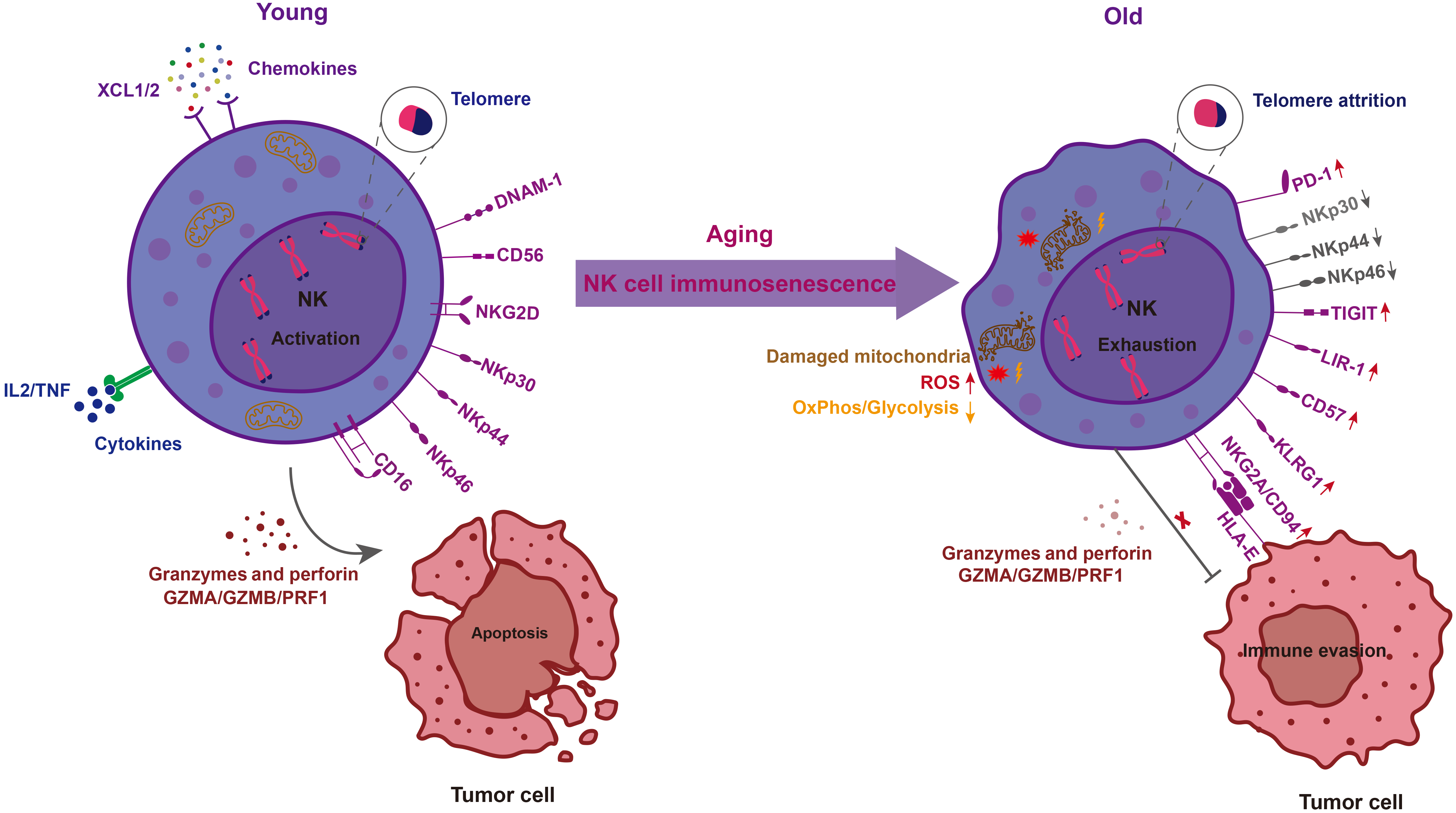
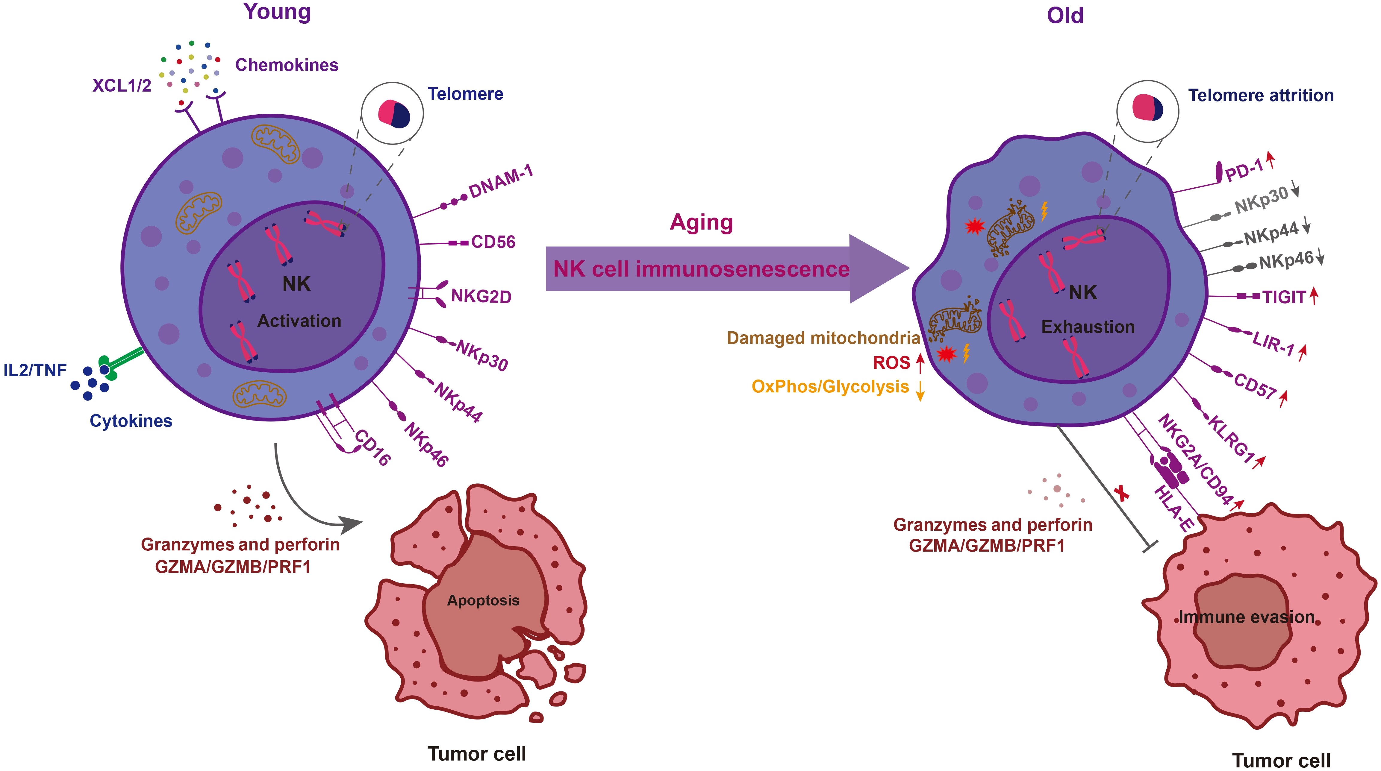
Figure 1. Comparison of phenotypic, metabolic, and functional hallmarks between young and aged NK cells. The schematic illustrates the multi-dimensional remodeling of NK cells during the transition from a functional state to immunosenescence. (Left) Young NK cells are characterized by an “Activation” profile, featuring preserved telomere length, high expression of activating receptors (e.g., CD16, NKG2D, DNAM-1, and NCRs: NKp30, NKp44, NKp46), and efficient metabolic plasticity. Upon tumor recognition, they secrete pro-inflammatory cytokines (IL-2, TNF) and chemokines (XCL1/2), and release potent cytotoxic effectors (GZMA/B, PRF1) to induce tumor cell apoptosis; (Right) Aged NK cells undergo “Exhaustion” and immunosenescence driven by biological aging. This state is defined by intrinsic hallmarks including telomere attrition, mitochondrial dysfunction (characterized by damaged mitochondria, increased ROS, and diminished OxPhos/glycolysis), and a shift toward bioenergetic rigidity. Phenotypic remodeling involves the downregulation of activating receptors (NKp30/44/46) and the dramatic accumulation of inhibitory/exhaustion markers (e.g., PD-1, TIGIT, LIR-1, CD57, KLRG1, and NKG2A/CD94). Functionally, the impaired secretion of granzymes and perforin, combined with inhibitory signals from the tumor microenvironment (e.g., HLA-E/NKG2A interaction), allows tumor cells to achieve immune evasion, establishing a self-reinforcing cycle of malignancy in the elderly. Created in Adobe Illustrator. NK: natural killer; NCRs: natural cytotoxicity receptors; IL: interleukin; TNF: tumor necrosis factor; ROS: reactive oxygen species; PD-1: programmed death 1; TIGIT: T-cell immunoglobulin and ITIM domain; LIR-1: leukocyte immunoglobulin-like receptor 1; KLRG1: killer cell lectin-like receptor G1; HLA-E: histocompatibility leucocyte antigen E.
2.1 Phenotypic remodeling and subset redistribution
The fundamental architecture of the NK cell compartment is defined by the balance between the immunoregulatory CD56brightCD16- subset and the cytotoxic CD56dimCD16+ population. While this balance remains relatively stable from birth (cord blood) through early adulthood, biological aging orchestrates a marked and progressive subset skewing. Unlike dwindling B-cell pools, NK cells often expand in the very old to maintain numerical parity. This numerical resilience is driven by the selective expansion of the mature CD56dim subset, which occupies an increasingly larger portion of the lymphocyte pool while masking a qualitative erosion of the immature reservoir[38]. Lifespan analyses reveal a progressive contraction of CD56bright frequency and absolute numbers, most notably in the “very old” (p < 0.001)[38-40].
Within the dominant CD56dim pool, the most conspicuous hallmark is the dramatic accumulation of CD57, which identifies NK cells with shortened telomeres and negligible proliferative capacity[38,41]. This senescent profile is further accentuated by the specific upregulation of leukocyte immunoglobulin-like receptor 1/immunoglobulin-like transcript receptor-2 (LIR-1/ILT-2), an inhibitory receptor whose expression doubles in very advanced age, serving as a persistent brake on major histocompatibility complex class I (MHC-I) recognition[38]. Concurrently, NK cells exhibit a hallmark “exhaustion signature” characterized by the increased expression of KLRG1 and TIGIT[32,33,42]. While KLRG1 serves as a terminal marker of replicative senescence, the surge in TIGIT represents a pivotal functional blockade that competes with the activating receptor DNAM-1 for their shared ligand, CD155, thereby elevating the activation threshold. Paradoxically, this gain in inhibitory signaling is accompanied by the coordinated loss of key maturation and activating markers, including the downregulation of CD161, NKp30, and NKp46[43,44]. This aged repertoire skewing is heavily confounded by latent cytomegalovirus (CMV) infection rather than reflecting chronological aging alone. Chronic, virus-driven clonal expansion of adaptive NK cells largely drives this senescent phenotype, an intricate interplay explored in Section 2.2.
2.2 CMV serostatus and biological heterogeneity of NK cell aging
While human long-lived NK cell generation remains poorly understood, CMV seropositivity is firmly linked to the expansion of memory-like NKG2C+CD57+ subsets[45,46]. Once primed, these cells expand further during subsequent viral infections, highlighting how CMV shapes the biological heterogeneity of the aging NK cell pool[47-49]. Indeed, the expansion of CD57+CD56dimCD16+ NK cells, which largely encompasses this adaptive population, is traditionally considered a hallmark of NK cell immunosenescence[50]. However, comparative studies reveal that this phenotypic shift is primarily initiated by CMV infection rather than chronological aging itself, as young and old CMV-seropositive donors display similar frequencies of these cells[51]. Instead of driving CD57 expression directly, aging acts as a progressive accumulator of these CMV-primed cells to maintain overall NK cell homeostasis as other subsets decline[45]. Indeed, while CMV selectively drives phenotypic receptor remodeling (such as CD94/NKG2C and CD57 expression), aging primarily drives subset redistribution and functional decline, marked by the expansion of a granzyme A/B-deficient CD56-CD16+ population[51]. This phenomenon highlights how persistent antigen stimulation, as a potent extrinsic factor, accelerates terminal differentiation and limits the functional lifespan of NK cells.
At the molecular level, this CMV-driven differentiation involves marked epigenetic and transcriptional rewiring[52,53]. Specifically, the loss of the transcription factor promyelocytic leukemia zinc finger protein (PLZF) precipitates the subsequent downregulation of key signaling adapter molecules, including SYK, EAT-2, and FcεRγ. This maturation is regulated by T cell immunoglobulin and mucin domain-containing protein 3 (TIM-3), whose decreased expression during chronic infections skews NK cells toward an exhausted yet cytotoxic phenotype, identifying this pathway as a promising checkpoint target[54]. Notably, effective CMV control is driven exclusively by either NKG2C+ NK cells or T cells, indicating a rheostat-like regulation of antiviral T-cell responses whose long-term impact on immunosenescence remains to be determined[55]. Beyond isolated CMV exposure, Epstein-Barr virus (EBV) co-infection in the elderly drives a mature CD56neg subset that, despite lacking classical senescent markers, exhibits reduced cytotoxicity and correlates with a T-cell-associated immune risk profile[56]. Additionally, identifying CMV-derived peptides that selectively activate NKG2C+ NK cells presents new opportunities for NK cell regulation. These activity-modulating peptides could directly guide future vaccine and therapeutic design[57]. Furthermore, highly functional adaptive NK cells can be efficiently expanded from CMV-seropositive donors using an anti-CD94 monoclonal antibody combined with interleukin (IL)-2 or IL-15. These expanded effectors maintain high antibody-dependent cellular cytotoxicity (ADCC) capacity without upregulating programmed death 1 (PD-1), providing a safe, cost-effective platform for adoptive immunotherapy[58].
Beyond these therapeutic aspects, natural aging also affects these specialized subsets. The CD56-CD16+ NK cell subset, described in human immunodeficiency virus (HIV) patients[59], increases in elderly donors[51]. Additionally, CMV infection in older individuals drives the accumulation of long-lived CD57+ NK cells expressing NKG2C[60,61]. In terms of tissue distribution, these adaptive NK cells accumulate in the blood, bone marrow, and lungs, but are not significantly detected in lymph nodes, tonsils, or the gut[62]. Similarly, aging causes mature NK cells to decrease in peripheral tissues but accumulate in the bone marrow[34,63].
2.3 Shifting profiles and functional imbalance of cytotoxicity receptors
Repertoire dysregulation and recalibrated signaling thresholds underpin the functional decline of senescent NK cells. While certain primary activating receptors, most notably NKG2D, maintain stable surface density and positivity across age groups, their functional output is qualitatively compromised. Evidence from Hazeldine et al.[64] suggests that the age-related decline in NK cell cytotoxicity is not necessarily a failure of target recognition, but rather a post-binding defect characterized by impaired polarization of lytic granules and reduced perforin secretion into the immunological synapse. This intrinsic functional failure is further compounded by the progressive attrition of natural cytotoxicity receptors (NCRs) such as NKp30 and NKp46. Beyond direct tumor lysis, the loss of these receptors disrupts critical NK-DC crosstalk and adaptive Th1 priming, thereby contributing to clinical vulnerabilities in older adults, including the reactivation of latent infections and delayed resolution of inflammatory responses[19]. In addition to these activatory deficits, the senescent NK cell repertoire is increasingly dominated by inhibitory signals. Terminal differentiation during aging promotes the stochastic expansion of Killer-cell Immunoglobulin-like Receptors (KIRs) and the dramatic accumulation of the maturity marker CD57, effectively locking the cells into a functionally rigid “chronic-off” state[61]. The aging microenvironment actively reinforces this inhibitory landscape through extrinsic programming. Recent findings by Chen et al.[65] demonstrate that SASP-driven cytokines (e.g., CCL2 and IL-6) upregulate the ligand histocompatibility leucocyte antigen E (HLA-E) (Qa-1b) on myeloid populations, which subsequently engages the CD94/NKG2A checkpoint to robustly suppress both antitumoral and vaccine-induced immunity in aged hosts.
NK cell immunosenescence is modulated by diverse cellular interactions within the microenvironment. These crosstalk pathways, though largely characterized in general tumor settings, intersect with the suppressive mechanisms that accumulate in the aging microenvironment[19,65]. For instance, NK-derived interferon gamma (IFN-γ) drives antitumoral M1 macrophage polarization[66]. Conversely, TGF-β1-secreting M2 macrophages suppress NK cell survival and function[67]. Neutrophils also suppress NK cells through direct contact, downregulating CCR1, NKp46, and NKG2D[68]. They cleave NKp46 using cathepsin G-containing NETs[69] and release reactive oxygen species (ROS) to reduce NK viability[70]. Furthermore, myeloid-derived suppressor cells (MDSCs) and Tregs inhibit NK cells via TGF-β1 release[71], IL-2 deprivation[72], and TIGIT signaling[73]. These suppressive signals decrease CD3ε expression[74]. Reciprocally, NK cells normally promote dendritic cell maturation and Th1 priming via IFN-γ and NCRs[75-77]. The age-related loss of NCRs (described in Section 2.3) therefore disrupts this helper axis. Finally, tumor-associated fibroblasts (TAFs) suppress NK cells by releasing PGE2 and IDO[78]. PGE2-EP4 signaling blocks IFN-γ production[79], while IDO-mediated tryptophan degradation prevents the recovery of NKp46 and NKG2D[80].
2.4 Metabolic dysregulation and mitochondrial impairment
Beyond receptor-mediated signaling, the energetic requirements for such dynamic membrane remodeling are further curtailed by metabolic reprogramming in aged NK cells. Efficient immunosurveillance is an inherently energy-intensive process; however, immunosenescence shifts the NK cell from a state of metabolic plasticity to one of bioenergetic rigidity. Reflecting a systemic hallmark of aging where mitochondria serve as the primary site of cumulative organelle damage across tissues[81-83], this bioenergetic rigidity is underpinned by the progressive impairment of PGC-1α, the central co-transcription factor orchestrating mitochondrial biogenesis and respiratory homeostasis[84,85]. In aged human NK cells, this is specifically manifested as a failure to upregulate PGC-1α upon IL-2 activation, leading to inhibited mitochondrial expansion and aberrant ROS accumulation[86]. This bioenergetic failure is further compounded by a deficiency in the NAD+ salvage pathway; specifically, the downregulation of the rate-limiting enzyme nicotinamide phosphoribosyltransferase (NAMPT) disrupts mitochondrial homeostasis and oxidative phosphorylation, thereby stripping tumor-infiltrating NK cells of the metabolic endurance required for sustained cytotoxicity[87]. Moreover, this bioenergetic crisis is further exacerbated within the tumor microenvironment (TME), where nutrient deprivation suppresses the glycolysis and OxPhos required for NK cell effector functions[88,89]. Specifically, the TME paralyzes NK cell glucose metabolism via lipid peroxidation-associated oxidative stress, revealing a lack of metabolic substrate flexibility in exhausted NK cells that leads to persistent bioenergetic insufficiency and functional exhaustion[90]. This systemic bioenergetic rigidity and the resulting functional exhaustion are epitomized by the accumulation of dysfunctional subsets like the CD56neg NK cells. The failure of these cells to execute cytotoxicity and produce IFN-γ provides a clinical reflection of the profound innate immune disturbance and impaired responsiveness to antiviral therapies during chronic viral infections[91]. Indeed, long-term exposure to the immunosuppressive TME is a key extrinsic driver of this metabolic failure. Factors like TGF-β and hypoxia suppress glycolytic enzymes and impair mitochondrial respiration. This chronic signaling forces a metabolic reprogramming that drives NK cells into exhaustion.
2.5 Telomere attrition and p16 p21 mediated cell cycle arrest
The immunosenescence of NK cells is primarily anchored in the progressive loss of physiological integrity, characterized by systematic telomere erosion and the induction of stable cell cycle arrest[83,92]. Although NK cells uniquely possess the capacity to upregulate telomerase, their telomeric repeats nonetheless undergo lineage-specific attrition that eventually reaches a critical minimum threshold, mandating irreversible cell cycle exit and orchestrating phenotypic remodeling[93]. Mechanistically, critically short telomeres activate the DNA damage repair (DDR) via ataxia telangiectasia mutated (ATM) and ataxia telangiectasia mutated Rad3-related (ATR) kinases[94]. Generally, this axis triggers p53/p21-mediated growth arrest and subsequent p16-mediated maintenance[95,96], though its downstream role in NK cells remains to be fully characterized. Although this classical pathway is widely accepted based on robust data from fibroblasts and T cells, direct evidence specifically validating p16/p21-mediated cell cycle arrest in NK cells remains relatively limited. Yet, genomic DNA damage is a major intrinsic trigger of NK cell aging. It also promotes a pro-inflammatory secretory phenotype, linking intrinsic genomic instability to extrinsic chronic inflammation. The chronic activation of this DDR pathway serves as a primary driver of NK cell exhaustion, triggering the downregulation of NKG2D and Eomesodermin while facilitating the enrichment of terminal markers such as KLRG1[94]. The resulting permanent loss of proliferative ability prevents the mobilization of a sufficient effector population. Clinically, shorter leukocyte telomere length is directly associated with a higher risk of malignant tumors, as demonstrated in a cohort of over 470,000 middle-aged and older adults[97,98].
2.6 Epigenetic alterations in NK cell immunosenescence
Epigenetic changes are a key hallmark of aging, regulating gene expression without altering the DNA sequence[99]. During immunosenescence, DNA methylation shows a bidirectional imbalance[100]. Specifically, age-associated hypomethylation disrupts functional pathways in immune cells[101]. Meanwhile, promoter hypermethylation silences critical genes, impairing DNA repair and cell cycle checkpoints[102]. Furthermore, histone modifications undergo major imbalances[103]. Aging cells lose repressive histone marks like H3K9me3, causing heterochromatin loosening and genomic instability[104,105]. Finally, chromatin remodeling alters chromatin architecture[106]. This chromatin relaxation permits the abnormal expression of silenced genes[107]. Concurrently, the downregulation of core histones impairs the chromatin barrier, increasing transcriptional noise in aging cells[108].
3. Impact of the Aging Microenvironment on NK Cell Function
Complementing these cell-intrinsic biological clocks, the progressive erosion of NK cell immunosurveillance is reshaped by the extrinsic pressures of the aged systemic and local environments.
3.1 Inflammaging: Driving NK cells from senescence to exhaustion
Inflammaging and cellular senescence orchestrate a self-reinforcing positive feedback loop, in which the accumulation of the SASP drives systemic inflammation and the bystander spread of senescence through p38 MAPK-mediated autophagy inhibition[109,110]. This reciprocal axis facilitates mitochondrial dysfunction and ROS accumulation, ultimately impairing immune surveillance and creating a permissive environment for the progression of diverse malignancies[109,111,112]. This persistent state is largely fueled by “sterile” damage-associated molecular patterns (DAMPs) released from damaged cells and environmental insults, which sustain low-grade chronic inflammation even in the absence of infection[113,114]. Such sustained inflammation has been intrinsically linked to the classic hallmarks of aging, regulating physiological aging and increasing susceptibility to malignancy[83,115,116]. Recent deep-learning analysis of human immunomes has integrated these systemic inflammatory patterns into a metric termed the inflammatory clock (iAge), identifying the interferon-related chemokine CXCL9 as a primary driver of this process[117]. Elevated CXCL9 levels not only track with multimorbidity but also correlate with the upregulation of multiple inflammatory pathways and the downregulation of proliferation-associated genes[117]. While these senescent effects of CXCL9 were originally characterized in vascular endothelial cells, this systemic inflammatory milieu is hypothesized to similarly impact immune cells. Exposure to such chronic inflammatory factors may reinforce the senescent phenotype of NK cells, potentially priming them for exhaustion under tumor-associated stress and compromising early tumor clearance[42].
3.2 SASP-mediated immune suppression
The molecular genesis of the SASP is linked to the cGAS/STING-mediated detection of cytosolic DNA arising from the transcriptional derepression of LINE-1 retrotransposons and heightened mitochondrial ROS production[118,119]. Although initially programmed as a two-step transient response to facilitate tissue repair through immune recruitment, the inherent heterogeneity of the SASP allows for the dominant secretion of immunosuppressive factors such as TGF-β, which neutralize NK cell surveillance and promote paracrine senescence in the surrounding niche[120,121]. This systemic failure to achieve senescence clearance results in the chronic persistence of these cells, which eventually fosters a fibro-inflammatory environment conducive to malignant progression[6,120]. Within this microenvironment, these persistent cells remain metabolically active and exert immunosuppressive effects primarily through the continuous release of the SASP[122,123]. Specifically, senescent myeloid subsets such as PMN-MDSCs exhibit a prototypical SASPome that promotes their persistence in the niche and reinforces the immunosuppressive landscape[124,125]. These senescent populations amplify the suppression by secreting key SASP-related cytokines, including IL-6, IL-8, and TGF-β, which directly impair the production of cytotoxic molecules such as granzyme B and perforin[97,126,127]. The functional output of the SASP is inherently pleiotropic, often shifting from a beneficial immune-recruiting signal to a potent mediator of immune evasion as malignancies progress. This transition is particularly evident in preneoplastic environments, where the SASP recruits immature myeloid populations that act as a cellular shield to directly inhibit NK cell-mediated surveillance, thereby facilitating tumor initiation and growth[128]. In addition to immune cell recruitment, the SASP actively enables senescent cells to evade immune clearance by secreting metalloproteases that induce the proteolytic shedding of activating NKG2D ligands, such as MICA and ULBP2, from their surface[129,130]. Furthermore, senescent cells in aged tissues, such as fibroblasts in the skin of older humans, upregulate the inhibitory ligand HLA-E, which binds to the NKG2A receptor on NK cells to robustly suppress their cytotoxic activity and promote senescent cell persistence[131]. Beyond these mechanisms, senescent cells exploit the upregulation of immune checkpoint proteins, most notably PD-L1 and PD-L2, creating a stable immunosuppressive environment that facilitates their escape from immune surveillance and contributes to malignant progression[132-134].
3.3 Bone marrow niche deterioration
Aging of the bone marrow (BM) niche is histologically characterized by increased adipogenesis and vascular rarefaction, establishing a metabolic stress environment that prioritizes myeloid-biased differentiation at the expense of lymphoid-primed progenitors, presumably limiting the precursor pool available for NK-cell replenishment[135,136]. This structural deterioration is further compounded by biomechanical rigidification; age-related matrix stiffening activates Yap/Taz signaling in stromal cells, suppressing the molecular cues[137] that are thought to support the proper maturation of the NK cell pool. Within this framework, the progressive attrition of critical factors like CXCL12 and Netrin-1 impairs hematopoietic maintenance, although the calvarial BM exhibits a unique microenvironmental resilience that partially preserves lymphoid potential in the elderly[138-140]. Beyond structural and biomechanical shifts, the erosion of the neural-hematopoietic axis, specifically the loss of sympathetic nervous system (SNS) innervation and impaired ADRβ3 signaling within arteriolar niches, serves as a potent driver of hematopoietic stem cell (HSC) immunosenescence. Strikingly, pharmacological restoration of these adrenergic signals via ADRβ3-selective sympathomimetics can significantly rejuvenate aged HSC function, offering a promising therapeutic strategy to recover the lymphoid output[141], thereby potentially restoring sustained NK cell immunosurveillance. Therefore, the multi-dimensional deterioration of the bone marrow microenvironment is proposed to restrict the homeostatic replenishment of functionally competent NK cells, thereby potentially contributing to the systemic decline in innate immunosurveillance observed during biological aging.
4. The Vicious Cycle between NK Cell Senescence and Tumorigenesis
The intersection of NK cell immunosenescence and malignant progression establishes a self-reinforcing vicious cycle that compromises the host’s anti-tumor defense. Clinically, the age-associated decline in NK cell activity is linked to increased susceptibility to infections and malignancy, highlighting the significant impact of NK cell dysfunction on older adults[142]. This clinical vulnerability is driven by reciprocal interactions within the TME, which induces functional alterations in resident NK cells to facilitate immune evasion[143]. This functional collapse is underpinned by DNA damage; persistent tumor-induced stimulation triggers the ATM-mediated DDR pathway, which drives aged NK cells into a state of functional exhaustion characterized by the loss of eomesodermin and the activating receptor NKG2D[94]. Specifically, intra-tumoral NK cells, such as the CD103+ subset, exhibit substantial functional impairment characterized by the upregulated expression of co-inhibitory molecules (e.g., Tigit and TIM-3) and a marked reduction in granzyme B production[143]. This cytolytic failure is further exacerbated by TME-derived cytokines, where elevated IL-6 suppresses the cytotoxic activity of NK cells while decreased perforin and granzyme B levels impair their function[144]. Such inhibitory signatures are pervasive across various malignancies, including lung and breast cancers, where the downregulation of activating receptors like NKp30 and NKG2D impairs cytolytic function[145-147], as well as hematological malignancies marked by proliferative arrest associated with CD57 expression[148-151]. These receptor alterations and immunosenescence markers manifest through distinct mechanisms across different malignancies, shaped by their distinct tissue origins and immunogenicity. Specifically, alterations in key activating receptors, such as NKp30, NKp46, and DNAM-1, compromise NK cell immunosurveillance against tumors of various origins, including haematological malignancies[152], melanoma[153], and ovarian carcinoma[154]. In acute myeloid leukaemia, contact with ligand-expressing blasts[151] drives NK cells to downregulate NKp46, NKp30[151,152,155], and DNAM-1[151,155], paralleling alterations found in healthy elderly donors[155]. Combined with reduced ligand expression on these blasts, this receptor downregulation impairs NK cell-mediated tumor immunosurveillance.
Alongside these receptor-level and contact-dependent defects, cytokine-driven inhibition remains a major suppressive barrier. Compounding this cytokine-mediated suppression, TGF-β, abundantly present in the aged TME, further paralyzes NK cell function by directly targeting the mTOR pathway, thereby blocking IL-15-induced metabolic activation, curtailing proliferation, and downregulating key cytotoxic receptors to reinforce the immunosuppressive landscape[127]. Reciprocally, the tumor itself actively accelerates NK cell senescence: chronic tumor-associated stress drives persistent DDR activation and telomere attrition, progressively locking NK cells into a terminally differentiated state marked by CD57 upregulation, TIGIT accumulation, and loss of NKG2D-mediated cytolytic capacity[42,94,144]. In the aging population, cellular senescence promotes tumor growth and metastasis through the SASP[156,157]. This secretome locks the microenvironment into a self-sustaining cycle of immune paralysis and oncogenic progression.
5. Breaking the Cycle: Therapeutic Opportunities
The establishment of a self-perpetuating vicious cycle between NK cell immunosenescence and malignant progression necessitates a multi-dimensional therapeutic framework to decouple this reciprocal relationship and restore the host’s anti-tumor integrity (Figure 2).
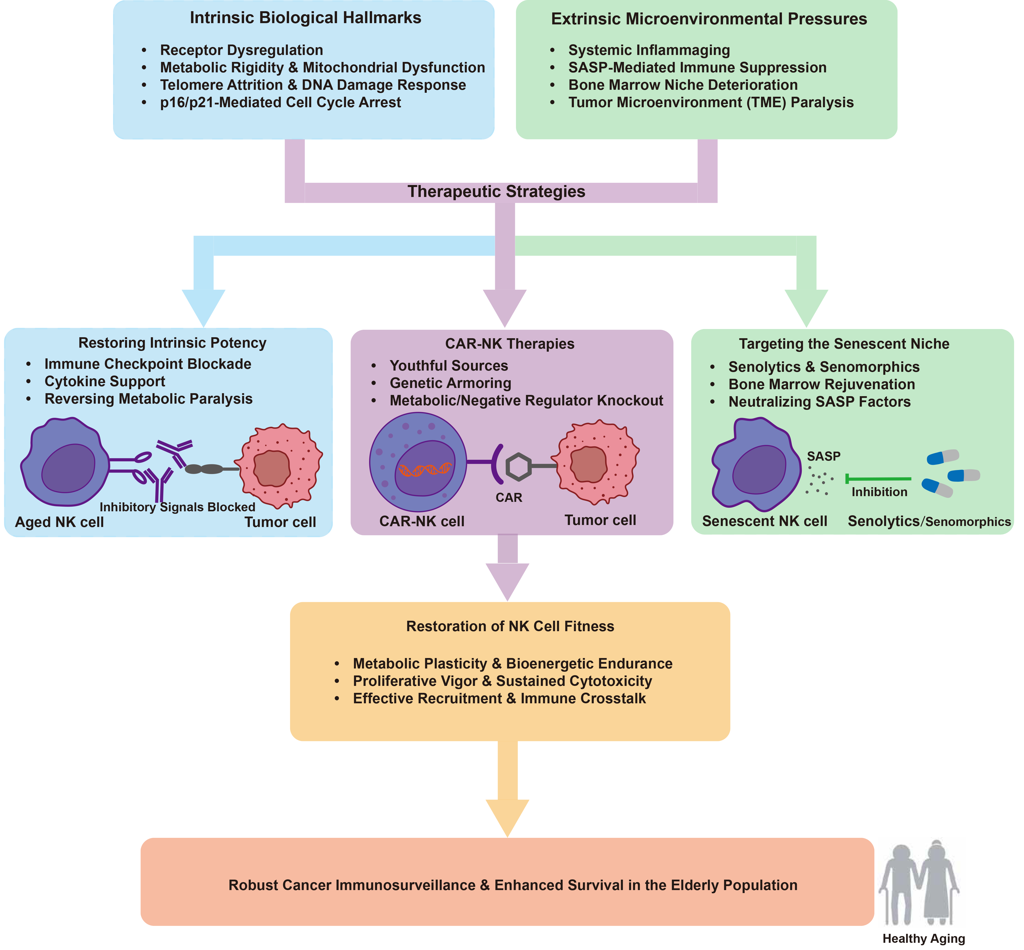
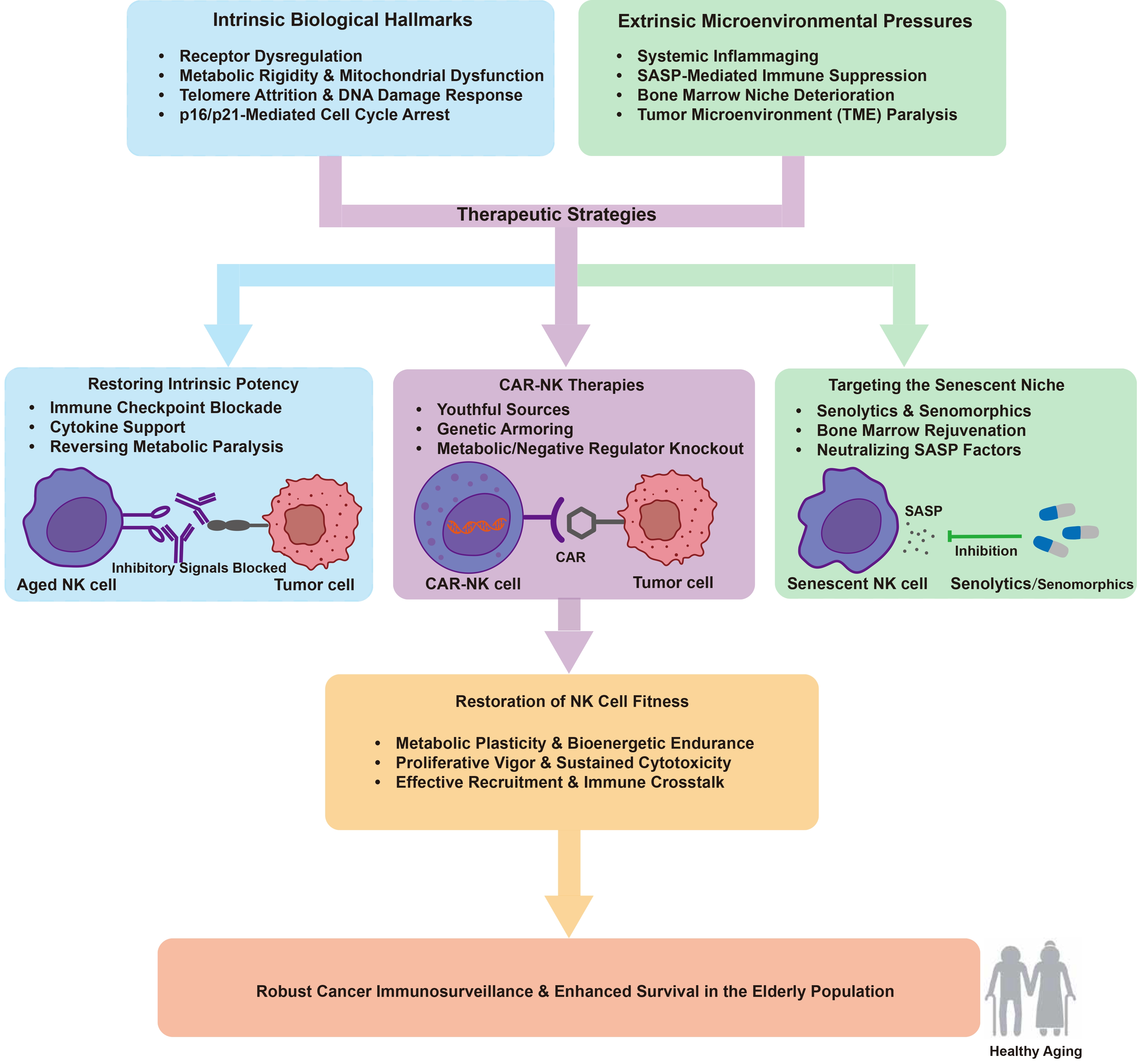
Figure 2. Integrated therapeutic framework for resuscitating NK cell function in the elderly. Biological aging impairs NK cell-mediated immunosurveillance through synergistic intrinsic hallmarks and extrinsic microenvironmental pressures, including receptor dysregulation, metabolic rigidity, and bone marrow niche deterioration. To counteract this decline, multi-dimensional therapeutic strategies are integrated to break the vicious cycle of immune paralysis. These approaches encompass the restoration of intrinsic potency via checkpoint blockade and cytokine support, the deployment of youthful or genetically armored CAR-NK cells to bypass host senescence, and the pharmacological remodeling of the senescent niche using senolytics. Collectively, these interventions facilitate the restoration of NK cell fitness and ensure sustained cytolytic vigor. This systemic rejuvenation achieves robust cancer immunosurveillance and promotes healthy aging in the elderly population. Created in Adobe Illustrator. NK: natural killer; CAR: chimeric antigen receptor; TME: tumor microenvironment; SASP: senescence-associated secretory phenotype.
5.1 Restoring intrinsic potency
TIGIT has emerged as a primary checkpoint specifically associated with NK cell exhaustion, serving as a more relevant mediator of NK dysfunction than traditional markers such as CTLA-4 or PD-1. Blockade of the TIGIT axis effectively prevents this functional collapse, not only restoring NK-mediated tumor surveillance but also potentiating adaptive T cell responses through essential immune crosstalk[33]. Complementing the TIGIT axis, therapeutic targeting of the inhibitory NKG2A receptor with monalizumab has demonstrated significant potential to simultaneously reinvigorate NK and CD8+ T cell effector functions. By alleviating inhibitory signaling, particularly from the HLA-E ligand, monalizumab has shown promising clinical efficacy in Phase II trials, providing a synergistic platform that may overcome resistance to first-generation immunotherapies in refractory settings[158]. Concomitant with the alleviation of these inhibitory brakes, the reinforcement of proliferative vigor via the IL-15 superagonist complex ALT-803 provides the necessary functional drive to overcome replicative senescence. Clinical evaluation has shown that ALT-803 triggers the robust activation and systemic expansion of NK and CD8+ T cells without the paradoxical induction of immunosuppressive regulatory T cells, thereby offering a potent platform to rejuvenate anti-tumor surveillance in the aged and immunocompromised host[159]. Building on these synergistic principles, the clinical combination of ALT-803 and nivolumab has demonstrated the ability to overcome resistance in refractory non-small cell lung cancer. By reinvigorating NK and T cell activity, this regimen provides a promising strategy to rescue anti-tumor responses in patients who previously progressed on standard checkpoint inhibitors[160]. Ultimately, these integrative strategies highlight that the simultaneous neutralization of inhibitory checkpoints and the reinforcement of stimulatory signaling can effectively resuscitate the latent anti-tumor potency of endogenous NK cell repertoires within the aging host.
5.2 Chimeric antigen receptor (CAR)-NK therapies
Compared to CAR-T cells, CAR-NK therapies show superior safety with a lower risk of graft-versus-host disease[161]. They are also less likely to trigger severe cytokine release syndrome or neurotoxicity[162]. This safety is due to different cytokine profiles; activated NK cells primarily secrete IFN-γ and granulocyte-macrophage colony-stimulating factor (GM-CSF)[163], whereas CAR-T cells release a wider range of inflammatory cytokines like IL-6 and tumor necrosis factor alpha (TNF-α)[164]. Additionally, CAR-NK cells retain their natural cytotoxicity through receptors like NKG2D and CD16-mediated ADCC[165,166]. This dual killing mechanism helps eradicate heterogeneous tumors that escape CAR-targeted antigen recognition. Furthermore, CAR-NK cells can be produced from allogeneic sources, including cord blood and induced pluripotent stem cells (iPSCs), as standardized, ready-to-use products[167]. This eliminates the need for patient-specific manufacturing[168]. For elderly patients, whose autologous T cells are often compromised by aging, this ready-to-use availability and lower toxicity represent a major clinical benefit.
Supporting these advantages, a landmark clinical trial demonstrated that cord blood-derived CAR-NK cells can yield high remission rates (73%) with minimal toxicity[169], proving the clinical feasibility of using youthful, allogeneic sources. Thus, these youthful cells may bypass the senescence and exhaustion inherent in older autologous therapies. The clinical translation of CAR-NK cells is further accelerated by the utilization of iPSCs as a limitless and homogeneous cell source[170,171]. Notably, iPSC-derived NK cells engineered with NK-optimized CAR constructs containing NKG2D and 2B4 signaling domains have demonstrated potent anti-tumor efficacy that rivals CAR-T cells. This strategy provides a standardized off-the-shelf platform that achieves robust tumor inhibition while maintaining a significantly superior safety profile[172]. To address the metabolic insufficiency and limited persistence typical of senescent immune cells, researchers have developed armored iPSC-NK cells through multiplexed genetic engineering[173,174]. The integration of membrane-bound IL-15 (mbIL-15) or IL-15/IL-15R fusion proteins into iPSCs provides essential autocrine survival signals that promote sustained in vivo persistence and metabolic fitness[173-175]. Furthermore, the introduction of high-affinity, non-cleavable CD16 (hnCD16) allows these engineered cells to maximize ADCC when combined with monoclonal antibodies[176,177]. Advanced platforms such as the iDuo-NK or quadruple-gene-edited CAR-iNK cells further incorporate the knockout of metabolic barriers like CD38 or the negative regulator cytokine-inducible SH2-containing protein (CISH)[175,178,179]. The therapeutic potential of these genetically modified platforms is supported by clinical data. Specifically, iPSC-derived NK cells offer a youthful cell source that avoids donor senescence. In a B-cell lymphoma trial, a single dose of 90 million or more cells induced objective responses in eight of eleven patients, with seven achieving complete remission[180]. These responses occurred without dose-limiting toxicities or severe neurotoxicity[180]. However, this trial lacked age-stratified data, and the aged recipient microenvironment may still suppress these cells. This favorable safety profile is vital for older patients with limited physiological resilience. Therefore, combining youthful cell origins with functional enhancements may help mitigate the effects of recipient immunosenescence, decoupling cell product potency from the patient’s chronological age.
5.3 Targeting the senescent niche
The clinical success of engineered natural killer cell therapies depends not only on cellular potency but also on the state of the surrounding microenvironment where these cells must operate. A primary strategy to remodel this niche involves the use of senolytics, which are pharmacological agents that selectively eliminate senescent cells by targeting their inherent pro-survival pathways[128]. Specifically, the combination of dasatinib and quercetin or Bcl-2 family inhibitors can significantly reduce the burden of senescent cells in aged tissues while improving overall healthspan parameters[181,182]. For instance, pharmacologically targeting specific cytokines such as TGF-β can alleviate the metabolic paralysis of natural killer cells by restoring the mTOR pathway, which is frequently impaired in the aged tumor microenvironment[127]. Niche remodeling also involves neutralizing the HLA-E and NKG2A axis, which allows senescent cells to evade immune surveillance[131]. Strategically blocking this checkpoint with monalizumab reinvigorates natural killer cell activity to enhance anti-tumor immunity within the aged host[158]. The long-term maintenance of natural killer cell surveillance also requires the systemic rejuvenation of the bone marrow niche as the primary site for lymphoid development. Research indicates that pharmacological activation of adrenoreceptor β3 signaling within the bone marrow niche via selective sympathomimetics significantly rejuvenates aged stem cell function to recover the lymphoid output required for sustained natural killer cell replenishment[141]. Engineering a soft bone marrow niche by modulating Yap Taz signaling can successfully reverse hematopoietic stem cell aging hallmarks and restore lymphopoiesis[137]. Supplementing the bone marrow niche with Netrin 1 rejuvenates aged niche cells and restores the regenerative potential of hematopoietic stem cells by activating essential DNA damage response pathways[139]. Additionally, the use of senomorphic agents such as metformin or rapamycin provides a non-cytotoxic strategy to suppress the secretory capacity of the niche without depleting resident cells, thereby neutralizing the chronic inflammatory state[128].
5.4 Clinical translation and evidence gaps in aging populations
Applying these immunotherapies and senolytics to older cancer patients presents distinct clinical challenges, particularly concerning age-related factors like frailty, polypharmacy, and reduced organ reserve. Physiologically, sarcopenia and increased adipose tissue lead to reduced endogenous IL-15 levels and higher levels of inflammatory adipokines[183]. This baseline inflammatory state can complicate treatment. Administering exogenous IL-15 superagonists (such as ALT-803) or CAR-NK cells in this inflammatory environment increases the risk of severe toxicities, including cytokine release syndrome (CRS). Similarly, new cell therapies like iPSC-derived NK cells are designed to bypass cellular aging. However, the therapeutic efficacy of these cells remains heavily constrained by the physiological state of the aged host. Aged tissues typically exhibit mitochondrial dysfunction, high levels of ROS, and altered lipid metabolism[184,185]. These factors can impair the metabolic fitness and immunological synapse formation of the transferred cells, even if the cell product itself is healthy. There is also a lack of clinical evidence for these therapies in older populations. Current CAR-NK trials do not include age-stratified analyses. Additionally, clinical data for senolytics, such as dasatinib plus quercetin (D + Q) or Bcl-2 inhibitors, remain very limited in older cancer cohorts. Future clinical trials must include older patients and report age-specific outcomes to establish safe treatment guidelines.
6. Conclusion and Future Perspectives
Biological aging remains the most significant challenge for cancer immunotherapy due to the systemic erosion of natural killer cell function known as immunosenescence. This review has delineated the multifaceted mechanisms underlying this decline, including phenotypic remodeling, receptor signaling dysregulation, and the metabolic rigidity driven by mitochondrial impairment. The intersection of natural killer cell immunosenescence and the suppressive aging microenvironment creates a self-reinforcing vicious cycle that facilitates tumor escape in the elderly population.
Breaking this cycle necessitates a multi-dimensional therapeutic framework that integrates cellular rejuvenation with niche remodeling. While innovative strategies such as iPSC derived CAR-NK cells offer a youthful and standardized platform to bypass host senescence, their long-term clinical efficacy will depend on the simultaneous transformation of the senescent niche. Combining intrinsic potency restoration with extrinsic interventions like senolytics or bone marrow rejuvenation provides a promising synergistic approach to resuscitate anti-tumor immunity.
Looking forward, several research directions are critical to advance this field. First, developing precise inflammatory clocks and spatial multiomic technologies will help map organ-specific NK cell senescence. Second, future engineering strategies should target epigenetic rejuvenation, such as restoring youthful DNA methylation patterns and histone modifications. Third, resolving technical hurdles like enhancing in vivo persistence and optimizing homing to specific tissue niches remains vital. Finally, clinical trials must implement age-stratified analyses and incorporate geriatric frailty indexes to guide treatment. Ultimately, targeting the biological hallmarks of natural killer cell immunosenescence represents a key frontier in geriatric oncology. This approach offers the potential to establish immune cell-based therapies as a new pillar of cancer treatment for the burgeoning elderly population.
Acknowledgements
The authors declare that Gemini 3.1 was used solely for language polishing and translation refinement during the manuscript preparation process. All research content, including study design, interpretations, and figures, is original and was not generated using any artificial intelligence tools.
Authors contribution
Gao Y: Methodology, writing-original draft, writing-review & editing.
Yu Y: Conceptualization, methodology, writing-review & editing.
Conflicts of interest
The authors declare no conflicts of interest.
Ethical approval
Not applicable.
Consent to participate
Not applicable.
Consent for publication
Not applicable.
Availability of data and materials
Not applicable.
Funding
This work was supported by the Frontiers Medical Center, Tianfu Jincheng Laboratory Foundation (Grant No. TFJCPI20250032).
Copyright
© The Author(s) 2026.
References
-
7. Félix J, de Toda IM, Cerro EDD, González-Sánchez M, De la Fuente M. Frailty and biological age. Which best describes our aging and longevity? Mol Asp Med. 2024;98:101291.[DOI]
-
8. Moqri M, Herzog C, Poganik JR, Ying K, Justice JN, Belsky DW, et al. Validation of biomarkers of aging. Nat Med. 2024;30(2):360-372.[DOI]
-
9. Tian YE, Cropley V, Maier AB, Lautenschlager NT, Breakspear M, Zalesky A. Heterogeneous aging across multiple organ systems and prediction of chronic disease and mortality. Nat Med. 2023;29(5):1221-1231.[DOI]
-
10. Walford RL. The immunologic theory of aging. Gerontologist. 1964;4(4):195-197.[DOI]
-
18. Marquardt N, Kekäläinen E, Chen P, Kvedaraite E, Wilson JN, Ivarsson MA, et al. Human lung natural killer cells are predominantly comprised of highly differentiated hypofunctional CD69−CD56dim cells. J Allergy Clin Immunol. 2017;139(4):1321-1330.e4.[DOI]
-
19. Cooper MA, Fehniger TA, Caligiuri MA. The biology of human natural killer-cell subsets. Trends Immunol. 2001;22(11):633-640.[DOI]
-
25. Moffett-King A. Natural killer cells and pregnancy. Nat Rev Immunol. 2002;2(9):656-663.[DOI]
-
27. Kim S, Iizuka K, Kang HSP, Dokun A, French AR, Greco S, et al. In vivo developmental stages in murine natural killer cell maturation. Nat Immunol. 2002;3(6):523-528.[DOI]
-
29. Bi J, Tian Z. NK cell exhaustion. Front Immunol. 2017;8:760.[DOI]
-
34. Dogra P, Rancan C, Ma W, Toth M, Senda T, Carpenter DJ, et al. Tissue determinants of human NK cell development, function, and residence. Cell. 2020;180(4):749-763.e13.[DOI]
-
37. Franceschi C, Garagnani P, Parini P, Giuliani C, Santoro A. Inflammaging: A new immune–metabolic viewpoint for age-related diseases. Nat Rev Endocrinol. 2018;14(10):576-590.[DOI]
-
40. Chidrawar SM, Khan N, Tracey Chan YL, Nayak L, Moss PA. Ageing is associated with a decline in peripheral blood CD56bright NK cells. Immun Ageing. 2006;3(1):10.[DOI]
-
42. Guo Z, Wu F, Chen Y, Xu J, Chen Z. Phenotypes, mechanisms, and therapeutic strategies of natural killer cell immunosenescence. Immun Ageing. 2025;22(1):38.[DOI]
-
43. Campos C, López N, Pera A, Gordillo JJ, Hassouneh F, Tarazona R, et al. Expression of NKp30, NKp46 and DNAM-1 activating receptors on resting and IL-2 activated NK cells from healthy donors according to CMV-serostatus and age. Biogerontology. 2015;16(5):671-683.[DOI]
-
51. Campos C, Pera A, Sanchez-Correa B, Alonso C, Lopez-Fernandez I, Morgado S, et al. Effect of age and CMV on NK cell subpopulations. Exp Gerontol. 2014;54:130-137.[DOI]
-
58. Giordano C, Carlomagno S, Falco M, Cantoni C, Vitale M, Caruana I, et al. CD94-driven in vitro expansion of highly functional adaptive NKG2C+ NKG2A- CD57+ NK cells from CMV+ healthy donors. Front Immunol. 2025;16:1481745.[DOI]
-
61. Solana R, Campos C, Pera A, Tarazona R. Shaping of NK cell subsets by aging. Curr Opin Immunol. 2014;29:56-61.[DOI]
-
70. Mellqvist UH, Hansson M, Brune M, Dahlgren C, Hermodsson S, Hellstrand K. Natural killer cell dysfunction and apoptosis induced by chronic myelogenous leukemia cells: Role of reactive oxygen species and regulation by histamine. Blood. 2000;96(5):1961-1968.[PubMed]
-
78. Li T, Yang Y, Hua X, Wang G, Liu W, Jia C, et al. Hepatocellular carcinoma-associated fibroblasts trigger NK cell dysfunction via PGE2 and IDO. Cancer Lett. 2012;318(2):154-161.[DOI]
-
87. Guo X, Tan S, Wang T, Sun R, Li S, Tian P, et al. NAD+ salvage governs mitochondrial metabolism, invigorating natural killer cell antitumor immunity. Hepatology. 2023;78(2):468-485.[DOI]
-
91. Gonzalez VD, Falconer K, Björkström NK, Blom KG, Weiland O, Ljunggren HG, et al. Expansion of functionally skewed CD56-negative NK cells in chronic hepatitis C virus infection: Correlation with outcome of pegylated IFN-α and ribavirin treatment. J Immunol. 2009;183(10):6612-6618.[DOI]
-
93. Kaszubowska L. Telomere shortening and ageing of the immune system. J Physiol Pharmacol. 2008;59(Suppl 9):169-186.[PubMed]
-
96. Gergues M, Bari R, Koppisetti S, Gosiewska A, Kang L, Hariri RJ. Senescence, NK cells, and cancer: Navigating the crossroads of aging and disease. Front Immunol. 2025;16:1565278.[DOI]
-
101. Roy R, Kuo PL, Candia J, Sarantopoulou D, Ubaida-Mohien C, Hernandez D, et al. Epigenetic signature of human immune aging in the GESTALT study. eLife. 2023;12:e86136.[DOI]
-
102. Cai Y, Song W, Li J, Jing Y, Liang C, Zhang L, et al. The landscape of aging. Sci China Life Sci. 2022;65(12):2354-2454.[DOI]
-
104. Zhang W, Qu J, Liu GH, Belmonte JCI. The ageing epigenome and its rejuvenation. Nat Rev Mol Cell Biol. 2020;21(3):137-150.[DOI]
-
105. Xu T, Shen Y, Guo R, Luo C, Niu Y, Luo Z, et al. Mutual regulation between histone methyltransferase Suv39h1 and the Wnt/β-catenin signaling pathway promoted cell proliferation and inhibited apoptosis in bone marrow mesenchymal stem cells exposed to hydroquinone. Toxicology. 2024;508:153932.[DOI]
-
106. Tsurumi A, Li W. Global heterochromatin loss: A unifying theory of aging? Epigenetics. 2012;7(7):680-688.[DOI]
-
107. Izadi M, Sadri N, Abdi A, Serajian S, Jalalei D, Tahmasebi S. Epigenetic biomarkers in aging and longevity: Current and future application. Life Sci. 2024;351:122842.[DOI]
-
110. da Silva PFL, Ogrodnik M, Kucheryavenko O, Glibert J, Miwa S, Cameron K, et al. The bystander effect contributes to the accumulation of senescent cells in vivo. Aging Cell. 2019;18:e12848.[DOI]
-
111. Salminen A, Kauppinen A, Kaarniranta K. Myeloid-derived suppressor cells (MDSC): An important partner in cellular/tissue senescence. Biogerontology. 2018;19(5):325-339.[DOI]
-
114. Goldberg EL, Dixit VD. Drivers of age-related inflammation and strategies for healthspan extension. Immunol Rev. 2015;265(1):63-74.[DOI]
-
121. Meyer K, López-Domínguez JA, Maus M, Kovatcheva M, Serrano M. Senescence as a therapeutic target: current state and future challenges. In: Serrano M, Muñoz-Espín D, editors. Cellular senescence in disease. Amsterdam: Elsevier; 2022. p. 425-442.[DOI]
-
122. Prattichizzo F, De Nigris V, La Sala L, Procopio AD, Olivieri F, Ceriello A. “Inflammaging” as a druggable target: A senescence-associated secretory phenotype: Centered view of type 2 diabetes. Oxi Med Cell Longev. 2016;2016:1810327.[DOI]
-
123. Ohtani N. Deciphering the mechanism for induction of senescence-associated secretory phenotype (SASP) and its role in ageing and cancer development. J Biochem. 2019;166(4):289-295.[DOI]
-
124. Zhou L, Ma B, Ruscetti M. Cellular senescence offers distinct immunological vulnerabilities in cancer. Trends Cancer. 2025;11(4):334-350.[DOI]
-
130. Majewska J, Krizhanovsky V. Immune surveillance of senescent cells in aging and disease. Nat Aging. 2025;5(8):1415-1424.[DOI]
-
137. Zhang X, Cao D, Xu L, Xu Y, Gao Z, Pan Y, et al. Harnessing matrix stiffness to engineer a bone marrow niche for hematopoietic stem cell rejuvenation. Cell Stem Cell. 2023;30(4):378-395.e8.[DOI]
-
138. Pinho S, Zhao M. Hematopoietic stem cells and their bone marrow niches. In: Zhao M, Qian P, editors. Hematopoietic stem cells. Singapore: Springer; 2023. p. 17-28.[DOI]
-
160. Wrangle JM, Velcheti V, Patel MR, Garrett-Mayer E, Hill EG, Ravenel JG, et al. ALT-803, an IL-15 superagonist, in combination with nivolumab in patients with metastatic non-small cell lung cancer: A non-randomised, open-label, phase 1b trial. Lancet Oncol. 2018;19(5):694-704.
-
161. Hu F, Li J, Wang Y, Lin Y, Zhang J, Xu J, et al. Large-scale generation of iNK and CAR-iNK cells from CD34+ haematopoietic stem and progenitor cells for adoptive immunotherapy. Nat Biomed Eng. 2026;10(4):765-784.[DOI]
-
164. Hunter BD, Jacobson CA. CAR T-cell associated neurotoxicity: Mechanisms, clinicopathologic correlates, and future directions. J Natl Cancer Inst. 2019;111(7):646-654.[DOI]
-
168. Ueda T, Kumagai A, Iriguchi S, Yasui Y, Miyasaka T, Nakagoshi K, et al. Non-clinical efficacy, safety and stable clinical cell processing of induced pluripotent stem cell-derived anti-glypican-3 chimeric antigen receptor-expressing natural killer/innate lymphoid cells. Cancer Sci. 2020;111(5):1478-1490.
-
172. Li Y, Hermanson DL, Moriarity BS, Kaufman DS. Human iPSC-derived natural killer cells engineered with chimeric antigen receptors enhance anti-tumor activity. Cell Stem Cell. 2018;23(2):181-192.e5.[DOI]
-
173. Woan KV, Kim H, Bjordahl R, Davis ZB, Gaidarova S, Goulding J, et al. Harnessing features of adaptive NK cells to generate iPSC-derived NK cells for enhanced immunotherapy. Cell Stem Cell. 2021;28(12):2062-2075.e5.[DOI]
-
177. Patel K, Bachanova V, Goodman AM, Pagel JM, Griffis K, Anderson M, et al. Phase I study of FT516, an off-the-shelf iPSC-derived NK cell therapy, in combination with rituximab in patients with relapsed/refractory B-cell lymphoma. Blood. 2021;138(Supplement 1):3873.[DOI]
-
179. Bjordahl R, Gaidarova S, Goodridge JP, Mahmood S, Bonello G, Robinson M, et al. FT576: A novel multiplexed engineered off-the-shelf natural killer cell immunotherapy for the dual-targeting of CD38 and Bcma for the treatment of multiple myeloma. Blood. 2019;134(Supplement_1):3214.[DOI]
-
180. Bachanova V, Ghobadi A, Patel K, Park JH, Flinn IW, Shah P, et al. Safety and efficacy of FT596, a first-in-class, multi-antigen targeted, off-the-shelf, iPSC-derived CD19 CAR NK cell therapy in relapsed/refractory B-cell lymphoma. Blood. 2021;138:823.[DOI]
-
184. Quinn KM, Linterman MA. Senescence blurs the line between innate and adaptive immune cells. Immunol Cell Biol. 2020;98(6):431-433.[DOI]
-
185. O’Brien KL, Finlay DK. Immunometabolism and natural killer cell responses. Nat Rev Immunol. 2019;19(5):282-290.[DOI]
Copyright
© The Author(s) 2026. This is an Open Access article licensed under a Creative Commons Attribution 4.0 International License (https://creativecommons.org/licenses/by/4.0/), which permits unrestricted use, sharing, adaptation, distribution and reproduction in any medium or format, for any purpose, even commercially, as long as you give appropriate credit to the original author(s) and the source, provide a link to the Creative Commons license, and indicate if changes were made.
Publisher’s Note
Science Exploration remains a neutral stance on jurisdictional claims in published
maps
and institutional affiliations. The views expressed in this article are solely those
of
the author(s) and do not reflect the opinions of the Editors or the publisher.
Share And Cite


Science Exploration Style
Gao Y, Yu Y. Targeting NK cell immunosenescence in cancer: Mechanisms, impact, and therapeutic opportunities. Ageing Cancer Res Treat. 2027;4:202622. https://doi.org/10.70401/acrt.2026.0037
Tips
Copy completed.
Submit a Manuscript
Author Instructions
Cite this Article
Article Metrics
0
View
0
Download
Cited
Article Updates

- Abstract
- Keywords
- 1. Introduction
- 2. Biological Hallmarks and Phenotypic Remodeling of NK Cell Immunosenescence
- 3. Impact of the Aging Microenvironment on NK Cell Function
- 4. The Vicious Cycle between NK Cell Senescence and Tumorigenesis
- 5. Breaking the Cycle: Therapeutic Opportunities
- 6. Conclusion and Future Perspectives
- Acknowledgements
- Authors contribution
- Conflicts of interest
- Ethical approval
- Consent to participate
- Consent for publication
- Availability of data and materials
- Funding
- References
- Copyright
 Get citation
Science Exploration Style

Gao Y, Yu Y. Targeting NK cell immunosenescence in cancer: Mechanisms, impact, and therapeutic opportunities. Ageing Cancer Res Treat. 2027;4:202622. https://doi.org/10.70401/acrt.2026.0037
copy
Share Link
copy