Navigating the aging brain: The interplay between brain malignancy and the aging microenvironment

Qianquan Li
1,#

,
Junyan Zhang
1,#
,
Jian Luo
2
,
Shi-Qing Cai
1,*
,
Xin Chen
1,*
*Correspondence to:
Xin Chen, Songjiang Research Institute, Shanghai Key Laboratory of Emotions and Affective Disorders, Songjiang Hospital Affiliated to Shanghai Jiao Tong University School of Medicine, Shanghai 201600, China.
E-mail: xinchen@shsmu.edu.cn
Shi-Qing Cai, Songjiang Research Institute, Shanghai Key Laboratory of Emotions and Affective Disorders, Songjiang Hospital Affiliated to Shanghai Jiao Tong University School of Medicine, Shanghai 201600, China. E-mail: sqcai@shsmu.edu.cn
Shi-Qing Cai, Songjiang Research Institute, Shanghai Key Laboratory of Emotions and Affective Disorders, Songjiang Hospital Affiliated to Shanghai Jiao Tong University School of Medicine, Shanghai 201600, China. E-mail: sqcai@shsmu.edu.cn
Ageing Cancer Res Treat. 2027;4:202614. 10.70401/acrt.2026.0038
Received: March 31, 2026Accepted: August 31, 2026Published: August 31, 2026
Abstract
The rising incidence of age-related brain pathologies, including brain malignancy, underscores an urgent need to understand the mechanistic interplay between aging and tumorigenesis. Historically viewed as distinct entities, brain tumors and neurodegenerative disorders are now recognized to share key biological processes, such as cellular senescence, chronic inflammation, and metabolic dysregulation. In this review, we deconstruct the existing knowledge at the intersection of cancer neuroscience and aging biology, and propose that age-related alterations in neuronal function, such as the accumulation of senescent cells, ion channel dysregulation, and neurotransmitter imbalance, are not inert background features but as active mediators of tumor progression and treatment resistance. We summarize the aged neural microenvironment, marked by a proinflammatory senescence-associated secretory phenotype (SASP) and blood-brain barrier dysfunction, underlies a permissive soil for malignancy. Moreover, we highlight the emerging concept that tumors can induce a pathological aging phenotype in surrounding neurons, which in turn potentiates the observed cognitive deterioration. By framing brain tumors as products of a dysfunctional aging ecosystem, we propose therapeutic strategies that target convergent aging mechanisms through senolysis, metabolic reprogramming, and neurotransmitter modulation may simultaneously achieve tumor control and preserve cognitive function. This integrated perspective opens new avenues for repurposing neuroactive drugs and designing interventions that address the sophisticated biology of the aging brain and malignancy.
Keywords
Aging, cellular senescence, brain tumor, cancer neuroscience, neuronal signaling, tumor microenvironment
1. Introduction
The global demographic shift toward an aging population has profound implications for the incidence and management of age-related neurological diseases. As a matter of fact, aging represents a critical risk factor for primary brain tumors, such as glioblastoma (GBM), as well as neurodegenerative disorders[1]. Historically, these conditions have been studied as distinct pathological entities with separate mechanistic frameworks and therapeutic strategies. However, a growing body of evidence indicates that they share fundamental biological processes, including cellular senescence, chronic inflammation, metabolic dysregulation, and impaired proteostasis[1-4]. While the association between advanced age and increased brain tumor incidence is well-documented, the underlying biological mechanisms remain incompletely understood. Aging is accompanied by profound structural, cellular, and molecular alterations in the brain, many of which may create a permissive or even tumor-promoting microenvironment.
The emergence of cancer neuroscience has revolutionized our understanding of tumor-nervous system interactions as a distinct landscape[5,6]. Malignant brain tumors do not simply occupy space within the brain parenchyma; rather, they actively engage in bidirectional communication with neurons, glia, and other cellular components of the brain tumor microenvironment[7-9]. Tumors secrete factors that modulate neuronal excitability, induce synaptic remodeling, and exploit neural activity to enhance their own proliferation and invasion. Conversely, neuronal activity can also drive tumor growth through paracrine signaling and direct electrochemical communication[10-13].
Aging is accompanied by behavioral and cognitive decline as the aging brain is characterized by low-grade chronic inflammation, accumulation of senescent cells, blood-brain barrier dysfunction, and alterations in neurotransmitter systems and ion channel expression[14-17]. Critical neuronal signaling pathways and molecules have been identified through genetic analysis[18-20], and these age-related molecular alterations may create a fertile soil for tumor development while simultaneously eroding the cognitive reserve necessary to cope with the neurological damage caused by disease progression.
In this review, we aim to summarize and deconstruct the existing knowledge at the intersection of aging, neuronal signaling, and brain tumor biology. We propose that age-related alterations in neuronal function, particularly those involving ion channels and neurotransmitter systems, are not merely passive background conditions but active drivers of brain tumor progression and treatment resistance. Furthermore, we put forward the idea that therapeutic strategies targeting neuronal aging mechanisms may offer unprecedented opportunities to achieve both tumor control and cognitive preservation.
2. The Biology of Neuronal Aging
2.1 Neuronal regulation of aging
Aging constitutes a major risk factor for a spectrum of human diseases, most notably neurodegenerative disorders, cancer, and diabetes[2]. The hallmarks of aging, including genomic instability, telomere attrition, epigenetic alterations, loss of proteostasis, mitochondrial dysfunction, cellular senescence, stem cell exhaustion, and altered intercellular communication, contribute to the progressive deterioration in physiological integrity that characterizes the aging process[2,3]. Within the nervous system, aging manifests as a complex phenotype involving multiple cell types and functional communications. As postmitotic cell types, neurons accumulate damage over decades and lead to impaired synaptic plasticity and neurotransmitter release, eventually inducing cell death in susceptible populations[21-23]. On the other hand, the aging brain is not simply a collection of failing neurons; rather, it represents a system-level reorganization in which glial cells, vascular elements, and immune cells all participate[24,25], sometimes accompanied by the progressive deterioration in both the structure and function of the body as a whole[26].
Notably, astrocytes, which play essential roles in ion homeostasis, neurotransmitter recycling, neuronal metabolic support, and synaptic modulation[27], undergo significant functional changes with age. Aged astrocytes exhibit altered expression of potassium channels (such as Kir4.1)[28], which are critical for buffering extracellular K+ following neuronal activity. Dysregulation of these channels can lead to impaired K+ homeostasis with neuronal hyperexcitability and excitotoxicity, which is involved in both neurodegeneration and tumor-associated epilepsy[29-31]. Other cell types such as microglia, which belong to the resident immune cells of the brain, also undergo age-related functional changes[32,33]. Comprehensive immune aging studies have revealed that aging is coupled with an imbalance in inflammatory signaling networks in the brain, which promotes the extravasation of monocyte-derived macrophages with suppressive phenotypes[34]. These triggering receptor expressed on myeloid cells 2 (TREM2+)/T-cell immunoglobulin and mucin-domain containing-3 (TIM-3+) suppressive myeloid cells can contribute to chronic inflammation and immune evasion, creating a microenvironment permissive for tumor development while exacerbating neurodegenerative processes[34] (Figure 1a).
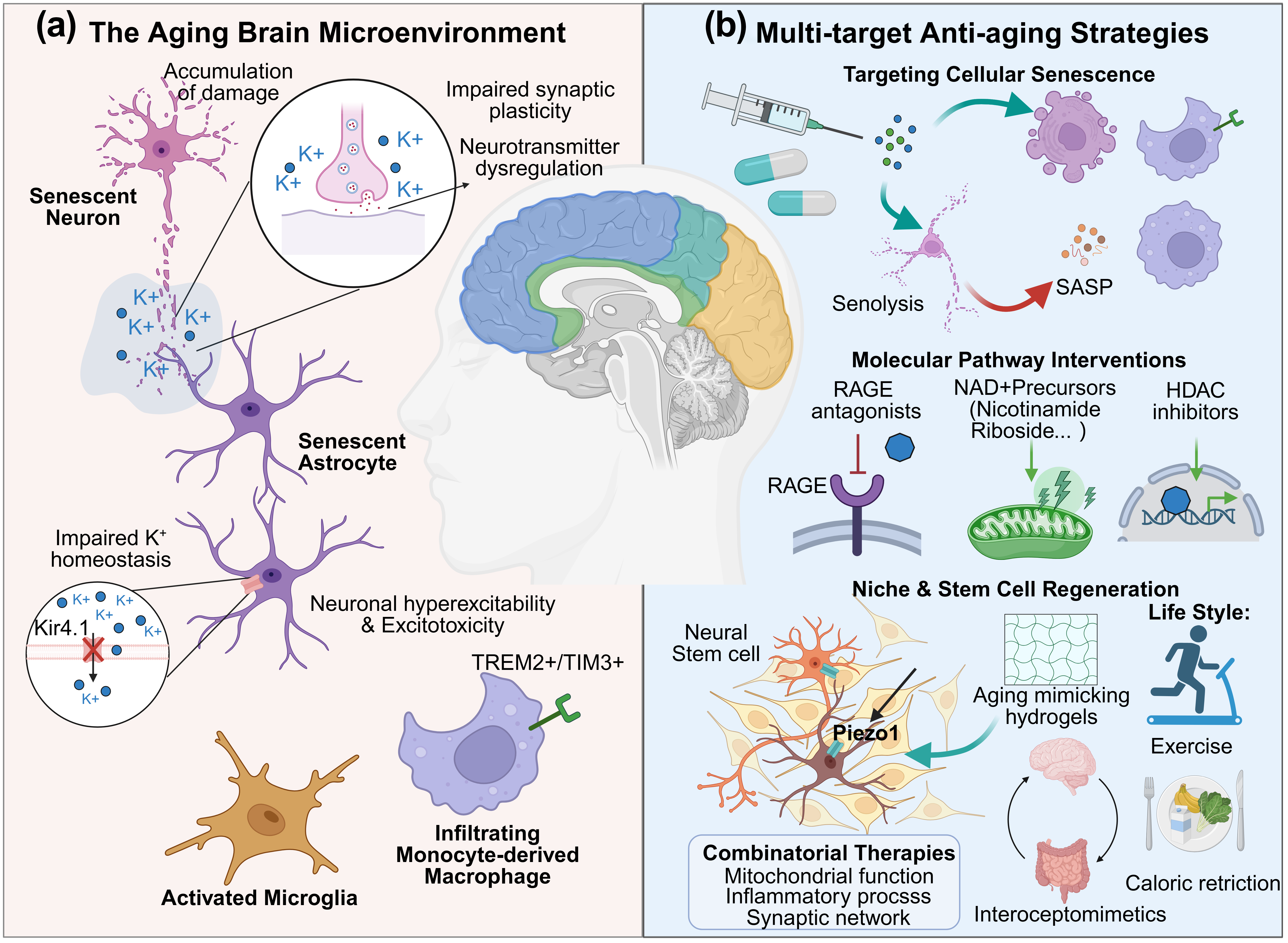
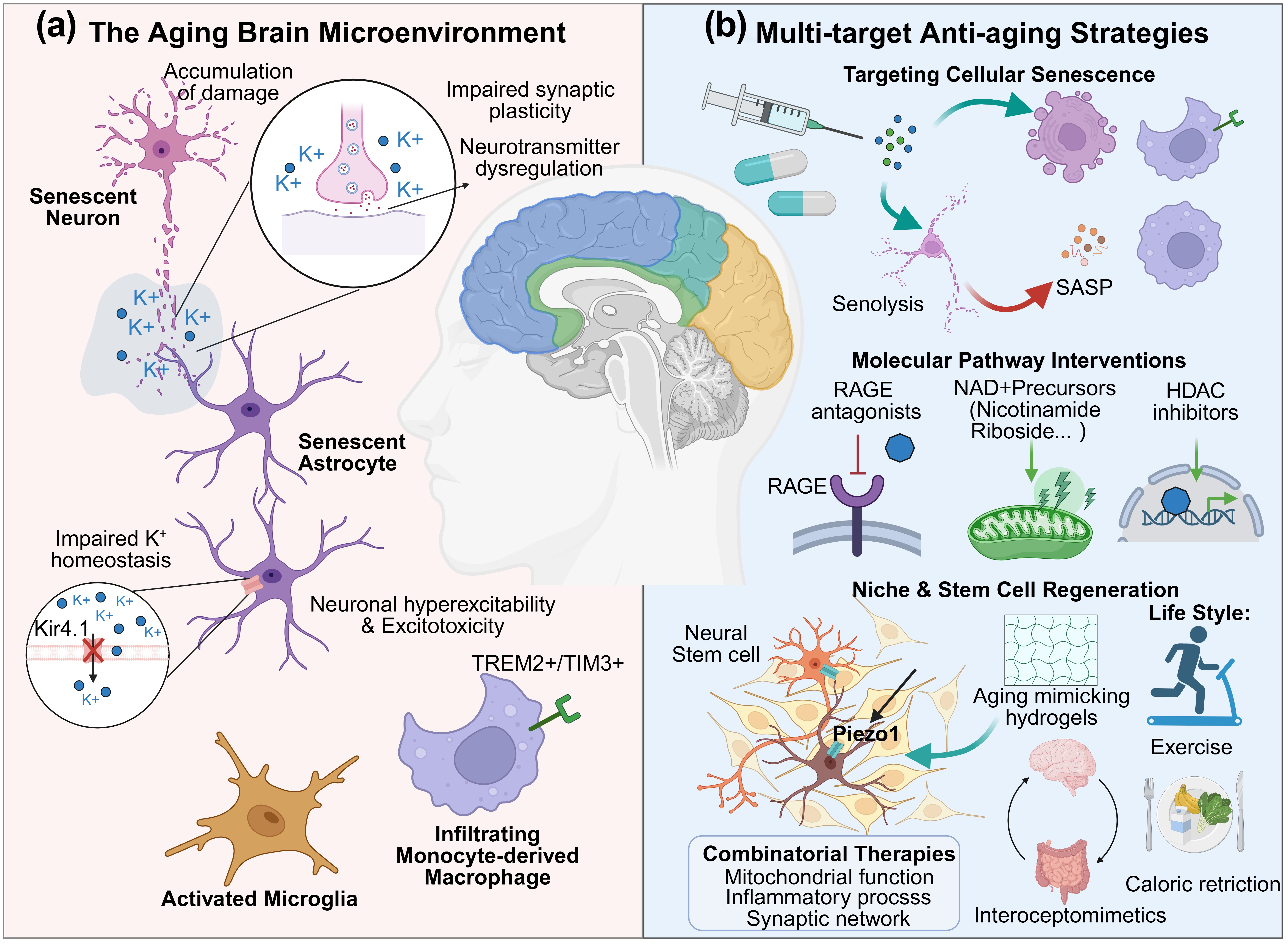
Figure 1. The cellular microenvironment of brain aging and targeted interventional strategies. (a) The microenvironment of the aging brain. Aging triggers a systemic reorganization in the nervous system. Neurons accumulate damage, leading to synaptic impairment. Aged astrocytes downregulate Kir4.1 channels, causing extracellular K+ accumulation and neuronal excitotoxicity. Simultaneously, microglia and TREM2+/TIM3+ suppressive macrophages create a pro-inflammatory niche via the SASP; (b) Multidimensional anti-aging strategies. Emerging therapeutics target specific vulnerabilities: Senolytics eliminate senescent cells; RAGE antagonists, NAD+ precursors, and HDAC inhibitors restore molecular homeostasis; and engineered hydrogels modulate Piezo1 channels to rejuvenate neural stem cells. Systemic approaches, including interoceptomimetics and lifestyle interventions, offer combinatorial benefits to preserve cognitive function. Created in BioRender. Chen, X. (2026) https://app.biorender.com/illustrations/69c2b92b82aae754449e3dfe. TREM2+: triggering receptor expressed on myeloid cells-2; TIM3+: T-cell immunoglobulin and mucin; SASP: senescence-associated secretory phenotype; RAGE: receptor for advanced glycation end-products; NAD+: nicotinamide adenine dinucleotide; HDAC: histone deacetylase.
2.2 Targeting molecular mechanisms to reverse brain aging
The recognition that aging is a modifiable biological process, rather than an unalterable chronological inevitability, has spurred intense interest in interventions to target fundamental aging mechanisms. Cellular senescence has emerged as a particularly attractive therapeutic target. Senescent cells accumulate with age in various organs including the nervous system, where they contribute to tissue dysfunction through the secretion of pro-inflammatory cytokines and chemokines, as well as growth factors and matrix-remodeling enzymes, which are collectively termed the SASP[35]; Representatively, the aging brain undergoes several changes associated with aging, including tissue atrophy, neurotransmitter alterations, and the accumulation of cellular damage. These phenomena are causatively connected to age-related transformations, which ultimately lead to cognitive decline[15].
The strategic elimination of senescent cells, which are termed senolysis, has demonstrated remarkable potential in preclinical models of aging and age-related diseases[36]. In the nervous system specifically, senolytic treatment has been associated with reduced neuroinflammation, preserved cognitive function, and decreased accumulation of age-related pathology[37]. Beyond cellular senescence signaling, several molecular pathways governing neuronal aging have emerged as promising therapeutic targets. The receptor for advanced glycation end-products (RAGE) represents one such node, which integrates signals from multiple pathogen-associated molecular patterns that accumulate with age to drive neuroinflammation, oxidative stress, and synaptic dysfunction[38]. Pharmacological RAGE antagonists have shown efficacy in brain disease control and cognitive protection[39], suggesting that targeting this pathway could simultaneously address multiple aspects of brain aging pathology. Other emerging molecular targets such as nicotinamide adenine dinucleotide (NAD+) precursors nicotinamide riboside[40,41], as well as epigenetic dysregulation targeted by histone deacetylase (HDAC) inhibitors (which have shown increasing therapeutic potential in cancer through regulation of chromatin remodeling, brain tumor progression, and resistance to conventional therapies[42,43]), represent promising strategies to restore cellular homeostasis and extend healthspan. In addition, age-related decline in brain tissue regeneration is driven by a loss of function in adult stem and progenitor cell populations[44]. A critical regulator of this process is the alteration of tissue mechanics, which is sensed by force-activated ion channels such as Piezo1[45]. This mechanistic insight offers a therapeutic opportunity, for example, using age-mimicking hydrogels to recapitulate the mechanical niche of the hippocampus could potentially modulate neural stem cell senescence[46]. Interestingly, Piezo1 can also serve as a mechanosensitive mediator linking noninvasive ultrasonic neuromodulation to the alleviation of age-associated neural dysfunction, thereby offering a potential therapeutic avenue for neurodegenerative disorders such as Parkinson’s disease[47] (Figure 1b).
The convergence of multiple aging-related molecular pathways in the nervous system suggests that combinatorial therapies targeting complementary mechanisms may yield superior outcomes. A compelling recent study has uncovered a critical role for impaired interoception in brain aging, and proposed that interoceptomimetics designed to enhance gut-brain communication could represent a viable approach to counteract age-associated cognitive decline[48]. Lifestyle interventions such as exercise and caloric restriction can activate overlapping longevity pathways and exert neuroprotective effects[49,50]. In light of the multifactorial nature of brain aging, a comprehensive therapeutic approach that concurrently targets senescent cells, mitochondrial function, inflammatory processes, and neurotransmitter systems may represent the most effective strategy for preserving cognitive function in the aging population (Figure 1b).
3. The Interplay Between Brain Tumors and the Aging Milieu
3.1 Aging regulates brain tumors
Aging is an intricate and inevitable biological process that commences in early adulthood, which is characterized by a progressive decline in physiological integrity and function[51]. This natural phenomenon leads to diminished organ reserve, cognitive alterations, and increased vulnerability to diseases, including cancer[52,53]. A recent study demonstrated that physiological aging does not merely create a permissive environment but actively reprograms the evolutionary trajectory of lung cancer, suppressing primary tumor growth while epigenetically driving a more lethal metastatic fate; this work provides a crucial mechanistic framework and highly actionable strategy for addressing metastasis in the older lung adenocarcinoma patient population[54]. Epidemiological data clearly demonstrate that the incidence of brain tumors (such as glioblastoma) increases with age, reflecting the biological complexity of addressing predictive aging biomarkers of brain neoplasms[1,55]. Actually, the molecular landscape of brain tumors demonstrates an age-dependent biological heterogeneity: pediatric gliomas are characterized by frequent alterations in the MAPK signaling cascade and recurrent gene fusions, whereas adult gliomas more often exhibit isocitrate dehydrogenase (IDH) mutations, telomerase reverse transcriptase (TERT) promoter mutations, and epidermal growth factor receptor (EGFR) amplification[56]. Interestingly, the activation of mitogen-activated protein kinase (MAPK) plays a key role in initiating cellular senescence[57]. This state of permanent growth arrest induces profound alterations in cell-cycle regulation and cellular metabolism, while promoting the secretion of diverse soluble factors through the SASP. Although MAPK-driven senescence has been identified as a therapeutic vulnerability in pediatric low-grade glioma models[58], these findings are largely derived from developmental tumor contexts and should be interpreted separately from aging-associated brain pathologies. In adult and aging brains, cellular senescence involves distinct mechanisms associated with chronic inflammation, metabolic dysfunction, and tissue remodeling.
Notably, at the cellular level, the senescent cells play a dual role in tumors as they may suppress tumor growth through permanent cell cycle arrest, while their SASP factors can promote tumor malignancy through paracrine effects on neighboring cells[59-61]. In the aging brain, the accumulation of senescent neurons, astrocytes, microglia, and endothelial cells creates a multicellular senescent niche rich in SASP factors, fueling tumor progression, recurrence, and therapeutic resistance[62]. Age-related blood-brain barrier dysfunction further compounds these effects by promoting the extravasation of peripheral immune cells into brain parenchyma and exposing brain tumor tissues to circulating inflammatory mediators[63,64] (Figure 2a). Within the brain tumor microenvironment, cellular senescence arises not only as a consequence of physiological aging but also in response to anticancer therapy[62,65]. In glioblastoma, accumulating evidence indicates that radiotherapy and temozolomide can induce therapy-induced senescence (TIS) in tumor cells, whereas radiotherapy can also induce senescence in stromal cells such as astrocytes, with persistent DNA damage responses contributing to the establishment of the senescent phenotype[66-68]. Rather than undergoing complete elimination, a subset of irradiated glioblastoma cells enters a therapy-induced senescent state, characterized by persistent proliferative arrest and a sustained SASP that can promote tumor recurrence and therapy resistance[66]. Importantly, despite extensive DNA damage, senescent cells can acquire resistance to apoptosis through the upregulation of anti-apoptotic BCL-2 family proteins, particularly BCL-XL. This BCL-XL-dependent survival promotes their persistence during and after treatment, thereby altering cellular turnover and sustaining SASP-mediated signaling within the tumor microenvironment as observed in other senescent cell models[67,69,70].
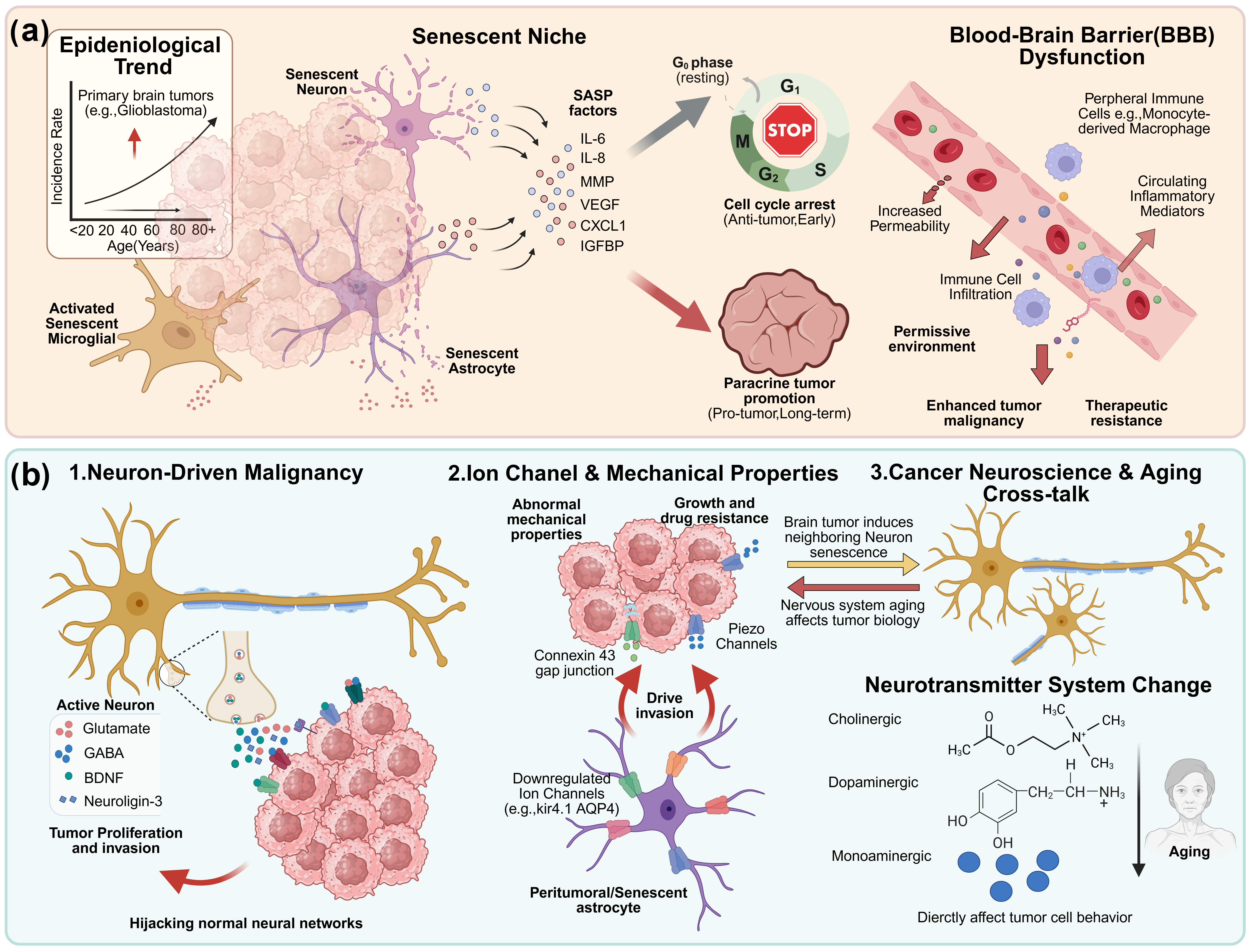
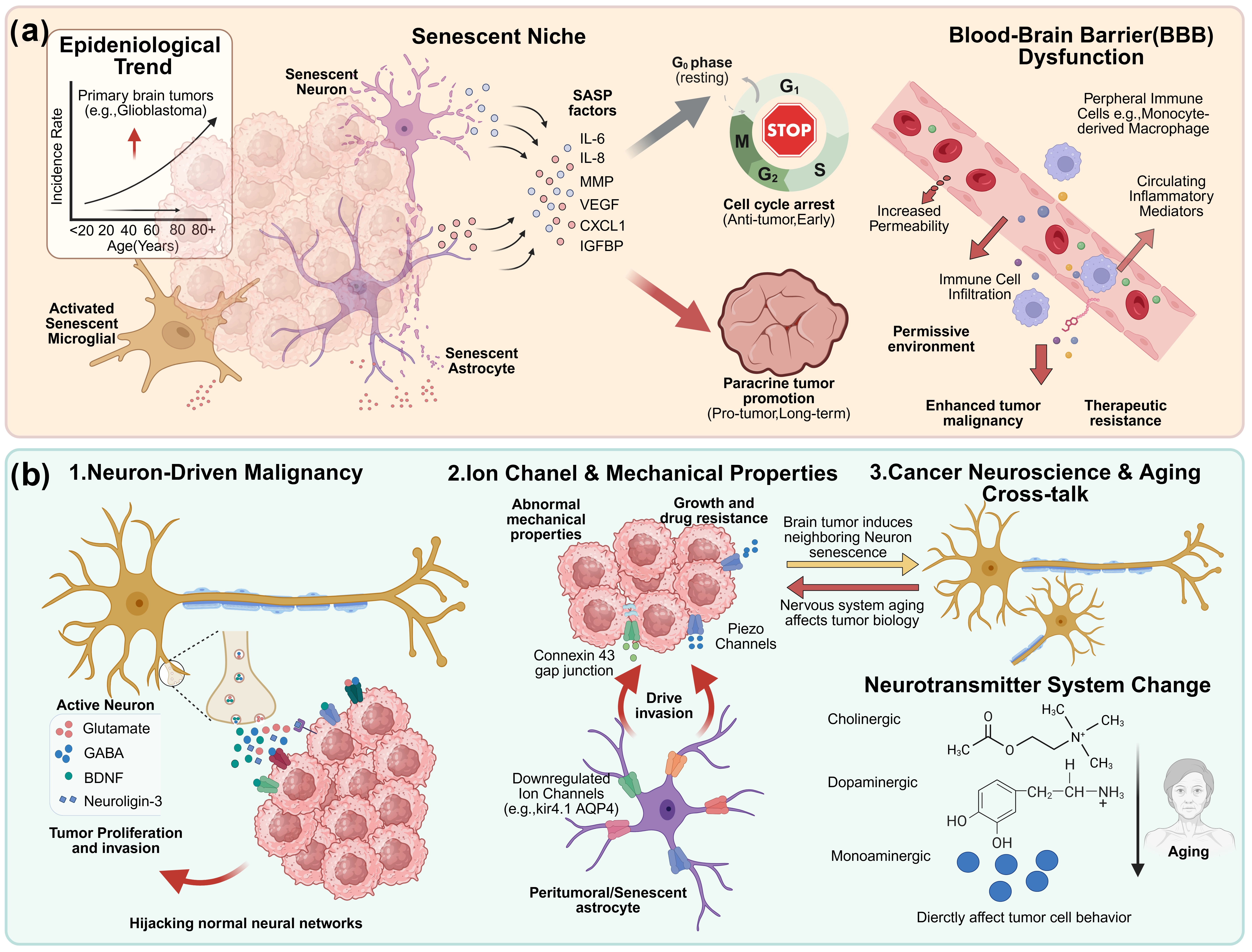
Figure 2. Bidirectional communication between brain tumors and the aging nervous system. (a) Aging as a primary driver of a tumor-permissive environment. The aging process establishes a fertile soil for glioma progression through a multicellular senescent niche and SASP secretion, which drive tumor malignancy and therapeutic resistance. Age-related BBB dysfunction further fuels this pro-inflammatory environment by allowing the extravasation of peripheral immune cells and inflammatory mediators, ultimately leading to a sharp, age-dependent increase in brain tumor incidence; (b) Neural-tumor crosstalk and age-related molecular reprogramming. The nervous system promotes tumor progression by establishing functional neuron-to-tumor synapses that “hijack” neural circuits via neurotransmitters and neurotrophins (e.g., Glutamate, NLGN3). Concurrently, tumor cells co-opt aberrant tissue mechanics and glial communication through Piezo channels and Connexin 43 gap junctions, which are integrated with age-dependent neurotransmitter shifts, synergistically drive tumor malignancy and invasion. Brain tumors establish a bidirectional feedback loop by inducing senescence in adjacent neurons, particularly after treatment. This convergence of senescence and dysregulated signaling creates a pathobiological hub, resulting in a self-sustaining cycle of tumor malignancy and cognitive decline. Created in BioRender. Chen, X. (2026) https://app.biorender.com/illustrations/69c2b92b82aae754449e3dfe. SASP: senescence-associated secretory phenotype; NLGN3: neuroligin-3; IL-6: interleukin-6; GABA: γ-aminobutyric acid; BDNF: brain-derived neurotrophic factor; MMP: matrix metalloproteinase; VEGF: vascular endothelial growth factor; IGFBP: insulin-like growth factor-binding protein.
3.2 Neuronal system in regulation of brain tumors
The nervous system actively regulates brain tumor biology through multiple integrated mechanisms that operate across distinct spatial and temporal scales, forming a complex bidirectional crosstalk central to disease progression[5,6,71,72]. Neuronal activity directly fosters brain tumor malignancy through paracrine signaling and the establishment of functional neuron-to-tumor synapses. Recent studies have established that glioma cells can form bona fide functional synapses with neurons[7,8]. In adult glioblastoma and other high-grade gliomas, glutamatergic neuronal inputs induce rapid α-amino-3-hydroxy-5-methylisoxazolepropionate (AMPA) receptor-dependent postsynaptic currents and calcium transients in a subset of tumor cells, thereby promoting tumor-cell proliferation and progression. These synaptically connected cells are further integrated into electrically coupled glioma networks that facilitate signal propagation, invasion, and treatment resistance. Emerging evidence also suggests that functional γ-aminobutyric acid (GABA) ergic neuron-to-glioma interactions occur in specific pediatric glioma subtypes, including diffuse midline glioma[73]. These interactions are mediated by a diverse array of molecular signals, including classical neurotransmitters (such as glutamate and GABA), neurotrophins (like brain-derived neurotrophic factor, brain-derived neurotrophic factor (BDNF)), and other secreted factors (such as neuroligin-3) that collectively drive tumor proliferation and invasion[7,73-76]. These studies highlight that tumors do not merely passively reside within the brain parenchyma but actively hijack and corrupt the extant neural architecture to fuel their own growth. This pathological integration indicates that intrinsic neuronal activity, whether associated with normal cognitive function or pathological hyperexcitability, can be reprogrammed to stimulate tumor progression (Figure 2b).
As key regulators of cellular excitability and signaling, ion channels represent critical nodes in brain tumor progression. Astrocytes, which intimately interact with both neurons and tumor cells, express a diverse repertoire of ion channels to not only maintain ionic homeostasis but also participate in signaling pathways that regulate astrocyte reactivity, gliotransmitter release, and neuroinflammation[27]. Interestingly, peritumor and aging astrocytes from human cerebral cortices have been demonstrated to downregulate some ion channel and membrane protein genes involved in astrocyte-synapse interactions[77]. Notably, alterations of tissue mechanical properties is a physical hallmark in both brain tumors and aged tissues, and force-activated ion channel Piezo can co-opt aberrant tissue mechanics to promote brain tumor growth and therapeutic resistance[78,79]. Other membrane transducers such as connexins, which form gap junctions, are now recognized as significant contributors to the brain tumor microenvironment[80,81]. For example, the brain tumor-astrocyte gap junction communication formed by Connexin 43 at the tumor border is a driving force for invasion[82,83] (Figure 2b).
3.3 Cancer neuroscience cross-talking with aging
The intersection of brain tumor biology and neuronal aging reveals complex bidirectional interactions that influence disease outcomes[1]. Brain tumors can induce senescence in adjacent neural cells, thereby creating a feedback loop that supports tumor survival and treatment resistance. Studies using glioblastoma organoid-neuron co-culture systems have shown that radiotherapy or chemotherapy can promote the recruitment and induction of pro-tumor senescent neurons[84]. Shared pathways linking brain tumors, aging may promote tumor progression and radioresistance. Through these shared mechanisms, the same pathways may also impair synaptic plasticity and cognitive function, contributing to cognitive decline akin to neurodegeneration[39].
A growing body of evidence implicates ion channels and neurotransmitters as key signaling agents in the communication between nerves and cancer, where they promote malignancy by directly influencing tumor cell behavior and dynamically remodeling the tumor microenvironment through actions on immune, vascular, and stromal components[30,85-87]. Since ion channel function and neurotransmitter signaling undergo progressive age-associated remodeling in the human and mammalian nervous systems[15,88,89], these alterations, together with age-related inflammatory and metabolic dysfunction, may contribute to the establishment of a permissive neural microenvironment for tumor development. The cholinergic system, which plays critical roles in attention, memory, and cortical plasticity, exhibits progressive decline with age due to degeneration of basal forebrain cholinergic neurons and reduced acetylcholine synthesis[90]. Similarly, dopaminergic signaling diminishes with age, contributing to motor slowing, reduced motivation, and impaired cognitive flexibility[91,92]. Monoaminergic systems, including noradrenergic and serotonergic pathways, also show age-related alterations that affect mood, arousal, and stress responsiveness[93]. Importantly, these neurotransmitter changes not only contribute to cognitive and behavioral symptoms of aging but can also influence brain tumor biology through direct effects on tumor cell proliferation, migration, and survival[12,94-96]. These observations point to a bidirectional relationship between aging of the nervous system and tumor biology. Elucidating the molecular links between neural activity, aging, and tumor behavior may therefore reveal novel therapeutic targets that simultaneously address both neuronal aging and oncological processes (Figure 2).
These aging-associated mechanisms form a hierarchical yet interconnected network, in which systemic and microenvironmental alterations create a permissive context for tumor progression, while more proximal neural signaling pathways directly regulate neuron-tumor interactions[97,98]. Cellular senescence and the senescence-associated secretory phenotype may act as broad, self-reinforcing processes that promote chronic inflammation, vascular dysfunction, extracellular-matrix remodelling, and immunosuppression. Blood-brain barrier disruption and neuroinflammation may further amplify these changes by altering immune-cell trafficking, cytokine exposure, and tissue homeostasis. Ion-channel dysregulation and neurotransmitter imbalance may represent more proximal components of neuron-tumor communication, directly influencing tumor-cell excitability, proliferation, invasion, and neural-circuit integration. Combining microenvironment-directed interventions with clinically tractable modulation of neural signaling may therefore represent a promising therapeutic direction.
4. Targeting Neuronal Aging to Combat Brain Tumors
4.1 Reversing senescent neuronal system for treating brain tumors
The recognition that cellular senescence, including neuronal components contributes to tumor progression opens new avenues for therapeutic intervention[59,99,100]. Senotherapy, the pharmacological targeting of senescent cells, has shown promise in diverse age-related diseases and is now being explored in the context of cancer. In fact, both aging and cancer share a set of partially overlapping hallmarks. Specifically, some aging hallmarks (such as epigenetic alterations, genomic instability, dysbiosis, and chronic inflammation) overlap with cancer hallmarks as shared “meta-hallmarks”, whereas others (such as telomere attrition, stem cell exhaustion) tend to suppress tumorigenesis, functioning as “antagonistic hallmarks”[101]. In preclinical GBM models, inhibiting tumor-derived prostaglandin E2 (PGE2) signaling or asparagine endopeptidase (AEP) activity reduces neuronal senescence and delays tumor progression, suggesting that interventions targeting the senescence program in neurons could complement conventional anti-tumor therapies[84]. Several classes of senotherapeutic agents warrant investigation in neuro-oncology. Given the context-dependent and sometimes tumor-suppressive functions of senescence, broad senolysis may remove senescent cells with potentially protective growth-arrest functions and disrupt neural or vascular homeostasis[65]. Senomorphics may therefore be particularly attractive because they suppress the pro-tumorigenic SASP without necessarily eliminating senescent cells[102], thereby limiting harmful paracrine signaling while preserving potentially protective aspects of senescence (Figure 3a). Beyond conventional pharmacological interventions aimed directly at tumor cells, strategies focused on normalizing the aged neuronal microenvironment present a complementary approach with benefits for tumor control. For instance, disrupted neural synchrony and altered neurovascular coupling in glioma models have been demonstrated to drive tumor aggressiveness[103], highlighting the importance of rejuvenating the neuron and vascular systems in the brain to alleviate malignancy. Furthermore, interventions that restore blood-brain barrier integrity or mitigate neuroinflammation during aging[63,64,104-106] can fundamentally modify the permissive soil in which tumors grow. Such microenvironmental reprogramming not only has the potential to enhance responses to standard chemoradiotherapy by improving drug delivery and immune surveillance but may also reduce treatment-related cognitive toxicity in aging patients. This dual benefit, impeding tumor progression while preserving neural function, underscores the therapeutic promise of rejuvenating the broader brain microenvironment rather than the tumor alone. Following this rationale, systemic rejuvenating interventions such as heterochronic parabiosis[107,108], cerebrospinal fluid (CSF) administration[109,110], platelet-rich plasma (PRP)[111], as well as stem cell therapy[112] can offer new perspectives to offer experimental strategies for revitalizing the aging brain, although their efficacy in the treatment of brain tumors remains to be confirmed (Figure 3a).
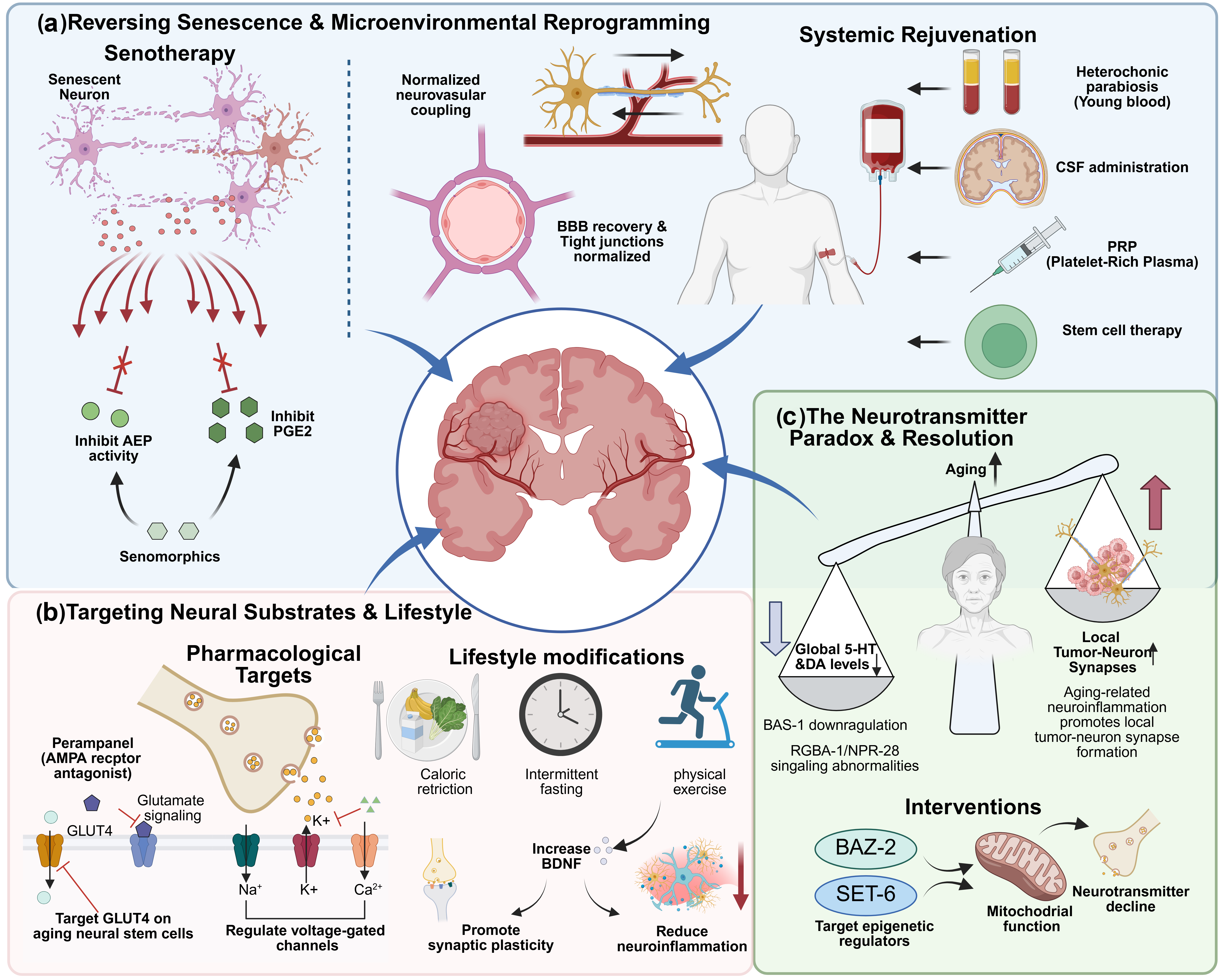
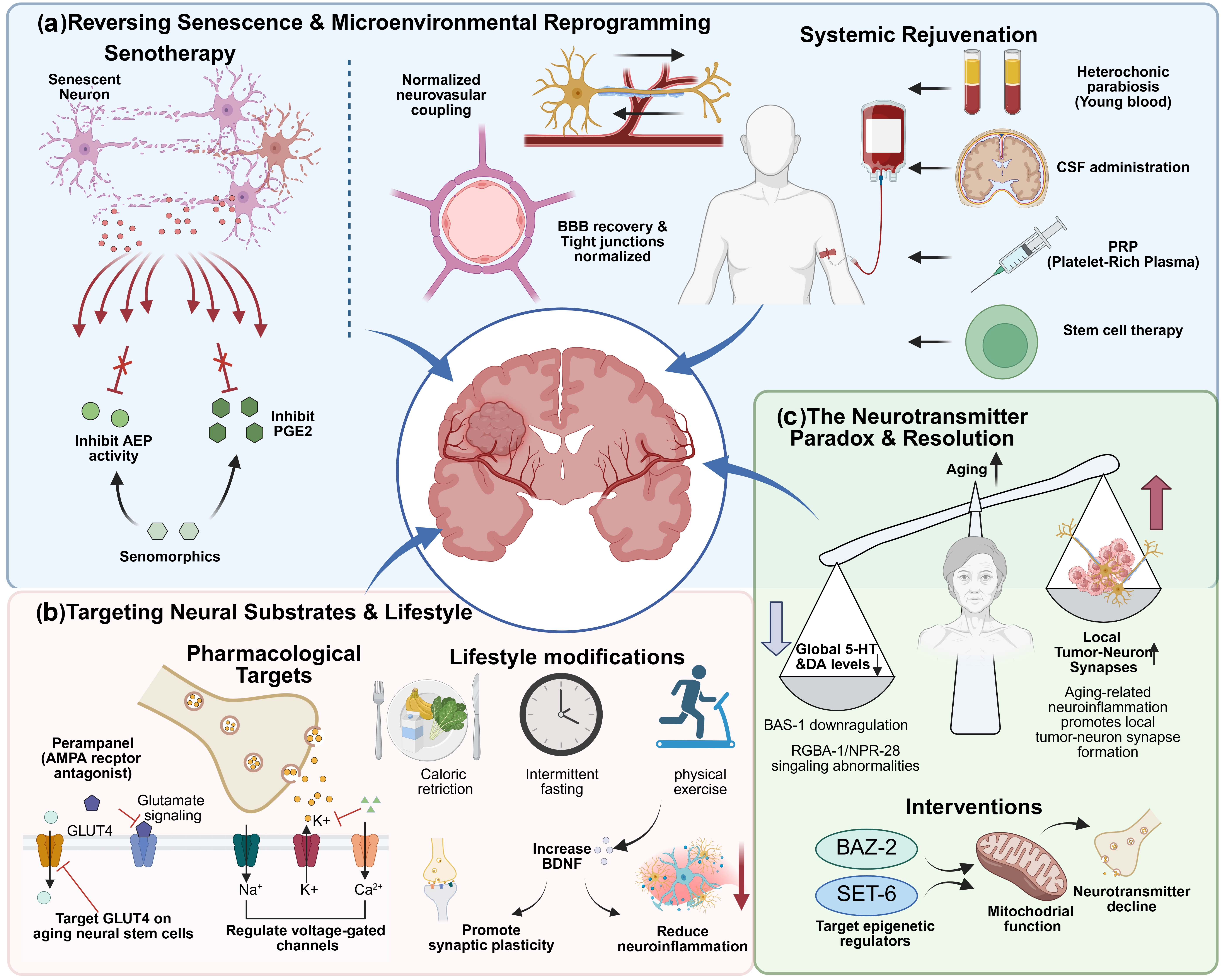
Figure 3. Therapeutic strategies targeting the aging nervous system to combat brain tumors. (a) Reversing neuronal senescence and microenvironmental reprogramming. Senotherapeutic approaches, particularly senomorphics, mitigate the pro-tumorigenic paracrine effects of the SASP by inhibiting pathways such as PGE2 and AEP, without eliminating the growth-arrested cells. Systemic rejuvenation interventions, including heterochronic parabiosis, CSF administration, PRP, and stem cell therapy, are intended to diminish the permissive soil that supports tumor growth; (b) Targeting neural substrates and lifestyle modifications. Brain aging biomarkers offer actionable targets for neuro-oncology. Pharmacological interventions include targeting metabolism-associated channels (e.g., GLUT4), voltage-gated ion channels, and using agents like Perampanel to block AMPA glutamate receptors, which otherwise drive glioma-related seizures and RTK-mediated tumor growth. Concurrently, lifestyle modifications such as exercise boost neurotrophic factors like BDNF, enhancing synaptic plasticity and reducing neuroinflammation; (c) Resolution of the neurotransmitter paradox. Aging is systemically characterized by a decline in neurotransmitters such as 5-HT and DA (regulated by BAZ2B). Paradoxically, some of these neurotransmitters promote high-grade glioma progression. This discrepancy is reconciled by age-related microenvironmental remodeling, which facilitates the localized formation of functional brain tumor-neuron synapses, granting tumor cells a competitive advantage despite the overall systemic neurotransmitter decline. Created in BioRender. Chen, X. (2026) https://app.biorender.com/illustrations/69c2b92b82aae754449e3dfe. SASP: senescence-associated secretory phenotype; PGE2: prostaglandin E2; AEP: asparagine endopeptidase; CSF: cerebrospinal fluid; PRP: platelet-rich plasma; GLUT4: glucose transporter type 4; BDNF: brain-derived neurotrophic factor; 5-HT: 5-hydroxytryptamine; DA: dopamine; BAZ2B: bromodomain adjacent to zinc finger domain protein 2B; BBB: blood-brain barrier; AMPA: α-amino-3-hydroxy-5-methylisoxazolepropionate; SET: suppressor of variegation 3-9 (Su(var) 3-9), Enhancer of Zeste (E(z)), and Trithorax; RGBA-1: regulator of behavioral aging-1.
4.2 Targeting neural substrates against brain tumor
Aging process exerts a multifaceted and interconnected impact on the nervous system[14,113], and a robust mechanistic understanding of the biomarkers for brain aging[114,115] offers avenues to treat age-related diseases such as brain tumors. High-throughput in vitro and in vivo genetic approaches with clustered regularly interspaced short palindromic repeats-associated protein 9 (CRISPR-Cas9) screens elucidated that aged neural stem cells exhibit significant alterations in the expression of metabolic candidates such as the glucose transporter type 4 (GLUT4). This finding highlights the dysfunction of metabolism-associated ion channels and transporters as a potentially conserved hallmark of nervous system aging[116], and these molecules can serve as candidate targets that warrant further investigation in brain tumor models. Biomarkers for brain aging such as neurotransmitter components and ion channels represent attractive therapeutic targets given their central role in neuron-tumor communication and their druggability with existing agents developed for neurological and psychiatric indications. For example, the neurotransmitter glutamate is the key for glioma-related seizures[117,118], and glutamate can also promote glioma growth through a non-excitable receptor tyrosine kinase-mediated signaling without altering the electrical properties of tumor cells, unveiling new understanding of how neurotransmitters enhance tumor growth[119]. The non-competitive AMPA glutamate receptor antagonist Perampanel, which was approved for epilepsy, has shown preclinical activity against GBM[120]. Additionally, targeting voltage-gated sodium, potassium, and calcium channels may affect both brain tumor behavior and neuronal function[86,121], yet achieving selective modulation between pathological and physiological signaling remains a key challenge.
Importantly, interventions targeting key aging-related molecules are not necessarily limited to pharmacological approaches. Lifestyle modifications, including caloric restriction, intermittent fasting, and regular physical exercise, activate many of the same longevity signaling pathways targeted by pharmacological interventions. These interventions exert particularly pronounced effects on brain aging and may have potential implications for brain tumor biology, though direct evidence is currently lacking[122-125]. Taking physical exercise as a prototypical example of healthy lifestyle interventions, we illustrate how behavioral modulation of the aged systemic milieu may reshape the brain tumor microenvironment and influence disease progression. Physical exercise may attenuate age-associated dysfunction through coordinated effects on the neural, immune, vascular, and metabolic compartments, although it has not been established as a direct senolytic intervention[125,126]. In the aging nervous system, exercise upregulates brain-derived neurotrophic factor (BDNF)/tropomyosin receptor kinase B (TrkB), insulin-like growth factor I (IGF-1), and other pro-survival pathways, thereby supporting neuronal survival, dendritic and synaptic plasticity, and long-term potentiation[127-129]. It also partially restores the age-related decline in hippocampal neural stem/progenitor-cell proliferation, neuronal differentiation, and survival, leading to enhanced adult neurogenesis and cognitive performance[130,131]. In parallel, exercise reduces microglial activation and inflammatory cytokine production, preserves blood-brain barrier tight-junction proteins, and improves neurovascular and glymphatic function, collectively establishing a less inflammatory and more regenerative neural milieu. At the systemic level, long-term physical activity is associated with preserved thymic output, higher frequencies of naive T cells and recent thymic emigrants, increased thymoprotective interleukin-7 (IL-7), and reduced IL-6, indicating attenuation of selected features of immunosenescence rather than complete immune rejuvenation[126]. Exercise-induced metabolic adaptations, including increased circulating IGF-1 and β-hydroxybutyrate and improved oxidative and vascular homeostasis, may further support neural resilience, although their direct effects on brain tumor-cell metabolism remain unclear. Direct evidence in brain tumors is currently limited to a small number of preclinical observations. In a GL261 glioma model implanted in the mouse motor cortex, voluntary wheel running reduced Ki67- and BrdU-positive tumor-cell proliferation and delayed motor dysfunction[132]; however, the causal molecular pathways were not determined. Exercise should therefore be regarded as a biologically plausible modifier of the aged brain-tumor ecosystem rather than an established glioblastoma-directed therapy[133,134] (Figure 3b).
It is worth noting that aging is associated with a decline in serotonin [5-hydroxytryptamine (5-HT)] and dopamine (DA) levels due to reduced expression of the shared synthetic enzyme BAS-1, leading to impaired neuronal activity and behavioral deterioration in C. elegans[18]. Novel underlying mechanisms include dietary restriction through PHA-4-mediated antioxidant pathways[18], while natural variation in glia-derived neuropeptide RGBA-1 signaling via NPR-28 in serotonergic/dopaminergic neurons accelerates behavioral aging through SIR-2.1-dependent mitochondrial unfolded protein response activation[19]. Interventions that elevate 5-HT/DA signaling or target conserved epigenetic regulators like BAZ-2 and suppressor of variegation 3-9 (Su(var)3-9), Enhancer of Zeste (E(z)), and Trithorax (SET)-6 in nematode (human orthologues: bromodomain adjacent to zinc finger domain protein 2B (BAZ2B) and euchromatic histone lysine methyltransferase 1 (EHMT1)) to enhance mitochondrial function represent promising strategies to mitigate neurotransmitter decline and promote healthy aging[20]. Besides, a high-throughput small molecule screen also identified novel aging modulators that target 5-HT/DA signaling pathway[135]. These findings, together with therapeutic strategies aimed at mitigating neuronal aging, may provide new opportunities for the treatment of brain tumors. Nevertheless, although several neurotransmitter systems undergo an overall decline during physiological aging, neuronal activity and neurotransmitter signaling can promote high-grade glioma progression[12,94-96,136]. This apparent discrepancy may reflect differences in spatial and biological scale, because regional neurotransmitter abundance does not necessarily predict local neurotransmitter release, receptor activation or synaptic signaling at the tumor-neural interface. Functional neuron-to-glioma synapses have been demonstrated; however, direct evidence that these synapses are more abundant or functionally more active in aged brains is currently lacking. Age-associated neuroinflammation, glial dysfunction, altered neuronal excitability and extracellular-matrix remodeling may modify local neuron-tumor communication, but this possibility remains to be tested experimentally (Table 1 and Figure 3c).
Table 1. Anti-Aging Therapeutic Strategies in Neuro-Oncology.
| Drug/Strategy | Molecular Target | BBB Penetration | Combination with Standard-of-Care | Mechanism | Ref. |
| Senomorphics | SASP suppression | Variable | TMZ/RT | Suppresses pro-tumorigenic SASP without eliminating cells | [84] |
| RAGE antagonists | RAGE signaling | Not established | N/A | Reduces neuroinflammation, oxidative stress | [39] |
| NAD+ precursors (NR, NMN) | NAD+ metabolism | Yes | N/A | Restores mitochondrial function, reduces senescence | [40,41] |
| HDAC inhibitors | Epigenetic regulation | Variable | TMZ | Restores cellular homeostasis | [42,43] |
| Perampanel | AMPA glutamate receptor | Yes | TMZ/RT | Blocks glutamate-driven tumor growth and seizures | [120] |
AMPA: α-amino-3-hydroxy-5-methylisoxazolepropionate; BBB: blood-brain barrier; SASP: senescence-associated secretory phenotype; RAGE: receptor for advanced glycation end-products; HDAC: histone deacetylase; NAD+ : nicotinamide adenine dinucleotide; TMZ: temozolomide; RT: radiotherapy; NR: nicotinamide riboside; NMN: nicotinamide mononucleotide.
For older patients with brain tumors, chronological age alone is insufficient for clinical decision-making. Instead, treatment planning should be guided by an integrated assessment of tumor type and molecular features, physiological fitness, frailty, baseline neurological function, cognitive reserve, comorbidities, polypharmacy, and treatment tolerance[137,138]. Clinically, older patients with cancer may be categorized as fit, vulnerable, or frail to guide treatment intensity, while biomarkers such as methylguanine-DNA methyltransferase (MGMT) promoter methylation can further inform therapeutic selection in older patients with glioblastoma[139,140]. However, interventions specifically targeting aging-related mechanisms or aging-neural interactions in brain tumors remain largely at the preclinical stage, and direct clinical evidence in older patients is currently lacking. Moreover, age-related disruption of the blood-brain barrier does not necessarily ensure effective drug delivery, because intracranial exposure is also influenced by regional barrier heterogeneity, cerebral perfusion, efflux transporters, systemic pharmacokinetics, and organ function[141,142]. Therefore, before therapies targeting aging-neural interactions can be translated clinically, these factors should be comprehensively evaluated together with cognition, functional independence, quality of life, and treatment burden to identify the patients most likely to benefit while minimizing neurotoxicity[143].
5. Summary and Future Perspective
The convergence of aging research, cancer neuroscience, and neurodegeneration studies reveals a previously underappreciated unity among age-related brain pathologies. Brain tumors do not exist in isolation but rather emerge from and actively remodel an aged neural microenvironment characterized by senescent cells, chronic inflammation, and dysregulated neuronal signaling[1]. The presence of tumor progression accelerates local or systemic aging processes, inducing senescence in adjacent neurons and glia while compromising cognitive function through mechanisms that overlap with those operating in classical neurodegenerative diseases[144,145]. In this review, we illustrate these integrated frameworks that contain important implications for therapeutic development. First, they indicate that effective treatments for brain tumors in aging populations must address not only the malignant cells themselves but also the aged microenvironment that supports them. Second, they underscore the potential of repurposing drugs originally developed for neurological and psychiatric disorders as anti-tumor agents, exploiting their favorable safety profiles and capacity to penetrate the blood-brain barrier. Third, they emphasize the importance of preserving cognitive function as a therapeutic endpoint co-equal with tumor control, as the two are mechanistically linked.
Notably, while historically conceptualized as isolated ensembles of “corrupted” normal cells[146], tumors are increasingly recognized as complex “outlaw organs”: within this aberrant architectural framework, malignant cells interact with components of the tumor microenvironment that serve as “accessories to the crime,” particularly senescent cells and neural innervation[147]. As the “retired yet disruptive instigators”, senescent cells leverage the SASP to paradoxically fuel adjacent tumor proliferation, drive angiogenesis, and orchestrate localized immunosuppression. Therefore, aging should not be viewed merely as a chronological progression, but as a profound structural and functional remodeling of host tissues. Advanced age coincides with the widespread accumulation of mutation-harboring cells, along with a progressive decline in immunosurveillance and a concomitant rise in chronic, low-grade systemic inflammation, which is defined as inflammaging[148]. Together, these systemic macroenvironmental shifts cultivate a highly permissive soil for tumorigenesis. Meanwhile, state-of-the-art imaging and genetic tools have recently revealed that neurons do not merely coexist with tumors; rather, they form bona fide physical contacts with malignant cells, culminating in the formation of pseudo-tripartite synapses[149]. In primary brain tumors and brain metastases, cancer cells exploit these synaptic interfaces to directly receive neurotransmitter signals, thereby gaining a substantial proliferative advantage[150]. Through this active innervation, the nervous system is effectively “hijacked” by the malignancy, thus becoming a critical accomplice in tumor expansion, apoptosis evasion, and metastasis. Elucidating the complex, tripartite crosstalk among the aging macroenvironment, the nervous system, and the brain tumor represents a critical scientific frontier. Deciphering these interconnected regulatory networks may not only be to unravel the fundamental biology of brain tumors, but also for the conceptualization and development of next-generation therapeutic modalities in both brain aging and malignancy.
Several key questions remain for future investigation. What are the relative contributions of different cell types, including neurons, astrocytes, microglia, endothelial cells, etc. to the age-related permissiveness for tumor growth? How do current therapeutic approaches for cancer influence neuronal aging? And can senotherapeutic interventions mitigate treatment-related cognitive toxicity in brain tumor patients? Furthermore, can we identify biomarkers that differentiate adaptive and reversible senescence from irreversible senescence within the nervous system, thereby enabling more precise therapeutic targeting in neuro-oncology treatment? Additionally, whether and how do genetic risk factors for neurodegeneration influence brain tumor biology and treatment response? Addressing these questions will require continued integration of disciplines and methodologies. Advanced model systems, such as patient-derived brain tumor organoids with neurons and glia components, are essential for dissecting cell-type-specific mechanisms[151-154]. As artificial intelligence (AI) based tools are emerging, AI-driven multi-omics integration with single-cell and spatial transcriptomic approaches applied to human tissue across the age spectrum can map the cellular ecosystems within which tumors arise and progress[155-158]. Longitudinal studies incorporating both tumor outcomes and cognitive endpoints will be necessary to validate the translational relevance of these mechanistic insights. Age is a major risk factor for cancer, cardiovascular disease, and dementia, yet how behavior changes across the lifespan and whether aging unfolds through discrete stages remain incompletely understood. Recent work using high-resolution behavioral tracking and machine-learning approaches has begun to define a vertebrate aging framework[159], offering new insights into biological aging and potential interventions for aging-related diseases, including brain tumors.
The therapeutic opportunities emerging from this integrated perspective are substantial. Targeting convergent core mechanisms of neuronal aging, including senescence, inflammation, ion channel dysfunction, and neurotransmitter imbalance, offers the potential to simultaneously inhibit tumor progression, enhance treatment efficacy, and preserve the cognitive function essential to quality of life. The aging brain is not merely a passive backdrop for tumor development but an active participant whose manipulation presents an opportunity for patients facing the burden of brain cancer and age-related neurological decline.
Acknowledgements
The authors declare that AI tools were used solely for language polishing during the manuscript preparation process. During the language polishing process, we utilized the following AI tools: DeepSeek-V3.2 and GPT-5.4. All content, including structural design, literature citation, and figures, is original and was not generated using AI tools. The authors reviewed, revised, and approved the final manuscript and take full responsibility for its content.
Authors contribution
Li Q, Zhang J: Investigation, writing-review & editing.
Luo J: Writing-review & editing.
Cai S: Conceptualization, writing-review & editing.
Chen X: Conceptualization, writing-original draft.
Conflicts of interest
The authors declare no competing interests.
Ethical approval
Not applicable.
Consent to participate
Not applicable.
Consent for publication
Not applicable.
Availability of data and materials
Not applicable.
Funding
This study was supported by the grants from the National Natural Science Foundation of China (Grant Nos. 32471019 to X.C., 82330047 to S.Q.C.), and the Shanghai Municipal Health Commission (Grant No. 202340045 to X.C.).
Copyright
© The Author(s) 2026.
References
-
2. López-Otín C, Blasco MA, Partridge L, Serrano M, Kroemer G. The hallmarks of aging. Cell. 2013;153(6):1194-1217.[DOI]
-
3. López-Otín C, Blasco MA, Partridge L, Serrano M, Kroemer G. Hallmarks of aging: An expanding universe. Cell. 2023;186(2):243-278.[DOI]
-
6. Mancusi R, Monje M. The neuroscience of cancer. Nature. 2023;618(7965):467-479.[DOI]
-
11. Sun Y, Wang X, Zhang DY, Zhang Z, Bhattarai JP, Wang Y, et al. Brain-wide neuronal circuit connectome of human glioblastoma. Nature. 2025;641(8061):222-231.[DOI]
-
12. Drexler R, Drinnenberg A, Gavish A, Yalçin B, Shamardani K, Rogers AE, et al. Cholinergic neuronal activity promotes diffuse midline glioma growth through muscarinic signaling. Cell. 2025;188(17):4640-4657.e30.[DOI]
-
16. Kitchigina VF. Mechanisms of cognitive aging: Health and pathology. Neurosci Behav Physiol. 2026;56(2):288-309.[DOI]
-
17. Yuan J, Cai SQ. The regulatory mechanisms of behavioral and cognitive aging. Hereditas. 2021;43(6):545-570. Chinese.[DOI]
-
19. Yin JA, Gao G, Liu XJ, Hao ZQ, Li K, Kang XL, et al. Genetic variation in glia–neuron signalling modulates ageing rate. Nature. 2017;551(7679):198-203.[DOI]
-
20. Yuan J, Chang SY, Yin SG, Liu ZY, Cheng X, Liu XJ, et al. Two conserved epigenetic regulators prevent healthy ageing. Nature. 2020;579(7797):118-122.[DOI]
-
22. Guskjolen A, Zirlinger M. The neuroscience of aging: Shining a candle in the dark. Neuron. 2025;113(1):1.[DOI]
-
24. Costa J, Martins S, Ferreira PA, Cardoso AMS, Guedes JR, Peça J, et al. The old guard: Age-related changes in microglia and their consequences. Mech Ageing Dev. 2021;197:111512.[DOI]
-
26. Jiang Q, Liu J, Huang S, Wang XY, Chen X, Liu GH, et al. Antiageing strategy for neurodegenerative diseases: From mechanisms to clinical advances. Signal Transduct Target Ther. 2025;10:76.[DOI]
-
27. Verkhratsky A, Nedergaard M. Physiology of astroglia. Physiol Rev. 2018;98(1):239-389.[DOI]
-
30. Zhang Y, Duan W, Chen L, Chen J, Xu W, Fan Q, et al. Potassium ion channel modulation at cancer-neural interface enhances neuronal excitability in epileptogenic glioblastoma multiforme. Neuron. 2025;113(2):225-243.e10.[DOI]
-
32. von Bernhardi R, Eugenín J. Ageing-related changes in the regulation of microglia and their interaction with neurons. Neuropharmacology. 2025;265:110241.[DOI]
-
36. Chaib S, Tchkonia T, Kirkland JL. Cellular senescence and senolytics: The path to the clinic. Nat Med. 2022;28(8):1556-1568.[DOI]
-
37. Aguado J, Amarilla AA, Taherian Fard A, Albornoz EA, Tyshkovskiy A, Schwabenland M, et al. Senolytic therapy alleviates physiological human brain aging and COVID-19 neuropathology. Nat Aging. 2023;3(12):1561-1575.[DOI]
-
38. Guvatova ZG, Vakhrusheva A, Moskalev A. A receptor for glycation end products (RAGE) is a key transmitter between garb-aging and inflammaging. Ageing Res Rev. 2026;113:102919.[DOI]
-
41. Li F, Wu C, Wang G. Targeting NAD metabolism for the therapy of age-related neurodegenerative diseases. Neurosci Bull. 2024;40(2):218-240.[DOI]
-
43. Lv Z, Ji T, Liu J, Sun X, Liang H. Synthetic approaches and clinical applications of representative HDAC inhibitors for cancer therapy: A review. Eur J Med Chem. 2025;283:117185.[DOI]
-
44. Goodell MA, Rando TA. Stem cells and healthy aging. Science. 2015;350(6265):1199-1204.[DOI]
-
52. Trastus LA, d’Adda di Fagagna F. The complex interplay between aging and cancer. Nat Aging. 2025;5(3):350-365.[DOI]
-
53. Wang L, Luo Y, Chen X, Wang Y, Zhang Y. The interplay of aging and cancer: Mechanisms, implications, and therapeutic strategies. MedComm. 2025;4(3):e70041.[DOI]
-
54. Patel AAH, Dzanan JJ, Ali KX, Eklund EA, Alvarez SW, Raj D, et al. Ageing promotes metastasis via activation of the integrated stress response. Nature. 2026;652(8112):1339-1348.[DOI]
-
56. Ashraf A, Ashraf A, Khan L, Shaikh S, Hanif F. Glioma in different life stages: A comparative analysis of adult and pediatric tumors. Hum Gene. 2025;46:201476.[DOI]
-
57. Anerillas C, Abdelmohsen K, Gorospe M. Regulation of senescence traits by MAPKs. Geroscience. 2020;42(2):397-408.[DOI]
-
58. Sigaud R, Stefanski A, Selt F, Kocher D, Usta D, Picard D, et al. Multi-omics dissection of MAPK-driven senescence unveils therapeutic vulnerabilities in KIAA1549:: BRAF-fusion pediatric low-grade glioma models. Sig Transduct Target Ther. 2025;10:197.[DOI]
-
59. Colucci M, Sarill M, Maddalena M, Valdata A, Troiani M, Massarotti M, et al. Senescence in cancer. Cancer Cell. 2025;43(7):1204-1226.[DOI]
-
60. Ma L, Yu J, Fu Y, He X, Ge S, Jia R, et al. The dual role of cellular senescence in human tumor progression and therapy. MedComm. 2024;5(9):e695.[DOI]
-
62. Liu Y, Feng Y, Cheng L, Xu Y, Wu A, Cheng P. Profiling with senescence-associated secretory phenotype score identifies GDC-0879 as a small molecule sensitizing glioblastoma to anti-PD1. Cell Death Dis. 2025;16:602.[DOI]
-
63. La Q, Baloch A, Lo DF. Aging-driven blood–brain barrier dysfunction and its impact on CNS cancer susceptibility: A comprehensive narrative review. Aging Cancer. 2025;6(2):46-53.[DOI]
-
64. Knox EG, Aburto MR, Clarke G, Cryan JF, O’Driscoll CM. The blood-brain barrier in aging and neurodegeneration. Mol Psychiatry. 2022;27(6):2659-2673.[DOI]
-
65. Gorgoulis V, Adams PD, Alimonti A, Bennett DC, Bischof O, Bishop C, et al. Cellular senescence: Defining a path forward. Cell. 2019;179(4):813-827.[DOI]
-
67. Ji J, Ding K, Cheng B, Zhang X, Luo T, Huang B, et al. Radiotherapy-induced astrocyte senescence promotes an immunosuppressive microenvironment in glioblastoma to facilitate tumor regrowth. Adv Sci. 2024;11(15):2304609.[DOI]
-
71. Monje M, Borniger JC, D’Silva NJ, Deneen B, Dirks PB, Fattahi F, et al. Roadmap for the emerging field of cancer neuroscience. Cell. 2020;181(2):219-222.[DOI]
-
73. Barron T, Yalçın B, Su M, Byun YG, Gavish A, Shamardani K, et al. GABAergic neuron-to-glioma synapses in diffuse midline gliomas. Nature. 2025;639(8056):1060-1068.[DOI]
-
74. Taylor KR, Barron T, Hui A, Spitzer A, Yalçin B, Ivec AE, et al. Glioma synapses recruit mechanisms of adaptive plasticity. Nature. 2023;623(7986):366-374.[DOI]
-
77. Krawczyk MC, Haney JR, Pan L, Caneda C, Khankan RR, Reyes SD, et al. Human astrocytes exhibit tumor microenvironment-, age-, and sex-related transcriptomic signatures. J Neurosci. 2022;42(8):1587-1603.[DOI]
-
81. Mulkearns-Hubert EE, Hajdari N, Hong ES, Jacobs AP, Gaboriau A, Giltner S, et al. Connexin 43 drives glioblastoma cancer stem cell phenotypes through a WNK lysine-deficient protein kinase 1-c-MYC signaling axis. Cell Rep. 2025;44(9):116303.[DOI]
-
86. Dong W, Fekete A, Chen X, Liu H, Beilhartz GL, Chen X, et al. A designer peptide against the EAG2–Kvβ2 potassium channel targets the interaction of cancer cells and neurons to treat glioblastoma. Nat Cancer. 2023;4(10):1418-1436.[DOI]
-
91. Berry AS, Shah VD, Baker SL, Vogel JW, O’Neil JP, Janabi M, et al. Aging affects dopaminergic neural mechanisms of cognitive flexibility. J Neurosci. 2016;36(50):12559-12569.[DOI]
-
93. Ossola P, Gerra ML, Luviè L, Piacente A, Marchesi C, Schoretsanitis G, et al. Effect of age on the response to serotonergic and noradrenergic antidepressants: A systematic review, meta-regression and individual participant data pooled analysis. J Psychiatr Res. 2025;183:133-143.[DOI]
-
94. Yang Y, Yang C, Chen X, Jiang Y, Lei X, Ma K, et al. Long-range cholinergic input promotes glioblastoma progression. Cancer Cell. 2025;43(11):2089-2105.e10.[DOI]
-
95. Mursaleen M, Tahir M, Suleman MU, Tabassum SN, Khalil U. The dopamine paradox in glioblastoma oncology: Methylxanthine therapy against nicotine-driven pathogenesis. Ann Med Surg. 2025;87(11):6928-6930.[DOI]
-
96. Karmakar S, Lal G. Role of serotonergic system in regulating brain tumor-associated neuroinflammatory responses. In: Ray SK, editor. Neuroprotection: Method and protocols. New York: Humana; 2024. p. 181-207.[DOI]
-
97. Li Y, Wang Y, Han X, Xu J, Liu E, Cheng J, et al. Glioma-derived SPARCL1 promotes the formation of peritumoral neuron-glioma synapses. J Neuro Oncol. 2025;173(3):515-526.[DOI]
-
98. Nejo T, Krishna S, Yamamichi A, Lakshmanachetty S, Jimenez C, Lee KY, et al. Glioma-neuronal circuit remodeling induces regional immunosuppression. Nat Commun. 2025;16:4770.[DOI]
-
99. Dong Z, Luo Y, Yuan Z, Tian Y, Jin T, Xu F. Cellular senescence and SASP in tumor progression and therapeutic opportunities. Mol Cancer. 2024;23(1):181.[DOI]
-
101. López-Otín C, Pietrocola F, Roiz-Valle D, Galluzzi L, Kroemer G. Meta-hallmarks of aging and cancer. Cell Metab. 2023;35(1):12-35.[DOI]
-
104. de Rezende VL, de Aguiar da Costa M, Martins CD, Mathias K, Gonçalves CL, Barichello T, et al. Systemic rejuvenating interventions: Perspectives on neuroinflammation and blood–brain barrier integrity. Neurochem Res. 2025;50(2):112.[DOI]
-
105. Sharma D, Kumar R. Breaking barriers: The role of NETosis in blood-brain barrier leakage and age-related cognitive decline. Explor Neurosci. 2024;3(5):375-381.[DOI]
-
106. Takata F, Nakagawa S, Matsumoto J, Dohgu S. Blood-brain barrier dysfunction amplifies the development of neuroinflammation: Understanding of cellular events in brain microvascular endothelial cells for prevention and treatment of BBB dysfunction. Front Cell Neurosci. 2021;15:661838.
-
112. Rando TA, Brunet A, Goodell MA. Hallmarks of stem cell aging. Cell Stem Cell. 2025;32(7):1038-1054.[DOI]
-
113. Higgins-Chen AT, Thrush KL, Levine ME. Aging biomarkers and the brain. Semin Cell Dev Biol. 2021;116:180-193.[DOI]
-
116. Ruetz TJ, Pogson AN, Kashiwagi CM, Gagnon SD, Morton B, Sun ED, et al. CRISPR–Cas9 screens reveal regulators of ageing in neural stem cells. Nature. 2024;634(8036):1150-1159.[DOI]
-
118. Simon M, von Lehe M. Glioma-related seizures: Glutamate is the key. Nat Med. 2011;17(10):1190-1191.[DOI]
-
120. Lange F, Hartung J, Liebelt C, Boisserée J, Resch T, Porath K, et al. Perampanel add-on to standard radiochemotherapy in vivo promotes neuroprotection in a rodent F98 glioma model. Front Neurosci. 2020;14:598266.[DOI]
-
121. Huang X, Jan LY. Targeting potassium channels in cancer. J Cell Biol. 2014;206(2):151-162.[DOI]
-
124. Lin Y, Li S, Xu X, Hu X, Li Y, Liang S, et al. Intermittent fasting inhibits Tp53-driven glioma through gut microbiota-mediated methionine-M6A regulation. Nat Commun. 2026;17(1):1804.[DOI]
-
126. Duggal NA, Pollock RD, Lazarus NR, Harridge S, Lord JM. Major features of immunesenescence, including reduced thymic output, are ameliorated by high levels of physical activity in adulthood. Aging Cell. 2018;17(2):e12750.[DOI]
-
128. Wrann CD, White JP, Salogiannnis J, Laznik-Bogoslavski D, Wu J, Ma D, et al. Exercise induces hippocampal BDNF through a PGC-1α/FNDC5 pathway. Cell Metab. 2013;18(5):649-659.[DOI]
-
133. Gehring K, Stuiver MM, Visser E, Kloek C, van den Bent M, Hanse M, et al. A pilot randomized controlled trial of exercise to improve cognitive performance in patients with stable glioma: A proof of concept. Neuro-Oncology. 2020;22(1):103-115.[DOI]
-
135. Ye SW, Song SD, Liu XJ, Luo Y, Cai SQ. A small-molecule screen identifies novel aging modulators by targeting 5-HT/DA signaling pathway. Aging Cell. 2025;24(3):e14411.[DOI]
-
137. Minniti G, Filippi AR, Osti MF, Ricardi U. Radiation therapy for older patients with brain tumors. Radiat Oncol. 2017;12(1):101.[DOI]
-
138. Weller M, van den Bent M, Preusser M, Le Rhun E, Tonn JC, Minniti G, et al. EANO guidelines on the diagnosis and treatment of diffuse gliomas of adulthood. Nat Rev Clin Oncol. 2021;18(3):170-186.[DOI]
-
142. Nayak L, Iwamoto FM. Primary brain tumors in the elderly. Curr Neurol Neurosci Rep. 2010;10(4):252-258.[DOI]
-
143. Vallet-Regí M, Manzano M, Rodriguez-Mañas L, Checa López M, Aapro M, Balducci L. Management of cancer in the older age person: An approach to complex medical decisions. Oncologist. 2017;22(3):335-342.[DOI]
-
146. Hanahan D, Weinberg RA. The hallmarks of cancer. Cell. 2000;100(1):57-70.[DOI]
-
147. Hanahan D. Hallmarks of cancer: Then and now, and beyond. Cell. 2026;189(8):2254-2277.[DOI]
-
148. Franceschi C, Garagnani P, Parini P, Giuliani C, Santoro A. Inflammaging: A new immune–metabolic viewpoint for age-related diseases. Nat Rev Endocrinol. 2018;14(10):576-590.[DOI]
-
149. Anastasaki C, Gao Y, Gutmann DH. Neurons as stromal drivers of nervous system cancer formation and progression. Dev Cell. 2023;58(2):81-93.[DOI]
-
150. Karreman MA, Winkler F. Cancer neuroscience of brain metastasis: When in Rome, do as the Romans do. Neuron. 2025;113(17):2740-2759.[DOI]
-
152. Peng T, Ma X, Hua W, Wang C, Chu Y, Sun M, et al. Individualized patient tumor organoids faithfully preserve human brain tumor ecosystems and predict patient response to therapy. Cell Stem Cell. 2025;32(4):652-669.e11.[DOI]
-
153. Watanabe F, Hollingsworth EW, Bartley JM, Wisehart L, Desai R, Hartlaub AM, et al. Patient-derived organoids recapitulate glioma-intrinsic immune program and progenitor populations of glioblastoma. PNAS Nexus. 2024;3(2):pgae051.[DOI]
-
154. Hu JL, Todhunter ME, LaBarge MA, Gartner ZJ. Opportunities for organoids as new models of aging. J Cell Biol. 2018;217(1):39-50.[DOI]
-
159. Bedbrook CN, Nath RD, Zhang L, Linderman SW, Brunet A, Deisseroth K. Lifelong behavioral screen reveals an architecture of vertebrate aging. Science. 2026;391(6790):eaea9795.[DOI]
Copyright
© The Author(s) 2026. This is an Open Access article licensed under a Creative Commons Attribution 4.0 International License (https://creativecommons.org/licenses/by/4.0/), which permits unrestricted use, sharing, adaptation, distribution and reproduction in any medium or format, for any purpose, even commercially, as long as you give appropriate credit to the original author(s) and the source, provide a link to the Creative Commons license, and indicate if changes were made.
Publisher’s Note
Science Exploration remains a neutral stance on jurisdictional claims in published
maps
and institutional affiliations. The views expressed in this article are solely those
of
the author(s) and do not reflect the opinions of the Editors or the publisher.
Share And Cite


Science Exploration Style
Li Q, Zhang J, Luo J, Cai SQ, Chen X. Navigating the aging brain: The interplay between brain malignancy and the aging microenvironment. Ageing Cancer Res Treat. 2027;4:202614. https://doi.org/10.70401/acrt.2026.0038
Tips
Copy completed.
Submit a Manuscript
Author Instructions
Cite this Article
Article Metrics
0
View
0
Download
Cited
Article Updates

- Abstract
- Keywords
- 1. Introduction
- 2. The Biology of Neuronal Aging
- 3. The Interplay Between Brain Tumors and the Aging Milieu
- 4. Targeting Neuronal Aging to Combat Brain Tumors
- 5. Summary and Future Perspective
- Acknowledgements
- Authors contribution
- Conflicts of interest
- Ethical approval
- Consent to participate
- Consent for publication
- Availability of data and materials
- Funding
- References
- Copyright
 Get citation
Science Exploration Style

Li Q, Zhang J, Luo J, Cai SQ, Chen X. Navigating the aging brain: The interplay between brain malignancy and the aging microenvironment. Ageing Cancer Res Treat. 2027;4:202614. https://doi.org/10.70401/acrt.2026.0038
copy
Share Link
copy