Designing bone adhesives for fracture repair across skeletal health and disease

Hailong Guo
1,#

,
Huihui Liu
3,#
,
Kexin Wang
4,#
,
Junrui Hu
4,#
,
Xudong Yao
6
,
Haijiao Mao
1,*
,
Xiaozhao Wang
1,2,5,7,*
*Correspondence to:
Xiaozhao Wang, Department of Orthopedic Surgery, The First Affiliated Hospital of Ningbo University, Ningbo 315010, Zhejiang, China; Liangzhu Laboratory, Zhejiang University School of Medicine, Hangzhou 311113, Zhejiang, China.
E-mail: xiaozhaowang@zju.edu.cn
Haijiao Mao, Department of Orthopedic Surgery, The First Affiliated Hospital of Ningbo University, Ningbo 315010, Zhejiang, China. E-mail: maohaijiao@nbu.edu.cn
Haijiao Mao, Department of Orthopedic Surgery, The First Affiliated Hospital of Ningbo University, Ningbo 315010, Zhejiang, China. E-mail: maohaijiao@nbu.edu.cn
BME Horiz. 2027;5:202627. 10.70401/bmeh.2026.0037
Received: June 18, 2026Accepted: September 28, 2026Published: October 08, 2026
Abstract
This review summarizes the research progress on bone adhesives as alternatives to traditional metal internal fixation, highlighting the differences in microenvironment and structure among bones with different bone qualities, which necessitate a shift from universal to personalized adhesive design. The article outlines four key clinical objectives: rapid and robust adhesion, sufficient adhesive and cohesive strength, adjustable degradation and good biocompatibility, biological promotion and microenvironment modulation. It evaluates systems such as cyanoacrylate, calcium phosphate cement, and biomimetic composite hydrogels, while analyzing adhesion mechanisms like catechol-mediated and phosphate-calcium coordination. For different bone qualities and anatomical sites, differentiated design strategies are proposed, emphasizing the synergistic optimization of adhesive chemistry, mechanical behavior, degradation, and bioactivity. This review provides direction for the development and clinical translation of next-generation bone adhesives.
Keywords
Bone adhesives, adhesive mechanisms, osteoporotic fracture repair, bioinspired biomaterials, bone regeneration
1. Introduction
Fractures are among the most common musculoskeletal injuries worldwide, with approximately 178 million new cases each year, imposing a heavy socioeconomic burden[1]. Internal fixation using plates, screws, and intramedullary nails in accordance with Arbeitsgemeinschaft für Osteosynthesefragen (AO) principles remains the clinical gold standard[2,3]. However, a clinically critical distinction is often overlooked: healthy bone with intact cortices and well-organized trabecular networks can tolerate rigid metal fixation well. Osteoporotic bone with low bone mass, porous cortices, and trabecular loss needs fundamentally different mechanical and biological requirements[4]. Applying a uniform fixation strategy to distinct bone conditions leads to impaired bone healing and elevated revision rates in osteoporotic patients[5-7].
Bone healing is a multistep cascade process involving vascular regeneration, immune regulation, osteoprogenitor cell recruitment, and matrix remodeling[8-10]. Fracture healing typically involves four key stages: the hematoma stage (0-3 days), the fibrocartilaginous callus stage (starting at 1-2 weeks and lasting until 3-6 weeks), the bony callus stage (starting at 4-6 weeks), and the bone remodeling stage (after 6 weeks). In osteoporotic bone, angiogenesis slows down or even stops, osteoblast activity decreases, osteoclast function increases, and the homeostatic balance of bone metabolism collapses as well as the interference of pathological fibrosis, thus damaging healing and forming a vicious cycle[11-14]. Therefore, for osteoporotic bone, what is needed is not only a strong internal fixation system, but also the correction of the local pathological microenvironment to restore the balance of bone metabolism. Fracture repair has long relied on metal implants such as plates and screws[15]. However, in complex fractures such as osteoporotic or comminuted fractures, problems such as poor bone quality often led to fixation failure[16]. In addition, internal fixation implantation requires extensive soft tissue dissection, which will destroy the blood supply around the fracture and may increase the occurrence of fracture nonunion. Therefore, researchers have been committed to exploring bone adhesive as an alternative to metal internal fixation devices[17,18].
Bone cement emerged in the 1950s as an early fixation material alternative to metal internal fixation devices. In 1958, Charnley first used polymethylmethacrylate (PMMA) in total hip replacement and then PMMA gained wide use in joint replacements, vertebroplasty, and bone defect filling due to its rapid setting and high compressive strength[19]. However, PMMA’s biological inertness, non-degradability, exothermic polymerization, and monomer toxicity limit its direct application in fracture adhesion[20]. Since the 1980s, calcium phosphate bone cement (CPC) has been developed, featuring a composition like bone minerals, along with osteoconductivity and degradability[21-23]. It was approved by the Food and Drug Administration (FDA) for non-weight-bearing bone defects, but its brittleness and insufficient wet-state bonding strength made it difficult to independently stabilize weight-bearing fracture blocks. To overcome material limitations, researchers have explored synthetic systems such as cyanoacrylate[24], fibrin glue[25], polyurethane[26], as well as novel approaches such as phosphoserine modified CPC[27] and worm inspired biomimetic adhesives[28] for constructing a new generation of bone adhesives. However, currently there is no truly approved bone adhesive for fracture fixation worldwide, and there is no bone adhesive specifically designed for local use in healthy fractures and osteoporotic fractures; due to the differences in the two bone healing mechanisms, there is no bone adhesive that can simultaneously promote their healing (Figure 1a,b). Therefore, there is an urgent need for an ideal bone adhesive to replace traditional internal metal fixation for the purpose of biological repair (Figure 1c).
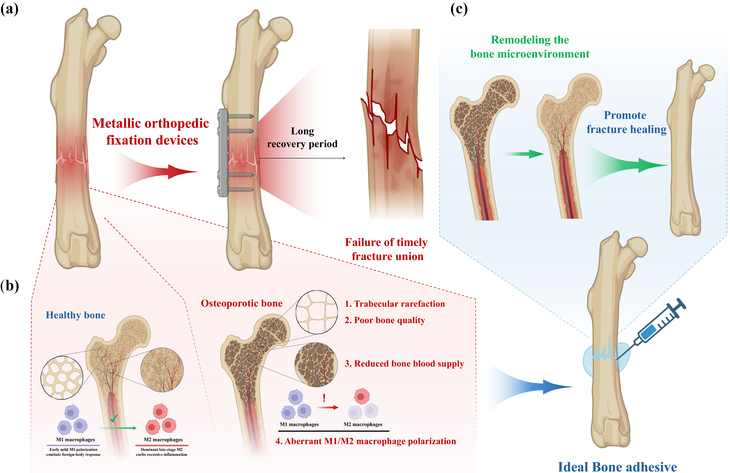
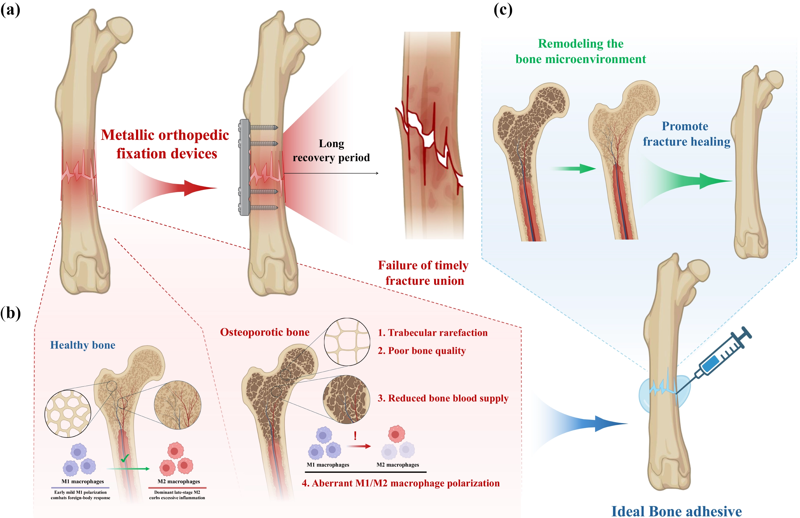
Figure 1. (a) Metal internal fixation can provide strong mechanical stability for the fracture site. Internal fixators are prone to causing implant infection of bone tissue and need removal after bone healing treatment; (b) The density and thickness of bone trabeculae in healthy bones are normal, and the bone healing ability is strong; the body has strong anti-inflammatory ability; the conversion ability of M1 and M2 is normal. Osteoporosis is characterized by reduced bone mass, changes in bone microstructure, and increased bone fragility; (c) The ideal bone adhesive that meets clinical needs not only provides robust fixation but also needs to have biological regulatory functions; it reshapes the bone microenvironment to promote different types of bone healing. Created in BioRender. Wang, X. (2026) https://BioRender.com/az75yxp.
In addition, bone bandages have also been used as one of the alternatives to metal internal fixation devices. These bandages can be wrapped around the fractured area like plaster, providing proteins for growth and repair to enhance the natural healing ability of the bone and accelerate the repair process. Lei et al. designed a pressure-sensitive bandage based on a citric acid-gelatin polymer. This bandage not only adheres firmly to bone tissue (with an adhesive strength of approximately 570 kPa) but also promotes regeneration through its material properties by inducing the differentiation of stem cells into osteoblasts and modulating the immune environment[29]. Tägil et al. developed a gelatin-nano-hydroxyapatite membrane (GM)-based bone bandage designed to control the release of bioactive molecules. The bandage incorporated recombinant human bone morphogenetic protein-2 (rhBMP-2) and zoledronic acid (ZA) to promote osteoinduction and prevent callus resorption, respectively[30]. However, all reports on this “active healing” bone bandage are still in the laboratory research stage; there is no mature product applied to clinical practice yet.
Bone adhesive has become a promising alternative to traditional internal fixation. Bone adhesive is used to fix periarticular fractures and diaphyseal fractures that do not require immediate weight-bearing after treatment[16]. Unlike screws and plates, bone adhesives can be directly bonded to the wet bone surface, fit irregular and comminuted fracture blocks, disperse the load on a wide range of contact areas, and promote fracture healing and bone regeneration without extensive soft tissue dissection and destruction of periosteal blood supply[31]. In addition, degradable bone adhesive can become a bridge structure between early fixation and late regeneration. There is no biological residue after degradation, and no need for secondary surgery[32,33].
Over the past two decades, numerous reviews have summarized bone adhesive research from various angles. Early works chronicled the historical development and clinical limitations of adhesives such as cyanoacrylates and PMMA[17,18], while more recent reviews categorized material classes, including calcium phosphate cements, hydrogels, and biomimetic composites, and discussed wet-state adhesion mechanisms[20,31,33]. Some focused on specific translational candidates like Tetranite and OsStic, highlighting the gap between bench and bedside[32]. Others addressed clinical needs and design criteria, such as rapid setting, mechanical strength, and biocompatibility[34,35]. However, these reviews predominantly treated bone adhesives as a single category, with limited systematic consideration of how adhesive design should be tailored to distinct bone conditions (e.g., healthy vs. osteoporotic) and anatomical sites (e.g., long tubular bones vs. flat/irregular bones). Moreover, the integration of adhesion chemistry, mechanical performance, degradation kinetics, and biological microenvironment modulation into a unified, clinically relevant framework has been lacking. This review distinguishes itself by providing such a unified framework: it explicitly links bone quality (from healthy to osteoporotic), anatomical site (load-bearing vs. non-load-bearing), adhesion chemistry, cohesive mechanics, degradation, and biological regulation. By comparing the differential requirements of healthy and osteoporotic bone, we advocate a shift from universal formulations to targeted, bone-quality-matched designs. This framework is consistently applied throughout the subsequent sections, from adhesive mechanisms to preclinical and clinical translation, and is reiterated in the Conclusions.
At present, the research and development of bone adhesive and its clinical transformation are still full of challenges. Existing designs mainly focus on achieving stable adhesion and strong mechanical strength but often ignore whether these properties match the unique biological and mechanical requirements of the target bone. In contrast, aged or osteoporotic bone is characterized by disrupted metabolic balance and decreased regenerative capacity, which requires not only strong and tough micro-anchoring, but also biological functions such as osteogenic induction and immune regulation to restore bone regeneration[36-38]. Therefore, the future bone adhesive must consider the unique biological and mechanical requirements of bone tissue under different pathological conditions, to achieve tailored healing strategies.
Building on the unified framework outlined above, this review summarizes the research status of adhesive chemistry, wet-surface adhesion mechanisms, and biomimetic design strategies. By comparing the differential requirements of healthy and osteoporotic bone, we argue that adhesive design must shift from a universal formula to an interface that matches bone quality and the local microenvironment, enabling targeted customization of adhesion chemistry, mechanical behavior, degradation kinetics, and bioactive functions. This framework provides a roadmap for the development of next-generation bone adhesives that can replace traditional metal internal fixation and achieve biological repair.
2. Clinical Needs-Driven Design Goals for Bone Adhesives
Metal internal fixation can provide strong mechanical stability for the fracture site and promote fracture healing[39]. However, there are still the following problems: the comminuted fractures in the joint need anatomical reduction, and the internal fixation device is difficult to fix fractured bone fragments[40]. Osteoporotic bone cannot be fixed firmly and is prone to nonunion[41,42]. The internal fixation device needs to be removed by secondary surgery and is prone to implant infection[43,44]. Therefore, bone adhesives that meet clinical needs need to have the following properties (Table 1): rapid and robust adhesion to wet bone surfaces, adequate cohesive strength to maintain the spatial positioning of fracture fragments, tunable biodegradability that matches the bone healing rate, excellent biocompatibility, and the capacity to promote osteogenesis[31]. Furthermore, for osteoporotic bone, such adhesives should possess the ability to modulate the local pathological microenvironment[11,45].
Table 1. Typical Properties of Bone Adhesives.
| Property | Detailed requirement |
| Rapid and Robust Adhesion | Form a stable interfacial adhesion;Suitable setting time |
| Adhesive and Cohesive Strength | Strong adhesion to bone surface;Maintaining structural integrity under physiological loads |
| Degradation and Biocompatibility | Degradation rate that matches bone healing;No toxicity; biocompatibility toward surrounding tissues |
| Biological Promotion and Microenvironment Modulation | Disrupt excessive osteoclast activity;Modulate chronic oxidative stress and inflammatory microenvironment;Impaired cell recruitment and defective neurovascular signaling |
2.1 Rapid and robust adhesion in wet bone surfaces
For any bone adhesive, achieving instant strong adhesion on wet, bleeding bone surfaces is the primary challenge. Unlike synthetic adhesives used under dry conditions, bone adhesives must form a stable interfacial adhesion despite the presence of physiological fluids, blood, and soft tissue residues. This requires adhesive chemistry to displace water from the mineralized surface or react chemically with bone components such as hydroxyapatite or collagen to form a lasting bond[46,47]. Bone adhesives of different systems exhibit significant variations in curing/adhesion time, which directly determines their clinical operability. Among synthetic polymers, cyanoacrylates complete curing within seconds on wet bone surfaces via anionic polymerization; polyurethane and PMMA systems, however, require anywhere from several minutes to 24 hours. For inorganic cements, phosphoserine-modified tetra-calcium phosphate (Tetranite®) initially sets in 10-15 minutes and reaches 90% of its final hardness after 24 hours, whereas phosphoserine-modified alpha tricalcium phosphate (α-TCP) (OsStic®) cures within minutes[34,35]. In addition, marine organisms such as mussels, barnacles, and sandcastle worms have evolved the ability to achieve rapid and robust adhesion on submerged surfaces enabling them to settle, attach, or build shelters under turbulent, fluid-rich conditions[48-50]. Their secreted adhesive proteins can displace water, bind to diverse substrates via ionic/covalent interactions, and maintain strong cohesion even against powerful currents[51,52]. This inherent capacity for instantaneous wet adhesion is precisely what bone adhesives urgently need when applied to bleeding, wet bone surfaces during orthopedic surgery. Thus, bioinspired adhesives derived from marine organisms offer a promising blueprint for developing next-generation bone adhesives that combine immediate fixation with long-term load-bearing performance.
2.2 Adhesive and cohesive strength: The mechanical basis for spatial positioning
The macro adhesive performance of bone adhesive depends on the synergy of interfacial adhesion and material cohesion. Cohesion strength not only directly affects the overall bonding strength but also determines the ability of materials to maintain structural integrity under physiological loads such as shear, compression, tension and impact, which is the core guarantee for the stable fixation of fracture blocks. At present, the strategies to enhance cohesion mainly include:
(1) Constructing double network/interpenetrating polymer networks (IPNs). Double-network hydrogels significantly improve strength and toughness through the synergistic effect of two polymer networks[53,54]. For example, Wistlich et al. introduced a six-arm isocyanate-functionalized prepolymer into a photocurable poly(ethylene glycol) dimethacrylate (PEGDMA) system, in situ forming an IPN and incorporating inorganic fillers, which increased the adhesive strength to cortical bone from 0.15-0.2 MPa to 0.3-0.5 MPa[55]. In addition, increasing the number of cross-linked functional groups (such as the introduction of tannic acid to enhance hydrogen bonding) or shortening the distance of side chain functional groups (such as carboxyl spacing) can also effectively improve the cross-linking strength and modulus[56-58].
(2) Constructing organic–inorganic composite structures. By mimicking the native composition of bone tissue, inorganic minerals such as bioactive glass (BG), calcium phosphate, or hydroxyapatite (HA) are incorporated into the adhesive matrix. Shahbazi et al. developed a BG-reinforced polypropylene fumarate adhesive with a compressive modulus of 80.49 MPa, which matches the range of human trabecular bone (50-500 MPa)[59]. Pinzón et al. incorporated calcium carbonate and HA into chitosan, where coordination bonds formed between Ca2+ and amino groups; the resulting composite exhibited an adhesive strength of 0.27 MPa to bone[60]. Liu et al. loaded zeolitic imidazolate framework-8 (ZIF-8) nanoparticles into catechol-modified chitosan, improving mechanical properties by 12.83-fold while simultaneously conferring antibacterial and osteogenic potential[61].
(3) Fiber reinforcement. Fiber reinforcement simultaneously enhances tensile strength and fracture toughness. For example, Kirillova et al. incorporated poly(lactic-co-glycolic) (PLGA) fibers into phosphoserine-based bone cement, which improved compressive strength, Young's modulus, and fatigue resistance[62]. Granskog et al. developed a fiber-reinforced adhesive patch (FRAP) based on thiolene coupling chemistry; after primer optimization, the maximum shear adhesive strength reached 9-11 MPa on bone[63].
Only by optimizing the adhesion and cohesion strength can the adhesive achieve accurate spatial positioning and stable fixation of the fracture block in comminuted fractures. However, the current “liquid steel plate” type adhesive with the highest performance has a strength of about 11 MPa, and the “liquid support” type is more than 1 MPa, which is still significantly lower than the mechanical level of traditional metal internal fixation, and is the key breakthrough direction of future research and development[33].
2.3 Tunable degradation and biocompatibility
In the design of bone adhesive, adjustable degradability and biocompatibility are the core bottom line that determines its clinical translational potential. At the same time, its degradation rate needs to be synchronized with the formation of new bone, which gradually degrades in the process of fracture healing, to avoid the risk of secondary surgical removal. The material selection must put the internal safety in the first place.
Current material systems exhibit significant variations in degradability and biocompatibility. Among natural polymers, fibrin glue offers “excellent biocompatibility and biodegradability,” but its application is limited to non-load-bearing fractures due to insufficient adhesive strength[33,64]. Poly(ethylene glycol) (PEG), polysaccharide-based adhesives (e.g., chitosan), and gelatin exhibit “widely recognized biocompatibility” or “reliable biocompatibility from natural origins,” yet they are constrained by mechanical or adhesive deficiencies, making them unsuitable for independent use in load-bearing sites[65-67]. Regarding synthetic polymers, cyanoacrylates provide rapid curing but present “potential toxicity” and “non-degradability”. Kryptonite was recalled by the FDA in 2012 (Recall Number Z-1496-2012) because its strength and stiffness fell below specification at body temperature. Within inorganic systems, phosphoserine-modified calcium phosphate cements (such as Tetranite® and OsStic®) demonstrate clinically relevant high adhesive strength; but more detailed clinical studies are needed to fully assess their safety and performance[68,69]. Furthermore, incorporating osteogenic agents through strategic matrix selection, such as citrate (a Krebs cycle intermediate that regulates energy metabolism to promote osteogenic differentiation), Mg2+, bioactive glass (BG, an FDA-approved osteoconductive material), and calcium phosphate (Ca-P) (e.g., β-TCP) or loading bioactive substances including BMP-2, the sclerostin antibody Romosozumab, and gingival/bone marrow mesenchymal stem cells, enables the simultaneous tuning of degradation kinetics and osteogenic activity while maintaining biocompatibility[59,70-73].
In general, when developing bone adhesives, safety and non-exclusion must be put in the first place. As long as the formula is adjusted well and the degradation speed of adhesive just catches up with the speed of bone growth, the two biggest problems of poor adhesion and bone growth can be solved. Only in this way can bone adhesive really go out of the laboratory and be used in the operating room, and it is even expected to completely replace metal plates and screws in the future.
2.4 Biological promotion and microenvironment modulation
In addition to mechanical functions, modern bone adhesives are increasingly expected to participate actively in the bone healing cascade. For healthy bone with balanced metabolism and normal osteogenic or osteoclastic coupling, the biological role of adhesives is primarily to provide moderate osteoconduction and osteoinduction and promote fracture healing through structural support and the controllable release of osteogenic factors such as Ca2+, Po43- or BMP-2[74-77]. In contrast, osteoporotic fractures present three local pathological factors: (1) disrupted osteogenic or osteoclastic coupling with excessive osteoclast activity; (2) modulate chronic oxidative stress and an inflammatory microenvironment; and (3) impaired cell recruitment and defective neurovascular signaling[79]. Therefore, bone adhesives for osteoporotic fracture patients must act as multifunctional microenvironment modulators rather than merely providing stable mechanical support. To address oxidative stress damage, bone adhesives must integrate antioxidant functionalities to reverse the differentiation fate of stem cells. The gypsogenin (GN)-based organic-inorganic composite hydrogel (CCT/nHA@GN) hydrogel clears reactive oxygen species by activating the AMPKα-FOXO3a pathway and increases regenerated bone mass by 2.85-fold, confirming that antioxidant activity is a prerequisite for correcting the pathological microenvironment and restoring osteogenic/osteoclastic coupling[80]. To overcome insufficient angiogenesis, bone adhesives should achieve dual-paracrine coupling of osteogenesis and angiogenesis. The teriparatide (PTH)/strontium-zinc phosphate (SrZnP)@ZnCu-phosphocreatine-grafted gelatin hydrogel (GGP) hydrogel synergistically releases Zn2+, Sr2+, and PTH to simultaneously activate the hypoxia-inducible factor 1-alpha (HIF-1α)-vascular endothelial growth factor (VEGF) and PI3K-Akt-BMP-2 pathways, significantly accelerating mineralization and vascular network formation in an ovariectomized model[81]. To combat chronic inflammation and excessive osteoclastic activity, bone adhesives must execute a unified therapeutic action of “immunomodulation and anti-resorption and osteogenic promotion”. The magnesium-manganese layered double hydroxide (MgMn-LDH)/calcium alendronate (CaALN) scaffold continuously scavenges reactive oxygen species (ROS) through multi-enzyme-mimicking catalysis and releases Mg2+ and alendronate, successfully restoring osteogenic/osteoclastic homeostasis in osteoporotic rats[82].
3. Existing Adhesive Systems and Adhesion Mechanisms
In recent years, the research strategy of bone adhesive has evolved from the early single “adhesion and fixation” to the direction of biomimetic structure, functional compounding, and controllable degradation. Its core development strategies include: (1) Mimicking the organic-inorganic composite structure of natural bone tissue, introducing bioactive glass, calcium phosphate, hydroxyapatite and other inorganic phases into the organic adhesive matrix to consider the interfacial adhesion and bulk cohesion strength[83-85]. (2) The introduction of bone targeting functional groups (phosphonic acid, carboxylic acid, bisphosphonic acid, catechol/dopamine, etc.) can produce strong interactions with bone mineral phase through coordination bonds, hydrogen bonds[32,86]. (3) Thiolene coupling, Schiff base dynamic crosslinking, photopolymerization and other double crosslinking methods were used to improve cohesion[87]. Integrating hemostasis, antibacterial, anti-inflammatory, bone regeneration and other functions, the adhesive can actively regulate the bone healing microenvironment while being fixed for a short time.
Based on material systems, current bone adhesives can be broadly categorized into five major classes (Table 2): cyanoacrylates (the earliest clinically widely used yet limited by high brittleness and slow degradation)[98]. Calcium phosphate cements (CPCs; excellent biocompatibility but low early-stage mechanical strength). PMMA and its 4-MEMA-functionalized derivatives (high mechanical strength but non-degradable)[18,99]. Natural/polysaccharide-based hydrogels (e.g., chitosan, gelatin, silk fibroin; superior biocompatibility but insufficient wet-state adhesion)[72,100]. And biomimetic organic–inorganic composite hydrogels (the current research mainstream, represented by systems such as L-DOPA-PVA-ZIF-8 (L-DPZ) and the dual-biomimetic adhesive)[72,101]. Among these, biomimetic organic-inorganic composite systems are widely recognized as the most promising avenue for breaking through the “impossible trinity” of wet-state adhesion, cohesive strength, and bioactivity. Some representative materials were shown in Table 2.
Table 2. Classification of Bone Adhesive Material Systems.
| Representative Materials | Key Characteristics and Mechanism | Advantages | Limitations | Ref. | |
| Cyanoacrylates | EthylButylOctyl-2-cyanoacrylate | Rapid curingAnionic polymerization Covalent bonding | Strong adhesion.Fast setting. | High brittleness.Slow degradation.Toxicity concerns. | [88-90] |
| Calcium Phosphate Cements | Unmodified CPCsPyrophosphate-substituted CPCsPhosphoserine-modified CPCs (Tetranite, OsStic) | High viscosity.Mechanical interlocking. Electrostatic/H-bonding (modified). | Excellent biocompatibility.Osteoconductive. | Low early strength.High brittleness | [27,68,69,91] |
| PMMA and Derivatives | PMMA bone cementPolylactic acid-methacrylate copolymers.β-TCP reinforced methacrylate adhesives | Mechanical interlocking.No chemical bonding. | High mechanical strength.Mature and commercialized. | Non-degradable.Exothermic reaction.Volume shrinkage. | [18,92,93] |
| Natural/Polysaccharide-based Hydrogels | Fibrin glue, gelatin, chitosan, starch, dextran, alginate; L-DOPA functionalized polysaccharide systems | Physical/chemical cross-linking. Protein/polysaccharide based. | Superior biocompatibility.Degradable. | Insufficient wet adhesion.Low mechanical strength | [94,95] |
| Biomimetic Organic–Inorganic Composite Hydrogels | Phosphoserine-modified calcium phosphate, polyphenol/silk fibroin/HA hybrid hydrogels, bioactive glass composite methacrylates | Bone-mimicking structure.Organic-inorganic hybrid. | Balanced wet adhesion and strength.Promotes bioactivity. | Mostly preclinical.Complex fabrication | [96,97] |
CPCs: calcium phosphate bone cements; HA: hydroxyapatite; β-TCP: β tricalcium phosphate; PMMA: polymethylmethacrylate.
Currently, the mechanical properties of bone adhesives have achieved systematic breakthroughs, with multiple indicators approaching or reaching the load-bearing requirements of metallic internal fixation (Figure 2a,b,c,d). In terms of wet-state adhesion strength, the L-DPZ hydrogel (L-dopamine-polyvinyl alcohol-ZIF-8) exhibits an adhesion strength of 9.31 ± 1.29 MPa to bovine bone, with a shear strength of up to 10 MPa, significantly surpassing that of most previously reported bio-adhesives and commercially available cyanoacrylates (Figure 2a)[101]. Designed by Liu et al., a homogeneous “hard-soft” biphasic bone adhesive (HB-PTN) employs phosphorylated polyglutamic acid-coated tetracalcium phosphate as the hard phase and aminated polyethylene glycol-modified poly (sebacate) hydrogel as the soft phase. It achieves a wet-state bone bonding strength of 269.7 kPa (lap-shear) and 206.0 kPa (end-to-end bonding), a compressive modulus of 1.02 MPa, strength retention of 94.1% after 100 compressive cycles, and 102% mechanical performance retention after immersion in simulated body fluid (SBF) (Figure 2b)[102]. Zhang et al. constructed a dual-biomimetic organic–inorganic network-reinforced hydrogel (caffeic acid-grafted collagen (CAC)/aminated laponite (ALAP)/N-hydroxysuccinimide ester-terminated polyethylene glycol (tetra-PEG-SC)) that maintains structural integrity under 98% compressive strain and 600% tensile strain. In rabbit radial comminuted fracture and porcine femoral fracture models, its biomechanical and radiological performance outperformed commercial cyanoacrylate adhesives by more than two-fold (Figure 2c)[102]. Furthermore, an injectable magnesium–alendronate metal-organic frameworks (MOF)/gelatin-dialdehyde starch hydrogel bone adhesive enhanced flexural strength by 27.8% in osteoporotic fracture repair, marking a leap from “passive fixation” to “active bone metabolism modulation” (Figure 2d)[14]. Due to the presence of blood and tissue fluid at fracture sites, bone adhesives are often required to achieve strong adhesion in wet environments. This relies on the synergy of multi-scale, multi-type interfacial interactions, which can be primarily categorized into the following four mechanisms:
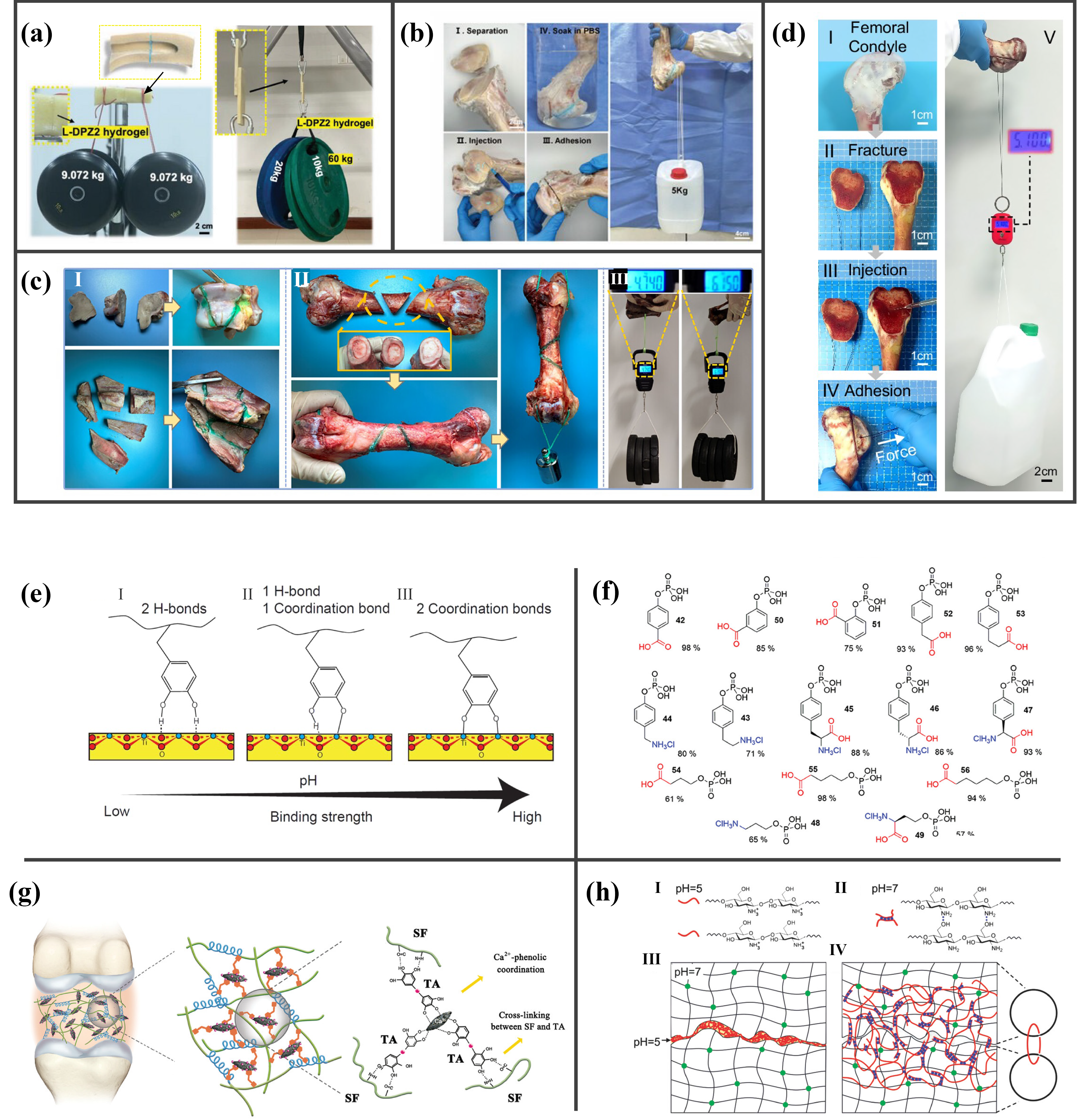
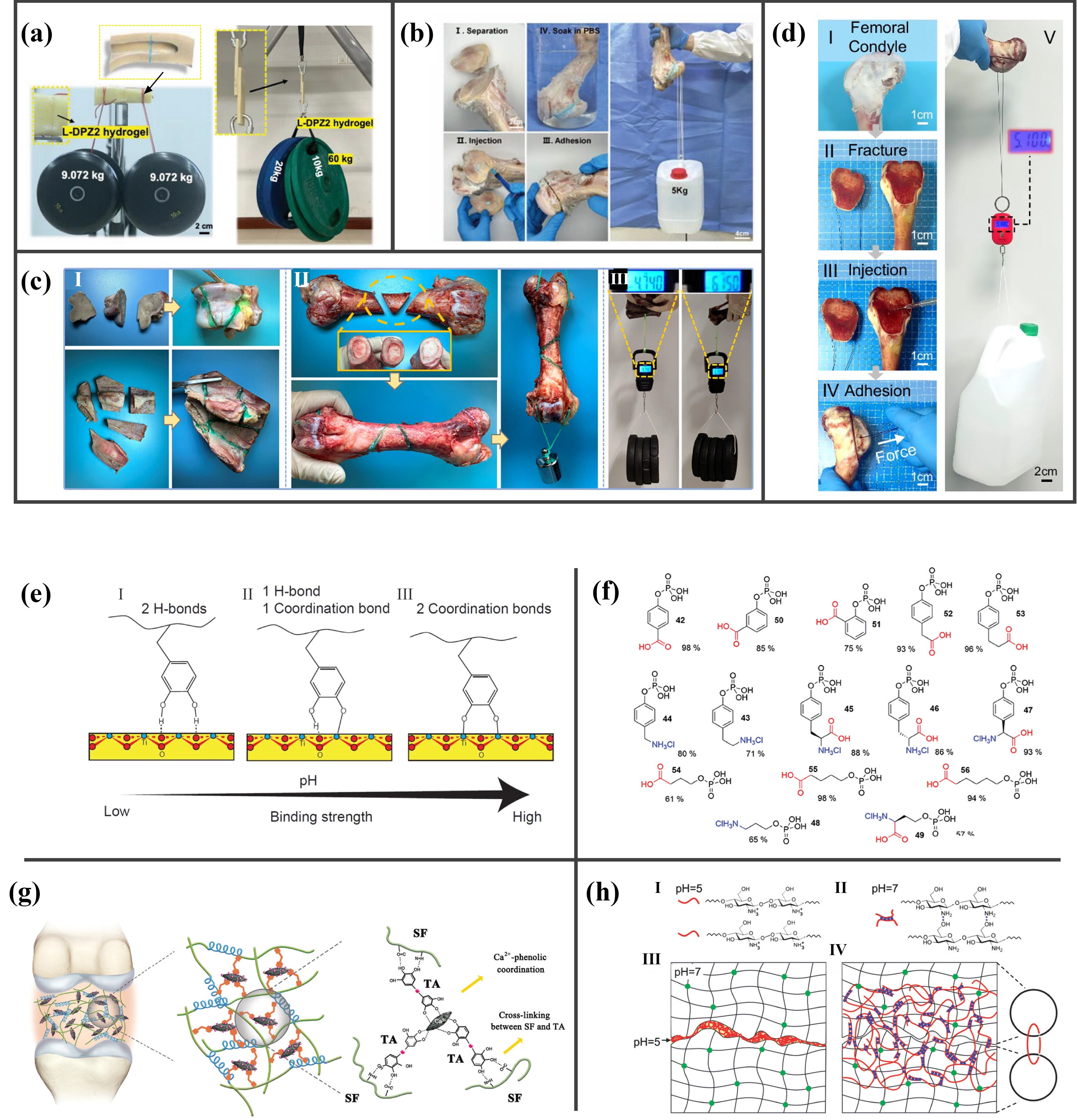
Figure 2. (a-d) Different types of bone adhesives exhibit superior mechanical properties in both dry and wet environments; they effectively bond fractured fragments together while maintaining good resistance. Reproduced from reference[14,101,103]. CC BY 4.0. Reproduced with permission from reference[102]. Copyright © 2025 Wiley; (e) Dopamine forms various binding modes with catecholamine groups, and its adhesion strength varies with changes in pH. Reproduced with permission from reference[104]. Copyright © 2013 American Chemical Society; (f) Synthesize phosphoserine analogues with different proportions. Reproduced with permission from reference[33]. Copyright © 2024 Wiley; (g) TA as a phenolic glue molecule to spontaneously co-assemble with SF and HA to fabricate the inorganic-organic hybrid hydrogel. The combination of the strong affinity between SF and TA significantly improves the toughness and adhesion strength of the hydrogel. Reproduced with permission from reference[72]. Copyright © 2018 Wiley; (h) Chitosan chains dissolve in water at pH 5 and form a network in water at pH 7. When two solutions with different pH were mixed, chitosan chains diffuse into the two hydrogels and form a network, which topologically entangles with the networks of the two hydrogels. Reproduced with permission from reference[105]. Copyright © 2018 Wiley. TA: tannic acid; SF: silk fibroin; HA: hydroxyapatite.
Catechol/polyphenol-mediated wet-state adhesion: Inspired by the adhesion mechanisms of marine mussels and barnacles, catechol groups can bind to bone surfaces through multiple interactions, including hydrogen bonding (with bidentate hydrogen bonds whose bond lifetime is approximately 106-fold that of monodentate bonds), metal-phenol coordination bonds, π-π stacking, and π-cation interactions. The phenolic hydroxyl groups of polyphenols (tannic acid, gallic acid, tea polyphenols, etc.) can also form metal-phenol coordination bonds with Ca2+, Mg2+, Fe3+, and other ions. Their aromatic rings displace the adsorbed water layer from the interface via hydrophobic effects, enabling phenolic hydroxyl groups to form strong covalent/non-covalent interactions with tissue functional groups (-NH2, -COOH, -SH). This constitutes the core strategy for achieving instant adhesion in blood environments. However, under alkaline conditions or in the presence of oxidizing agents, catechol can be readily oxidized to quinones, thereby compromising adhesive strength. Therefore, such scenarios should be carefully considered and mitigated when designing these bone adhesives (Figure 2e)[104,106].
Phosphate/phosphonate-calcium ion coordination and in situ mineralization: Polymers containing phosphate groups, such as phosphorylated polyglutamic acid (P-PGA), can form stable phosphate-calcium electrostatic coordination with Ca2+ in the bone mineral phase. In a SBF environment, tetracalcium phosphate (TTCP) transforms into nano-needle HA and undergoes in situ deposition and mineralization, forming a continuous organic–inorganic transitional hybrid layer (Figure 2f)[33].
Synergy of dynamic covalent bonds and physical cross-linking: Dynamic covalent bonds, such as Schiff bases (aldimine condensation), N-hydroxysuccinimide (NHS)-ester-amine reactions, and borate ester bonds, combined with physical cross-linking (hydrogen bonding, electrostatic interactions, and β-sheet protein conformational transitions), construct dynamic networks that possess both high adhesion and high cohesion[72,107,108]. For instance, in a dual-biomimetic bone adhesive, metal-phenol coordination bonds formed between tannic acid and Ca2+/Mg2+ impart favorable mechanical performance to the dynamic network; meanwhile, the conformational transition of silk fibroin from random coil to β-sheet further enhances cohesion (Figure 2g)[72].
Mechanical interlocking and topological anchoring: Certain systems (e.g., cyanoacrylates, two-component instant-curing systems) achieve adhesion by monomer infiltration into surface micropores of bone, forming microscopic mechanical interlocking[46,109]. Novel topological adhesion strategies exploit polymer chain entanglement and pH-triggered self-adaptive networks to realize “instantaneous interaction” rather than rigid locking at wet interfaces, effectively dissipating external stress and avoiding brittle fracture. Chitosan chains dissolve in water at pH 5 and form a network in water at pH 7. When two solutions with different pH were mixed, chitosan chains diffuse into the two hydrogels and form a network, which topologically entangles with the networks of the two hydrogels (Figure 2h)[105]. Inspired by topological adhesion mechanisms, there is promise for overcoming the wet-state interfacial brittleness of bone adhesives[103].
4. Differentiated Bone Adhesive Design: Targeted Strategies for Specific Bone Quality and Skeletal Classifications
Bone tissue not only has significant differences in health and pathological status between individuals but also presents different mechanical bearing requirements due to different anatomical sites in the same body[110-112]. Clinically, dual-energy X-ray absorptiometry (DXA) is recognized as the gold standard for diagnosing osteoporosis. It enables quantitative assessment by measuring bone mineral density (BMD) and calculating the T-score, which represents the number of standard deviations from the peak bone mass of a healthy young adult. Based on the World Health Organization (WHO) diagnostic criteria: a T-score ≥ -1.0 is defined as normal bone; a T-score between -2.5 and -1.0 indicates osteopenia; and a T-score ≤ -2.5 confirms osteoporosis[113]. Therefore, the design of bone adhesives should also consider the heterogeneity of bone quality classification and anatomical site characteristics. Guided by the unified framework introduced in the Introduction, this section categorizes design strategies according to bone quality and anatomical site.
4.1 Healthy bone: High mechanical strength and reliable immediate fixation
Healthy bone has abundant mineral phases and complete structure. The primary task of an adhesive in this context is to provide sufficient interfacial adhesion and cohesive strength to replace metal plates and screws for the immediate stabilization of fractured fragments[63,110]. Thermosetting materials employing thiolene/yne coupling exhibit a maximum bone adhesion strength of up to 11 MPa, significantly outperforming most commercial glues. Phosphoserine-modified calcium phosphate bone cements (Tetranite, OsStic) demonstrate high wet-state adhesive strength to cortical bone, with shear strength several to hundreds of times greater than that of conventional Ca-P and PMMA bone cements (Figure 3a,b)[32,110]. Similarly, SF@TA@HA (silk fibroin/tannic acid/hydroxyapatite) achieves an adhesive strength of 607.18 kPa on wet porcine bone, maintaining 922.83 kPa even in blood[115]. Therefore, when designing bone adhesives for healthy bone tissue, the key is to strengthen the interfacial chemical bonds and cohesive networks and ensure that they do not fail in the mechanical environment of healthy bone.
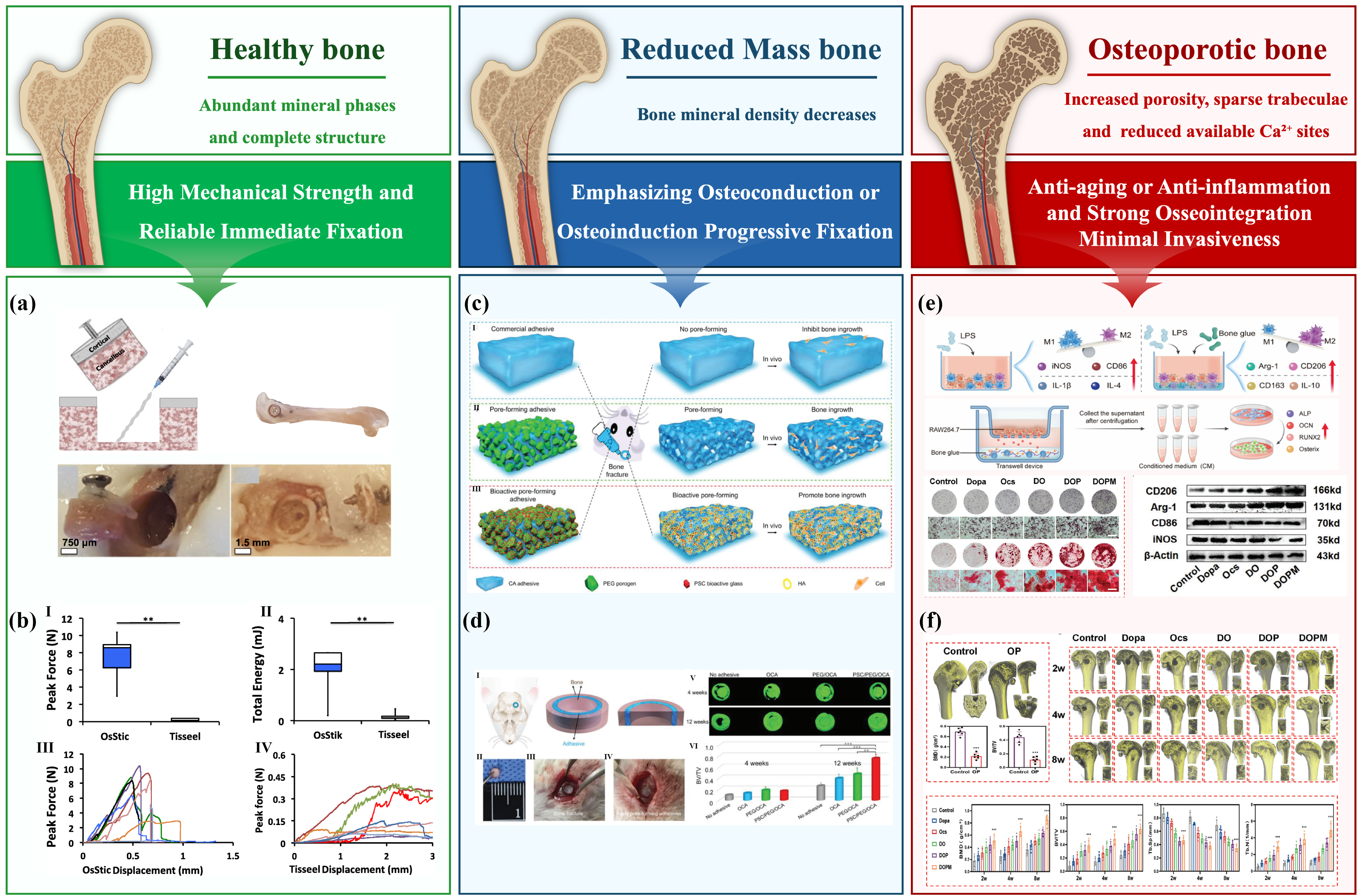
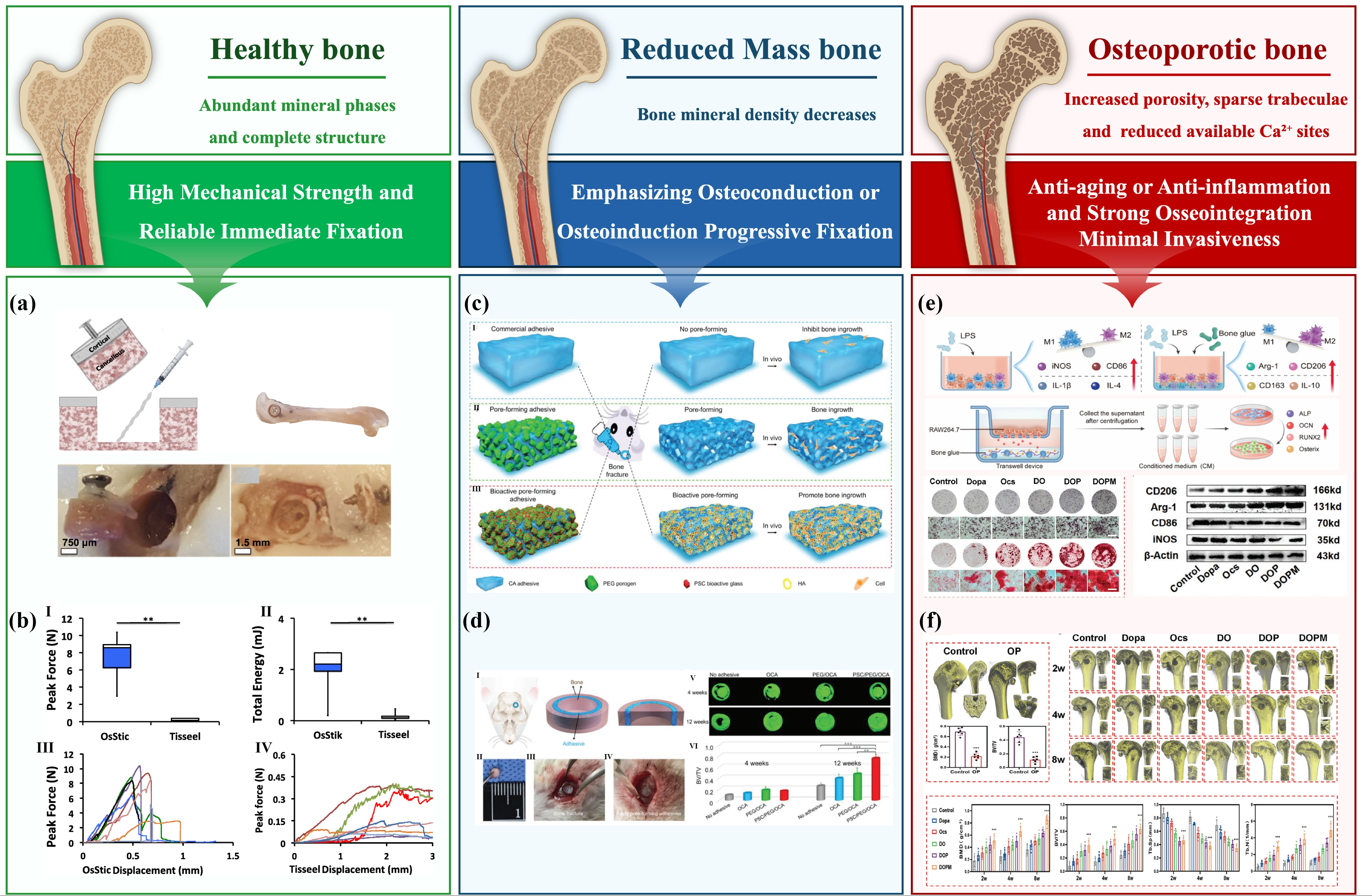
Figure 3. (a) The 2 mm deep bone core was created in the distal femoral metaphysis of ex vivo rat femurs using a trephine. The core was then extracted with the aid of a micro-screw, injected with Tisseel or OsStic. This process constructs a reconstructed “bone-adhesive-bone” interface suitable for pull-out testing. Reproduced from reference[110]. CC BY 4.0; (b) The pullout testing data show that the average peak force at failure and total energy of OsStic-treated metaphyseal bone cores were significantly greater than those of Tisseel. Reproduced from reference[110]. CC BY 4.0; (c) Bioactive pore-forming adhesives incorporate PSC/PEG composite porogens. These adhesives can create pores together with a bioactive HA layer (yellow) to offer a suitable environment for new bone generation. Reproduced with permission from reference[66]. Copyright © 2020 Wiley; (d) BV/TV of the PSC1/PEG4/OCA5 group was significantly higher than that of the PEG5/OCA5 group and the OCA group; it was suggested that the adhesive with pores for bone ingrowth and BG was essential for new bone formation. Reproduced with permission from reference[66]. Copyright © 2020 Wiley; (e) DOPM bone adhesive exhibits strong osteogenic differentiation ability and promotes the expression of osteogenic markers in cells. Furthermore, DOPM can also conditionally convert senescent macrophages from M1 to M2, exerting an anti-inflammatory effect. Reproduced from reference[114]. CC BY 4.0; (f) DOPM exhibits excellent bone healing capabilities in a bone defect model of osteoporotic rats. While the bone defects were completely covered by connective tissue in the presence of DOPM by Week 2, they were completely covered by bone-like tissue by Week 8. Reproduced from reference[114]. CC BY 4.0. PEG: poly(ethylene glycol); HA: hydroxyapatite; IL-4: interleukin-4; IL-1β: interleukin-1 beta; NOS: nitric oxide synthase; iNOS: inducible nitric oxide synthase; ALP: alkaline phosphatase; LPS: lipopolysaccharide; DOPM: levodopa/oxidized chitosan/PLGA/mof@nHA.
4.2 Bone tissue with reduced mass: Emphasizing osteoconduction or osteoinduction progressive fixation
In regions of reduced bone mass (early osteoporosis, local bone atrophy), bone mineral density decreases, making simple mechanical fixation prone to loosening. Adhesives are required to provide initial fixation while actively promoting bone regeneration to restore bone mass[14]. Introducing different monomers can address this issue. Furthermore, in addition to using organic-inorganic composite formulations, the design should balance adequate adhesion with high osteogenic activity[116]. The curing process must be mild to avoid heat-induced damage to the surrounding fragile bone tissue. For instance, incorporating inorganic phases such as BG, Ca-P, and HA imparts osteoconductivity, guiding bone cell ingrowth[117]. For instance, a class of bioactive glass-involved macropore-embedded bone adhesives is developed, which is capable of facilitating the migration of bone-derived mesenchymal stromal cells into the adhesive layer and differentiation into osteocytes (Figure 3c,d)[66]. A one-step solid phase reaction was developed to produce HA/magnesian whitlockite (MWH) dual-phase bioceramics, and the more favorable functional Mg2+ release kinetics of MWH resulted in a marked enhancement of osteogenic properties[118]. Citric acid-based matrices (injectable citrate-based mussel-inspired bioadhesive (iCMBA)/HA) promote the osteogenic differentiation of mesenchymal stem cells through metabolic regulation, while simultaneously providing injectable, rapidly cross-linking adhesive fixation[119].
4.3 Osteoporotic bone: Anti-aging or anti-inflammation and strong osseointegration minimal invasiveness
Osteoporotic bone is characterized by increased porosity, sparse trabeculae, and reduced available Ca2+ sites in the mineral phase, making anchorage difficult for traditional adhesives. It is necessary to design adhesive systems capable of penetrating micropores, releasing osteogenic/anti-osteoclastic ions, and possessing anti-inflammatory and antibacterial functions (Figure 3e,f)[114,120,121]. Magnesium phosphate cements (MPCs) have been reported to possess superior bone affinity and faster degradation rates compared to Ca-P[122,123]. Moreover, Mg2+ stimulates osteoblast differentiation and inhibits osteoclast formation, while also exhibiting intrinsic antibacterial activity, making MPCs strong candidates for osteoporotic bone repair[124,126]. Dynamic covalent (Schiff base) cross-linked oxidized dextran-gelatin/mesoporous bioactive glass hydrogels promote bone regeneration through degradation-mediated mineralization in a rabbit radius fracture model, making them suitable for gradual replacement in poor bone quality scenarios[127]. For open fractures (common in elderly osteoporotic patients), the antibacterial and anti-inflammatory functions of bone adhesives should be emphasized, such as antibiotic-loaded bone cements (those loaded with vancomycin/amikacin)[128,129].
In recent years, the use of bisphosphonates (BPs) effectively inhibits the activity of bone-resorbing osteoclasts, thereby significantly reducing bone loss[130]. However, it also activates fibroblasts and inhibits callus formation, resulting in the accumulation of fibrous tissue that hinders bone healing. Long-term administration of BPs has been found to cause a new type of fracture known as atypical femur fractures (AFFs)[131]. Its essence lies in not only inhibiting the formation of osteoclasts but also promoting the differentiation of myeloid progenitor cells into fibroblasts, ultimately leading to excessive fibrosis and impeding the formation of effective bone callus bridges. Injectable magnesium-bisphosphonate MOF-based bone adhesive binds firmly to irregular bone surfaces and degrades under acidic osteoporotic conditions, gradually releasing Mg2+ ions (Figure 4a). These ions interfere with the sclerostin (SOST)/transforming growth factor β (TGF-β) signaling pathway that promotes fibroblast activation and abnormal collagen deposition (Figure 4b)[14]. This dual-action mechanism significantly enhances fracture healing, resulting in a 27.8% improvement in flexural strength. Meanwhile, this promotes the predominance and orderly arrangement of Type I collagen, prevents the disordered mineralization induced by the fine and disorganized Type III collagen, and thereby redirects the collagen assembly at the fracture site back to the normal repair pathway (Figure 4c).
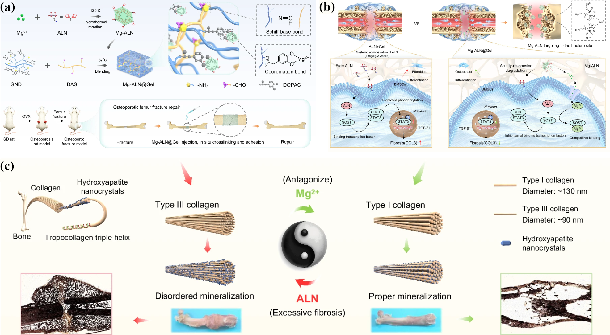
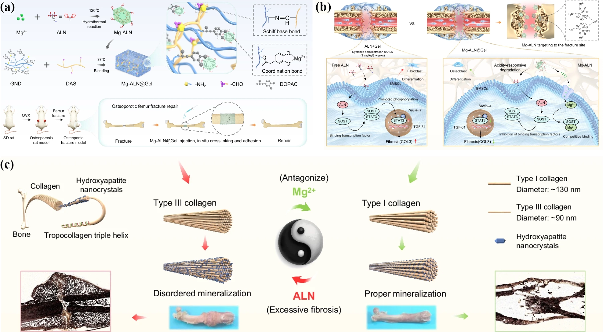
Figure 4. (a) MgCl2 solution and ALN solution were mixed and subjected to a hydrothermal reaction (120 °C, 24 h) to obtain Mg-ALN, which was then incorporated into catechol- and amine-rich highly branched gelatin/dialdehyde starch networks to synthesise Mg-ALN hydrogel bone adhesive (Mg-ALN@Gel), where Mg2+ in Mg-ALN enhanced the cross-linking network of Schiff base-bonded hydrogel. Mg-ALN@Gel was injected locally into a rat model of osteoporotic fracture and promoted osteoporotic fracture repair. Reproduced from reference[14]. CC BY 4.0; (b) Mg-ALN targets the fracture end and releases Mg2+ and ALN in response to acidity. The Mg2+ can competitively bind to SOST and attenuate the SOST/TGF-β signaling pathway, inhibiting fibrogenic differentiation and subsequent disordered mineralization. Reproduced from reference[14]. CC BY 4.0; (c) Mg-ALN@Gel promotes the predominance and orderly arrangement of Type I collagen, prevents the disordered mineralization induced by the fine and disorganized Type III collagen, and thereby redirects the collagen assembly at the fracture site back to the normal repair pathway. It not only can promote the healing of osteoporotic fractures but also inhibit fibrosis and improve the bone morphology at the healing site. Reproduced from reference[14]. CC BY 4.0. SOST: sclerostin; TGF-β: transforming growth factor β; ALN: alendronate sodium. MgCl2: magnesium chloride; DOPAC: 3,4-dihydroxyphenylacetic acid.
Notably, the bone quality-based strategies must ultimately be implemented in specific anatomical sites. The mechanical environment and functional requirements of different skeletal categories further determine the priority of adhesive formulations and structural design directions. Some representative bone adhesives were summarized by anatomical site and bone interface, which were shown in Table 3. Long tubular bones (e.g., femur, tibia), as highly load-bearing sites, demand adhesives with extremely high wet-state adhesive strength and moduli matching those of cortical/cancellous bone (Table 3-the section of Long tubular bones). Strategies such as fiber reinforcement and organic-inorganic composites (e.g., poly(propylene fumarate) (PPF)/2-hydroxyethyl methacrylate (HEMA)/nano bioactive glass systems are often employed to withstand physiological loads. In contrast, non-load-bearing or weakly load-bearing bones (e.g., skull, articular surfaces) prioritize fixation precision over absolute strength. Fibrin glue, cyanoacrylate, and sandcastle worm-inspired adhesives can achieve precise fixation and good biocompatibility with moderate adhesion, without interfering with postoperative imaging assessments (Table 3-the section of Non-load-bearing or weakly load-bearing bones). Flat, irregular bones (e.g., sternum, pelvis) are often accompanied by substantial cancellous bone bleeding; thus, “non-compressive hemostasis and flexible fixation” becomes the core requirement. Cationic hydrogels (e.g., catechol-conjugated chitosan (CHI-C)/dialdehyde cellulose nanocrystal (DACNC)) achieve rapid hemostasis by aggregating platelets through positive charges, while simultaneously supporting bone regeneration (Table 3-the section of Flat or irregular bone).
Table 3. Design strategies and performance of representative bone adhesives categorized by anatomical site and bone quality.
| Bone Category | Representative Adhesive | Key Performance | Ref. |
| Non-load bearingweakly load-bearing bones | Fibrin glue | • Adhesive strength: 5-17 kPa• Biodegradable, excellent biocompatibility• For non-load bearing/comminuted fractures | [18,132] |
| Cyanoacrylates (N-butyl, 2-octyl, etc.) | • Adhesive strength: 1,080-12,100 kPa• For craniofacial & mandibular fracture fixation | [88,133] | |
| OsStic (phosphoserine-modified α-TCP) | • Wet shear strength: 2.5-4 MPa•Fixation of the distal femoral metaphysis | [35,110] | |
| Complex coacervates (gelatin/polyphosphodopamineamide/Ca2+) | • Shear strength: 760 kPa•Fixation of cranial fractures in a rat model | [28,134] | |
| Long tubular bones(femur, tibia, radius) | DOPA/citrate-functionalized PEG/HA | • Wet/dry shear: 110/740 kPa• For rabbit radius comminuted fracture | [119] |
| Lactide-methylacrylate / inorganic particle-reinforced | • Dry/wet shear: 3,100-13,900/300-7,700 kPa• For femoral and tibial replantation models | [135,136] | |
| PPF/HEMA/nanobioactive glass | • Tensile shear resistance: 9-59 MPa• Promotes bone mineralization | [59] | |
| FRAP (thiolene coupling patch) | • Shear strength: 9 MPa• Simulates plate fixation on wet bone | [63] | |
| iCMBA/HA (citrate-based mussel-inspired) | • Crosslinks within 5 min; fully degrades in 30 days• Osteoinduction; for comminuted fractures | [119] | |
| DOPA-functionalized peptides/chitosan-peptides | • Tensile strength: 600-790 kPa• For rat tibial osteotomy | [137] | |
| Flat bone irregular bone (sternum) | Kryptonite (polyurethane/CaCO3) | • For sternal and vertebral fracture fixation• Clinical translation stage | [138,139] |
| Polyurethane-based adhesives | • Tensile strength: up to 1,340 kPa• Tested in canine, rabbit and frog models | [26,71,140,141] | |
| Tendon-to-bone interface | TN hydrogel | • Interfacial adhesion >800 J/m2• Mimics ligament-bone structure | [142] |
| Janus hydrogel | •High stretchability and extensibility•Restored tendon mechanics | [143] | |
| Frog mucous protein glue | • Enhances ultimate load in rotator cuff repair• Tested on ovine cadaver model | [144] | |
| Osteoporotic bonereduced bone mass (cross-site) | MPCs | • Shear strength: 1,220 ± 410 kPa• Mg2+ promotes osteogenesis and inhibits osteoclasts; antibacterial | [145-147] |
| Tetranite (phosphoserine/tetracalcium phosphate) | • Injectable; cures in 10-15 min• Osteo-integrative and osteoconductive | [62,148] |
α-TCP: alpha tricalcium phosphate; MPCs: magnesium phosphate cements; TN: triple-network; iCMBA: injectable citrate-based mussel-inspired bioadhesive; HA: hydroxyapatite; FRAP: fiber-reinforced adhesive patch; PPF: poly(propylene fumarate); HEMA: 2-hydroxyethyl methacrylate; PEG: poly(ethylene glycol); DOPA: 3,4-dihydroxyphenylalanine.
For special categories such as tendon-to-bone interfaces, it is necessary to move beyond the single evaluation metric of traditional shear strength and construct gradient adhesive systems (Table 3-the section of Tendon-to-bone interface). For example, triple-network (TN) hydrogels achieve an interfacial toughness of > 800 J/m2 through multiple hydrogen bonds and are supplemented with controlled drug release to reduce postoperative fibrosis, mimicking the mechanical transition of natural ligament-to-bone structures[149,150].
The design of bone adhesives is essentially a multi-parameter cross-optimization problem[33,151]. On one hand, it is necessary to progress from “simple mechanical fixation” to “ion-factor synergistic regulation” based on bone quality[152,153]. On the other hand, it is essential to expand from “high-strength load bearing” to “precise interface adaptation” according to anatomical sites[84,154]. Only by incorporating bone quality grading and site characteristics into a unified design framework and promoting the synergistic integration of multiple mechanisms (chemical bonding, physical cross-linking, in situ mineralization, and controlled drug release) can bone adhesives truly advance from the laboratory to clinical personalized applications.
5. Overcoming Translational Barriers: Lessons from Clinical Frontrunners
Currently, most systems remain at the proof-of-concept or preclinical stage. Although cyanoacrylates cure rapidly, they are limited to non-load-bearing applications due to their brittleness and potential toxicity. Most biomimetic hydrogels (e.g., L-DPZ, SF@TA@HA) and bioactive glass composites are evaluated only in small-animal models or in vitro[66]. Advanced strategies involving MOFs, exosomes, or dual-biomimetic networks are still in the early exploratory stage, and even bioactive bone bandages are confined to laboratory research[14].
In contrast, a few systems are approaching clinical translation. OsStic has entered a first-in-human study for periarticular fractures, with the index indication being closed tibial plateau fractures requiring plate-and-screw fixation plus defect filling (ISRCTN94484905; HRA IRAS 360651). Tetranite is being evaluated in a pilot clinical trial augmenting standard fixation of AO/Orthopaedic Trauma Association (OTA) 2R3C distal radius fractures, with primary endpoints of serious device-related adverse events at 52 weeks and fracture union at 26 weeks (NCT07452536). Kryptonite entered early clinical use but was recalled due to strength and stiffness fell below specification at body temperature, highlighting the translational barriers beyond the laboratory. Unlike cyanoacrylates (toxic/brittle) or PMMA (non-degradable/exothermic), Tetranite avoids monomer toxicity and combines fixation with regeneration, supported by ex vivo human bone and early clinical data. These bone adhesives stand out because they are designed around clinical and surgical realities, whereas most studies optimize these parameters in isolation rather than in combination.
6. Conclusions
The article elaborates on the adhesion mechanism and design strategies of bone adhesives, and discusses the design requirements for future bone adhesives. Given the significant differences in structure and microenvironment between healthy bones and osteoporotic bones, the design of bone adhesives in the future should shift from a universal approach to a personalized matching design based on bone quality. By implementing differentiated mechanical fixation strategies and biological activity regulation, targeted bone regeneration can be achieved for different bone qualities, promoting clinical translation and replacing traditional metal internal fixation.
Acknowledgements
All authors declare that Kimi k3 was used solely for language polishing during the manuscript preparation process. All research content, including study design, data analysis, interpretations, figures, and tables, is original and was not generated using AI tools. The authors reviewed, revised, and approved the final manuscript and take full responsibility for its content.
Authors contribution
Guo H: Writing-original draft, investigation, data curation, formal analysis.
Liu H: Writing-original draft, investigation, data curation, formal analysis, visualization.
Wang K, Hu J: Visualization.
Yao X: Writing-review & editing.
Mao H, Wang X: Conceptualization, methodology, supervision.
Conflicts of interest
Xiaozhao Wang is a Youth Editorial Board member of BME Horizon. The other authors declare no conflicts of interest.
Ethical approval
Not applicable.
Consent to participate
Not applicable.
Consent for publication
Not applicable.
Availability of data and materials
Not applicable.
Funding
This work was supported by the National Science Foundation of China (Grant Nos. 32371411 and 32671745), Zhejiang Provincial Natural Science Foundation (Youth Fund, Category B; Grant No. LQK26C100001), Hangzhou Natural Science Foundation Youth Project (Grant No. 2025SZRJJ0700) and the Key Research and Development Program of Zhejiang Province (Grant No. 2024SSYS0028).
Copyright
© The Author(s) 2026.
References
-
1. Wu AM, Bisignano C, James SL, Abady GG, Abedi A, Abu-Gharbieh E, et al. Global, regional, and national burden of bone fractures in 204 countries and territories, 1990-2019: A systematic analysis from the Global Burden of Disease Study 2019. Lancet Healthy Longev. 2021;2(9):e580-e592.[DOI]
-
2. Helfet DL, Haas NP, Schatzker J, Matter P, Moser R, Hanson B. AO philosophy and principles of fracture management: Its evolution and evaluation. J Bone Joint Surg Am. 2003;85(6):1156-1160.[PubMed]
-
3. Jebson PJL, Hayden RJ. AO principles of fracture management. JAMA. 2008;300(20):2432-2433. Available from: https://www.ovid.com/journals/jama/abstract/00005407-200811260-00040~ao-principles-of-fracture-management
-
6. Broderick JM, Bruce-Brand R, Stanley E, Mulhall KJ. Osteoporotic hip fractures: The burden of fixation failure. ScientificWorldJournal. 2013;2013(1):515197.[DOI]
-
7. Karaali E, Çiloğlu O, Keklikçioğlu B, Uslu H, Sarı B, Pazarcı Ö, et al. Using medial column support instead of screw density to predict radiological failure requiring revision in osteoporotic proximal humerus fractures. J Orthop Surg Res. 2026;21(1):399.[DOI]
-
11. Fischer V, Haffner-Luntzer M. Interaction between bone and immune cells: Implications for postmenopausal osteoporosis. Semin Cell Dev Biol. 2022;123:14-21.[DOI]
-
12. Liang K, Zhao L, Zhang S, Zheng L, Zhang Z, Wang S, et al. Spatiotemporal-adaptive nanotherapeutics promote post-injury regeneration in ageing through metabolic modulation. Nat Nanotechnol. 2025;20(11):1700-1712.[DOI]
-
17. Donkerwolcke M, Burny F, Muster D. Tissues and bone adhesives: Historical aspects. Biomaterials. 1998;19(16):1461-1466.[DOI]
-
18. Weber SC, Chapman MW. Adhesives in orthopaedic surgery. A review of the literature and in vitro bonding strengths of bone-bonding agents. Clin Orthop Relat Res. 1984;191:249-261.[PubMed]
-
19. Vaishya R, Chauhan M, Vaish A. Bone cement. J Clin Orthop Trauma. 2013;4(4):157-163.[DOI]
-
20. Sánchez-Fernández MJ, Hammoudeh H, Félix Lanao RP, van Erk M, van Hest JCM, Leeuwenburgh SCG. Bone-adhesive materials: Clinical requirements, mechanisms of action, and future perspective. Adv Mater Interfaces. 2019;6(4):1802021.[DOI]
-
22. Ambard AJ, Mueninghoff L. Calcium phosphate cement: Review of mechanical and biological properties. J Prosthodont. 2006;15(5):321-328.[DOI]
-
26. Schreader KJ, Bayer IS, Milner DJ, Loth E, Jasiuk I. A polyurethane-based nanocomposite biocompatible bone adhesive. J Appl Polym Sci. 2013;127(6):4974-4982.[DOI]
-
27. Zhong W, Sun L, Yu T, Zhou C. Preparation and characterization of calcium phosphate cement with enhanced tissue adhesion for bone defect repair. Ceram Int. 2021;47(2):1712-1720.[DOI]
-
29. Lei P, Wang S, Chen K, Wang L, Shen Y, Zhong H, et al. Pressure-sensitive composite bandages with high toughness, self-healability and strong tissues adhesion for rapid bone fixation and accelerated osteogenesis via immunomodulation and neovascularization. Mater Today Bio. 2026;36:102730.[DOI]
-
33. Liu C, Sha D, Zhao L, Zhou C, Sun L, Liu C, et al. Design and improvement of bone adhesive in response to clinical needs. Adv Healthc Mater. 2024;13(30):2401687.[DOI]
-
34. Norton MR, Kay GW, Brown MC, Cochran DL. Bone glue - The final frontier for fracture repair and implantable device stabilization. Int J Adhes Adhes. 2020;102:102647.[DOI]
-
36. Wang H, Wang J, Xiang J, Liu Y. VEGFA is essential for the potentiation of ATRA on BMP9-induced osteogenesis of preadipocytes and osteoporotic fracture healing. Vitro Cell Dev Biol Anim. 2026;62(5):713-726.[DOI]
-
38. Lerbs T, Cui L, Muscat C, Saleem A, van Neste C, Domizi P, et al. Expansion of bone precursors through Jun as a novel treatment for osteoporosis-associated fractures. Stem Cell Rep. 2020;14(4):603-613.[DOI]
-
39. Qian J, Qin H, Zeng P, Hou J, Mo X, Shen G, et al. Metal-organic Zn-zoledronic acid and 1-hydroxyethylidene-1, 1-diphosphonic acid nanostick-mediated zinc phosphate hybrid coating on biodegradable Zn for osteoporotic fracture healing implants. Acta Biomater. 2023;166:685-704.[DOI]
-
42. Yang S, Liu Y, Yang T, Zou J, Yang H. Early clinical efficacy comparison study of Gamma3 nail, percutaneous compression plate (PCCP) and femoral head replacement (FHR) treatment on senile unstable intertrochanteric fractures. J Investig Surg. 2018;31(2):130-135.[DOI]
-
48. Bhagat V, Becker ML. Degradable adhesives for surgery and tissue engineering. Biomacromolecules. 2017;18(10):3009-3039.[DOI]
-
49. Narayanan A, Dhinojwala A, Joy A. Design principles for creating synthetic underwater adhesives. Chem Soc Rev. 2021;50(23):13321-13345.[DOI]
-
51. Li X, Li S, Huang X, Chen Y, Cheng J, Zhan A. Protein-mediated bioadhesion in marine organisms: A review. Mar Environ Res. 2021;170:105409.[DOI]
-
52. Rathi S, Saka R, Domb AJ, Khan W. Protein-based bioadhesives and bioglues. Polym Adv Technol. 2019;30(2):217-234.[DOI]
-
55. Wistlich L, Rücker A, Schamel M, Kübler AC, Gbureck U, Groll J. A bone glue with sustained adhesion under wet conditions. Adv Healthc Mater. 2017;6(3):1600902.[DOI]
-
59. Shahbazi S, Moztarzadeh F, Sadeghi GMM, Jafari Y. In vitro study of a new biodegradable nanocomposite based on poly propylene fumarate as bone glue. Mater Sci Eng C. 2016;69:1201-1209.[DOI]
-
60. Pinzón LM, Cedano FJ, Castro CI, Briceño JC, Casas JP, Tabima DM, et al. Formulation and characterization of chitosan-based biocomposites with potential use for bone adhesion. Int J Polym Mater Polym Biomater. 2017;66(14):697-707.[DOI]
-
63. Granskog V, García-Gallego S, von Kieseritzky J, Rosendahl J, Stenlund P, Yuning Z, et al. High-performance thiol-ene composites unveil a new era of adhesives suited for bone repair. Adv Funct Mater. 2018;28(26):1800372.[DOI]
-
69. Goldberg MA, Krokhicheva PA, Sadovnikova MA, Gafurov MR, Komlev VS. Calcium and magnesium phosphates bone cements for biovisualization and theranostic applications. Biophys Rev. 2025;17(5):1319-1332.[DOI]
-
70. Krtička M, Michlovská L, Nekuda V, Poláček P, Valová K, Žídek J, et al. Ex-vivo biomechanical testing of pig femur diaphysis B type fracture fixed by novel biodegradable bone glue. J Mech Behav Biomed Mater. 2021;115:104249.[DOI]
-
71. Erken M, Tevlek A, Hosseinian P, Topuz B, Aydin HM. Effects of ceramic particle size on cell attachment and viability in polyurethane-based bone adhesive composites. J Compos Mater. 2020;54(15):2013-2022.[DOI]
-
72. Bai S, Zhang X, Lv X, Zhang M, Huang X, Shi Y, et al. Bioinspired mineral-organic bone adhesives for stable fracture fixation and accelerated bone regeneration. Adv Funct Mater. 2020;30(5):1908381.[DOI]
-
73. Su S, Xie J, Gao J, Liu S, Dong X, Li J, et al. A bone adhesive enhances osteoporotic fracture repair by regulating bone homeostasis. NPG Asia Mater. 2024;16:21.[DOI]
-
74. Marsell R, Einhorn TA. The biology of fracture healing. Injury. 2011;42(6):551-555.[DOI]
-
75. Williams T, Adler T, Smokoff L, Kaur A, Rodriguez B, Prakash K, et al. Bone cements used in vertebral augmentation: A state-of-the-art narrative review. J Pain Res. 2024;17:1029-1040.[DOI]
-
80. Zheng Y, Sun R, Han M, Yu C, Gu T, Wang Z, et al. Injectable gypsogenin-based composite hydrogel enhances osteoporotic bone regeneration by alleviating oxidative injury via promoting AMPKα phosphorylation. Adv Funct Mater. 2025;35(34):2424326.[DOI]
-
81. Qin H, Guan Z, Wang Y, Zhao J, Xie Z, Li H, et al. An injectable phosphocreatine-grafted hydrogel incorporating hierarchically structured teriparatide/SrZnP-functionalized Zn-Cu particles for osteogenesis-angiogenesis coupling and osteoporotic bone regeneration. Mater Today Bio. 2025;35:102272.
-
82. Wang Z, Gu C, Tang Y, Su X, Xia D, Zhou Q, et al. Bioactive multifunctional hydrogel scaffolds remodel the inflammatory microenvironment and osteogenic-osteoclastic homeostasis to advance osteoporotic bone defect repair. ACS Nano. 2025;19(50):42538-42555.[DOI]
-
83. Babel S, Babel S, Peeran SR, Pratap B. Nanohydroxyapatite and bioactive glass composites in bone regeneration: A systematic review. Cureus. 2025;17(12):e100455.[DOI]
-
84. Heilig P, Ritzmann S, Heilig M, Jordan MC, Meffert RH, Gbureck U, et al. A magnesium calcium phosphate-based cement as a bone adhesive: Characterization and biomechanical evaluation. BMC Musculoskelet Disord. 2025;26(1):258.[DOI]
-
86. Olofsson K, Granskog V, Cai Y, Hult A, Malkoch M. Activated dopamine derivatives as primers for adhesive-patch fixation of bone fractures. RSC Adv. 2016;6(31):26398-26405.[DOI]
-
89. García Cerdá D, Ballester AM, Aliena-Valero A, Carabén-Redaño A, Lloris JM. Use of cyanoacrylate adhesives in general surgery. Surg Today. 2015;45(8):939-956.[DOI]
-
90. Mehdizadeh M, Jian Y. Design strategies and applications of tissue bioadhesives. Macromol Biosci. 2013;13(3):271-288.[DOI]
-
91. Grover LM, Gbureck U, Farrar D, Barralet JE. Adhesion of a novel calcium phosphate cement to cortical bone and several common biomaterials. Key Eng Mater. 2006;309-311:849-852.[DOI]
-
92. Vainio J, Kilpikari J, Törmälä P, Rokkanen P. Experimental fixation of bone cement and composite resins to bone. Arch Orthop Trauma Surg. 1979;94(3):191-195.[DOI]
-
94. Hoffmann B, Volkmer E, Kokott A, Augat P, Ohnmacht M, Sedlmayr N, et al. Characterisation of a new bioadhesive system based on polysaccharides with the potential to be used as bone glue. J Mater Sci Mater Med. 2009;20(10):2001-2009.[DOI]
-
95. Serrano FJC, Pinzón LM, Narváez DM, Paéz CIC, Moreno-Serrano CL, Tabima DM, et al. Evaluation of a water-resistant and biocompatible adhesive with potential use in bone fractures. J Adhes Sci Technol. 2017;31(13):1480-1495.[DOI]
-
96. Sreena R, Raman G, Manivasagam G, Nathanael AJ. Bioactive glass–polymer nanocomposites: A comprehensive review on unveiling their biomedical applications. J Mater Chem B. 2024;12(44):11278-11301.[DOI]
-
99. Yamamuro T, Nakamura T, Iida H, Kawanabe K, Matsuda Y, Ido K, et al. Development of bioactive bone cement and its clinical applications. Biomaterials. 1998;19(16):1479-1482.[DOI]
-
100. Vargas Villanueva JG, Sarmiento Huertas PA, Galan FS, Esteban Rueda RJ, Briceño Triana JC, Casas Rodriguez JP. Bio-adhesion evaluation of a chitosan-based bone bio-adhesive. Int J Adhes Adhes. 2019;92:80-88.[DOI]
-
102. Zhou C, Liu C, Sha D, Sun L, Liu C, Yuan Y. Homogeneous “hard-soft” biphasic bone adhesives promote comminuted fracture healing through interfacial adaptation and mechanical property maintenance. Adv Mater. 2025;37(34):2502598.[DOI]
-
103. Cheng J, Wang H, Li M, Gao J, Liu X, Zhang C, et al. Dual-biomimetic bone adhesive with osteoimmunomodulatory capabilities for anatomical reconstruction of comminuted fractures. Adv Sci. 2025;12(32):e01108.[DOI]
-
105. Yang J, Bai R, Suo Z. Topological adhesion of wet materials. Adv Mater. 2018;30(25):1800671.[DOI]
-
106. Lee H, Scherer NF, Messersmith PB. Single-molecule mechanics of mussel adhesion. Proc Natl Acad Sci U S A. 2006;103(35):12999-13003.[DOI]
-
109. Huang SQ, Gao H. Cyanoacrylate as a bone adhesive: Its potential and superiority in fracture repair. Chin J Tissue Eng Res. 2016;20(43):6501-6506. Chinese. Available from: https://kns.cnki.net/kcms2/article/abstract?v=xiNUFhx2xxZ4Us7u0EDcEGud8ZZxDgOZCKBBwPRTrBKGMnPvGj-EyHWztaCJcwOrqPi-SsjuLsMynVw32coNoc6CNDZLaTBxImga3-rwkP40ujSSpPfSG-SzWDFAYh_mLDa40BHP01goXYBRGJUlSsz9H_Z_Kc74vYoce19LykbVCX4SUVvyXA==
-
110. Procter P, Pujari-Palmer M, Hulsart-Billström G, Wenner D, Insley G, Larsson S, et al. A biomechanical test model for evaluating osseous and osteochondral tissue adhesives. BMC Biomed Eng. 2019;1(1):11.[DOI]
-
116. Liu J, Kang H, Wei W, Tu R, Goto T, Li F, et al. Bioactive calcium and mangnesium phosphate bone adhesive for enhanced vascularization and bone regeneration. J Mater Sci Technol. 2023;164:246-257.[DOI]
-
117. Wang YZ, Gu JT, Li ZT, Bai Q, Dang GP, Wang YF, et al. A study on the preparation of a BGN-loaded thermosensitive adhesive and its performance in barrier membrane fixation. J Prev Treat Stomatol Dis. 2026;34(1):41-53. Chinese. Available from: https://kns.cnki.net/kcms2/article/abstract?v=xiNUFhx2xxbwqrUubBJFQQJlnx-3glhx9BJGNnr_HFYFZynWTL5bRFQExcaOvs5LbaIr7EM0W9JwpM64f7iTMDuwGkUzZdjGE6CPrLDZEqr_kevEJfAM0IcZBUA_1CDPz6EsoSSCcSKpGrNuaVqNVR9OiCBbIeYbZlsBrJNRTjFS15F1RkgNZQ==
-
118. Mao R, Yang Y, Li D, Huang Y, Luo F, Ge X, et al. One-step solid phase reaction-induced hydroxyapatite/magnesian whitlockite bioceramics for enhanced bone regeneration. Acta Biomater. 2025;205:672-686.[DOI]
-
120. Wang W, Li Z, Zhang S, Ma Y, Yu L, Zhang Q, et al. A pseudo- Mytilus edulis foot protein-based hydrogel adhesive with osteo-vascular-immune coupling effects for osteoporotic bone-implant integration. Adv Mater. 2026;38(6):e11840.[DOI]
-
121. Gong Z, Xiao T, Wu D, Yu H, Xing X, Li Y, et al. Injectable plant phosphate coordination compound-based adhesive hydrogel accelerates osteoporotic fracture healing by restoring osteoclast/osteoblast imbalance. ACS Nano. 2025;19(37):33601-33619.[DOI]
-
122. Jiang J, Wang J, Fan P, Zhao Z, Deng H, Li J, et al. Biomaterial-based strategies for bone cement: Modulating the bone microenvironment and promoting regeneration. J Nanobiotechnology. 2025;23(1):343.[DOI]
-
124. Qu X, Yin M, Sun Z, Liu Z, Ru J, Liu H, et al. An all-in-one injectable biocement: Self-setting magnesium phosphate for bone repair, fracture adhesion and osteoporotic fixation. Regen Biomater. 2026;13:rbaf133.[DOI]
-
125. Krokhicheva PA, Goldberg MA, Fomin AS, Khayrutdinova DR, Antonova OS, Sadovnikova MA, et al. Gadolinium-doped injectable magnesium-calcium phosphate bone cements for noninvasive visualization. J Magnes Alloys. 2024;12(9):3698-3716.[DOI]
-
126. Shan Y, Qiao B, Ouyang S, Du C, Zhao L, Wang G, et al. Biodegradable Mg-Ca/Mg-Cu bilayer membranes with enhanced mechanical, osteogenesis and antibacterial performances for GBR applications. J Magnes Alloys. 2025;13(2):792-809.[DOI]
-
127. Tang J, Xi K, Chen H, Wang L, Li D, Xu Y, et al. Flexible osteogenic glue as an all-In-one solution to assist fracture fixation and healing. Adv Funct Mater. 2021;31(38):2102465.[DOI]
-
128. Pradeep A, Varma PK, Arumugam T, Vijayan AM, Vasudevan AK, Rangasamy J. Antibacterial bone adhesive cement for preventing sternal infections after cardiac surgery. Ceram Int. 2023;49(10):16110-16122.[DOI]
-
130. Bellido T. Bisphosphonates for osteoporosis: From bench to clinic. J Clin Investig. 2024;134(6):e179942.[DOI]
-
132. Chivers RA, Wolowacz RG. The strength of adhesive-bonded tissue joints. Int J Adhes Adhes. 1997;17(2):127-132.[DOI]
-
133. Kandalam U, Bouvier AJ, Casas SB, Smith RL, Gallego AM, Rothrock JK, et al. Novel bone adhesives: A comparison of bond strengths in vitro. Int J Oral Maxillofac Surg. 2013;42(9):1054-1059.[DOI]
-
135. Ishihara K, Arai J, Nakabayashi N, Morita S, Furuya K. Adhesive bone cement containing hydroxyapatite particle as bone compatible filler. J Biomed Mater Res. 1992;26(7):937-945.[DOI]
-
136. Bauer NB, Brinke N, Heiss C, Skorupa AB, Peters F, Kraus R, et al. Biodegradable beta-Tri-Calciumphosphate/hydroxyethyl methacrylate enhanced three component bone adhesive demonstrates biocompatibility without evidence of systemic toxicity in a rabbit model. J Biomed Mater Res B Appl Biomater. 2009;90(2):767-777.
-
138. Guarnieri G, Tecame M, Izzo R, Vassallo P, Sardaro A, Iasiello F, et al. Vertebroplasty using calcium triglyceride bone cement (kryptoniteTM) for vertebral compression fractures. A single-centre preliminary study of outcomes at one-year follow-up. Interv Neuroradiol. 2014;20(5):576-582.
-
141. Brandeis E, Katz D, Silbermann M, Zinman C. A new bioadhesive for in vivo bone adhesion. J Mater Sci Mater Med. 1993;4(6):543-546.[DOI]
-
144. Millar NL, Bradley TA, Walsh NA, Appleyard RC, Tyler MJ, Murrell GAC. Frog glue enhances rotator cuff repair in a laboratory cadaveric model. J Shoulder Elb Surg. 2009;18(4):639-645.[DOI]
-
145. Mestres G, Abdolhosseini M, Bowles W, Huang SH, Aparicio C, Gorr SU, et al. Antimicrobial properties and dentin bonding strength of magnesium phosphate cements. Acta Biomater. 2013;9(9):8384-8393.[DOI]
-
149. Freedman BR, Kuttler A, Beckmann N, Nam S, Kent D, Schuleit M, et al. Enhanced tendon healing by a tough hydrogel with an adhesive side and high drug-loading capacity. Nat Biomed Eng. 2022;6(10):1167-1179.[DOI]
-
151. Shokri M, Dalili F, Kharaziha M, Baghaban Eslaminejad M, Ahmadi Tafti H. Strong and bioactive bioinspired biomaterials, next generation of bone adhesives. Adv Colloid Interface Sci. 2022;305:102706.[DOI]
-
154. Hoelscher-Doht S, Zufall N, Heilig M, Heilig P, Jordan MC, Meffert RH, et al. Bone filler and adhesive at the same time: in-vitro analysis in a porcine fracture model. BMC Musculoskelet Disord. 2025;26(1):524.[DOI]
Copyright
© The Author(s) 2027. This is an Open Access article licensed under a Creative Commons Attribution 4.0 International License (https://creativecommons.org/licenses/by/4.0/), which permits unrestricted use, sharing, adaptation, distribution and reproduction in any medium or format, for any purpose, even commercially, as long as you give appropriate credit to the original author(s) and the source, provide a link to the Creative Commons license, and indicate if changes were made.
Publisher’s Note
Science Exploration remains a neutral stance on jurisdictional claims in published
maps
and institutional affiliations. The views expressed in this article are solely those
of
the author(s) and do not reflect the opinions of the Editors or the publisher.
Share And Cite


Science Exploration Style
Guo H, Liu H, Wang K, Hu J, Yao X, Mao H, et al. Designing bone adhesives for fracture repair across skeletal health and disease. BME Horiz. 2027;5:202627. https://doi.org/10.70401/bmeh.2026.0037
Tips
Copy completed.
Submit a Manuscript
Author Instructions
Cite this Article
Article Metrics
0
View
0
Download
Cited
Article Updates

- Abstract
- Keywords
- 1. Introduction
- 2. Clinical Needs-Driven Design Goals for Bone Adhesives
- 3. Existing Adhesive Systems and Adhesion Mechanisms
- 4. Differentiated Bone Adhesive Design: Targeted Strategies for Specific Bone Quality and Skeletal Classifications
- 5. Overcoming Translational Barriers: Lessons from Clinical Frontrunners
- 6. Conclusions
- Acknowledgements
- Authors contribution
- Conflicts of interest
- Ethical approval
- Consent to participate
- Consent for publication
- Availability of data and materials
- Funding
- References
- Copyright
 Get citation
Science Exploration Style

Guo H, Liu H, Wang K, Hu J, Yao X, Mao H, et al. Designing bone adhesives for fracture repair across skeletal health and disease. BME Horiz. 2027;5:202627. https://doi.org/10.70401/bmeh.2026.0037
copy
Share Link
copy