Selenium metabolism, lipid peroxidation, and ferroptosis sensitivity: Lessons from selenium deficiency

Yoshiro Saito
*

*Correspondence to:
Yoshiro Saito, Laboratory of Molecular Biology and Metabolism, Graduate School of Pharmaceutical Sciences, Tohoku University, Sendai 980-8578, Japan.
E-mail: yoshiro.saito.a8@tohoku.ac.jp
Ferroptosis Oxid Stress. 2026;2:202627. 10.70401/fos.2026.0036
Received: June 07, 2026Accepted: July 21, 2026Published: July 21, 2026
Abstract
Selenium is an essential trace element that maintains cellular redox homeostasis through its incorporation into selenoproteins, including glutathione peroxidase 4, a key regulator of ferroptosis. Although selenium deficiency has long been linked to oxidative stress and lipid peroxidation, its significance in the context of ferroptosis has not been fully appreciated. In this review, building on our previous studies, we revisit selenium deficiency and impaired selenoprotein biosynthesis through the lens of ferroptosis. We summarize our findings that selenium deficiency induces a non-apoptotic, vitamin E- and deferoxamine-sensitive form of cell death associated with lipid peroxidation, and that selenium-deficient cells accumulate not only phospholipid oxidation products but also cholesterol oxidation products. We also discuss the possibility that cholesterol oxidation may represent a broader feature of membrane lipid peroxidation, as similar products are observed in cells exposed to the radical initiator. In addition, we highlight SECIS-binding protein 2 deficiency as a human model of impaired selenoprotein biosynthesis, in which increased linoleic acid and cholesterol oxidation products, together with their responsiveness to vitamin E, indicate progressive lipid oxidative damage in vivo. Finally, we discuss recent findings on peroxiredoxin 6 and arsenite, which suggest that toxicological disruption of selenium metabolism can suppress selenoprotein biosynthesis and increase ferroptosis sensitivity. By integrating these findings with recent advances in ferroptosis research, this review provides a perspective on how impaired selenium metabolism contributes to lipid oxidative damage and ferroptotic vulnerability.
Keywords
Selenium metabolism, lipid peroxidation, cholesterol oxidation, SECIS-binding protein 2, peroxiredoxin 6, arsenite
1. Introduction
Ferroptosis is a form of regulated cell death characterized by excessive iron-dependent lipid peroxidation in cellular membranes. Since its recognition as a distinct mode of cell death, ferroptosis has attracted considerable attention because of its mechanistic links to oxidative stress, membrane damage, and a wide range of pathological conditions, including cancer, neurodegeneration, ischemia-reperfusion injury, and metabolic disorders[1-3]. At the molecular level, the detoxification of lipid hydroperoxides and the suppression of lipid peroxidation have been recognized as central determinants of ferroptosis execution, placing redox-active metabolic pathways at the core of ferroptosis regulation.
Among these pathways, selenium biology has received considerable attention in relation to glutathione peroxidase 4 (GPx4), a selenoprotein that directly reduces phospholipid and cholesterol hydroperoxides and acts as a key suppressor of ferroptosis[4,5]. This GPx4-centered view has provided important insights into how selenium availability shapes susceptibility to ferroptotic cell death. However, selenium is not only a structural component of GPx4 but also an essential element that supports a broader metabolic network governing selenoprotein biosynthesis, selenium transport, redox homeostasis, and antioxidant defense[6,7]. In addition, other selenoproteins, such as glutathione peroxidase 3 (GPx3) and selenoprotein P, are also known to reduce lipid hydroperoxides, although their catalytic efficiencies differ[8,9]. From this perspective, selenium metabolism itself may represent a broader determinant of ferroptotic vulnerability.
Before ferroptosis was formally defined, selenium deficiency had already been linked to enhanced oxidative stress, tissue injury, and lipid peroxidation[10,11]. In this context, vitamin E was found to partially compensate for the antioxidant defects caused by selenium deficiency, whereas combined selenium and vitamin E deficiency markedly exacerbated oxidative damage. Previous studies further suggested that selenium and selenoprotein deficiency affect not only general membrane lipid peroxidation but also cholesterol oxidation[12,13]. These earlier observations deserve renewed attention in light of current ferroptosis research, which has largely focused on phospholipid peroxidation and GPx4 dysfunction. Reconsidering selenium deficiency in this context may therefore broaden our understanding of how impaired selenium metabolism regulates cellular defense against lipid peroxidation and susceptibility to ferroptosis-related injury.
In this mini-review, we revisit selenium deficiency in the context of ferroptosis and discuss both its similarities to ferroptosis and its biological significance. We examine how impaired selenium metabolism alters selenoprotein-dependent lipid peroxide defense, how this may increase susceptibility to ferroptosis-like membrane damage, and how cholesterol oxidation may contribute to this process. By integrating previous work on selenium deficiency with recent advances in ferroptosis research, we propose that selenium metabolism should be viewed not merely as a nutritional background factor, but as an active regulator of lipid peroxidation and ferroptotic vulnerability.
2. Past Evidence from Our Studies: Selenium Deficiency-Induced Non-Apoptotic Cell Death and Lipid Peroxidation Products
Our studies using selenium-deficient cultured cells have provided evidence for how impaired selenium metabolism promotes oxidative membrane injury[12-15]. Selenium is an essential trace element for humans and many other forms of life, and it is also well known that selenium is essential for cell culture when serum-free medium is used[12,16]. In our cell culture studies, the selenium-sufficient condition refers to serum-free medium supplemented with 100 nM sodium selenite, whereas the selenium-deficient condition refers to the corresponding selenium-free medium lacking sodium selenite supplementation. Because defined serum-free media for immune cells and neurons commonly include sodium selenite as an essential trace component, removal of this supplement results in progressive impairment of cellular selenium status and selenoprotein-dependent antioxidant defense. We have previously observed that selenium deficiency induces a non-apoptotic form of cell death accompanied by marked oxidative damage and lipid peroxidation[12]. Importantly, this selenium deficiency-induced cell death is attenuated by vitamin E and related lipophilic radical-scavenging antioxidants as well as the iron chelator deferoxamine. This observation indicates that free radical-mediated lipid peroxidation is not merely a secondary event accompanying cell death. It is also notable that these properties of selenium deficiency-induced cell death resemble those of ferroptosis.
The detailed characterization of the oxidized lipid species generated under selenium-deficient conditions indicated that in addition to phospholipid oxidation, cholesterol oxidation products were prominently increased in selenium-deficient cells[13,14]. Polyunsaturated fatty acids (PUFA) and cholesterol are oxidized both enzymatically and nonenzymatically to produce several types of oxidation products[17,18]. Because free radical-mediated lipid peroxidation generates characteristic products, its progression as well as its prevention can be evaluated by measuring these products (Figure 1)[13,19,20]. In the case of linoleate, 12/15-lipoxygenase-dependent oxidation characteristically yields 13S-hydroperoxy-9Z,11E-octadecadienoic acid in the relevant enzymatic context, whereas singlet oxygen-mediated oxidation produces mainly 10- and 12-(Z,E)-HPODE together with 9- and 13-(E,Z)-HPODE isomers. In contrast, free radical-mediated oxidation of linoleate generates 9- and 13-(Z,E)- and (E,E)-HPODE isomers (Figure 1A). After reduction in vivo, these are detected as the corresponding hydroxyoctadecadienoic acids (HODEs), and 9- and 13-(E,E)-HODEs are regarded as useful markers of radical-mediated oxidation[17,21]. Free radical-mediated oxidation of arachidonate produces numerous isomers of F2-isoprostanes[22].
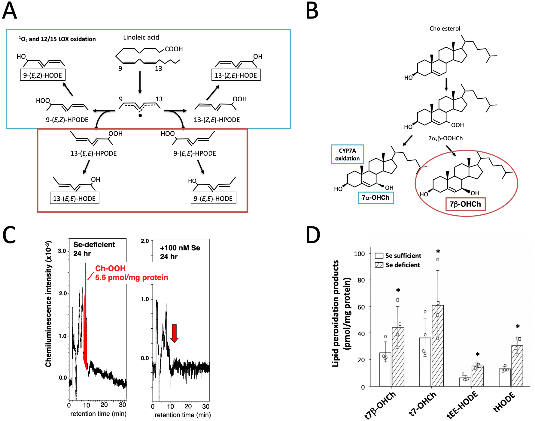
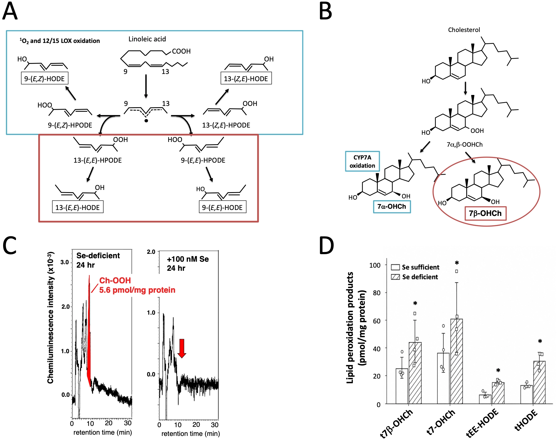
Figure 1. Selenium deficiency induces free radical-mediated oxidation of linoleic acid and cholesterol. (A-B) Schematic representation of free radical-mediated lipid peroxidation products derived from (A) linoleic acid and (B) cholesterol. Adapted with permission from reference[19]. Copyright © 2021 Elsevier; (A) (Z,E)-H(P)ODEs are generated by singlet oxygen and 12/15-lipoxygenase oxidation, whereas free radical oxidation of linoleate induces the formation of all H(P)ODE isomers. (E,E)-HODEs are stable markers of free radical-mediated oxidation of linoleate; (B) Free radical-mediated cholesterol oxidation produces 7α- and 7β-O(O)HCh, whereas enzymatic oxidation by CYP7A generates 7α-OHCh. 7β-OHCh is a stable marker of free radical-mediated oxidation of cholesterol; (C-D) Jurkat cells were cultured with (selenium-sufficient) or without (selenium-deficient) 100 nM sodium selenite for 24 h, and lipid oxidation products were measured. Adapted from reference[13]. CC BY 4.0; (C) Formation of 7-OOHCh in selenium-deficient Jurkat cells, but not in selenium-sufficient cells, was determined using a chemiluminescence HPLC system; (D) Effect of selenium deficiency on the levels of lipid peroxidation products (pmol/mg protein). The letter “t” indicates total amounts measured after reduction and saponification. *Statistically significant difference versus Se sufficient cells, as determined by Student’s t-test (P < 0.05). t7-OHCh: sum of 7α- and 7β-OHCh; tEE-HODE: sum of 9- and 13-[E,E]-HODEs; tHODE: the sum of 9- and 13-[Z,E]-HODEs and 9- and 13-[E,E]-HODEs; HODEs: hydroxyoctadecadienoic acids; HPLC: high-performance liquid chromatography.
In the case of cholesterol, free radical-mediated oxidation produces several oxysterols, among which 7α- and 7β-hydroperoxycholesterol (7a- and 7b-OOHCh) are representative primary products[21]. These 7-OOHCh species are reduced by GPx4 to yield 7α- and 7β-hydroxycholesterol (7a- and 7b-OHCh), whereas enzymatic oxidation by CYP7A predominantly yields 7α-OHCh. Thus, 7β-OHCh is widely used as a marker of free radical-mediated cholesterol oxidation, although it should be interpreted in the context of other oxidation products[21].
In selenium-deficient cells, ChOOH was readily detected, whereas phospholipid hydroperoxides remained below detectable levels[12]. ChOOH was not detected in the presence of sodium selenite, α-tocopherol (α-T), or fetal bovine serum. Furthermore, after reduction and saponification, the amounts of E,E-HODEs and 7β-OHCh were significantly increased under selenium-deficient conditions[13], indicating enhanced free radical-mediated lipid peroxidation. It is also notable that the increase in 7β-OHCh was greater than that of HODE[11,13,23], suggesting that cholesterol oxidation is a prominent feature of oxidative membrane injury in this setting.
In addition, cholesterol oxidation products were also detected when cells were exposed to the radical initiator 2,2′-azobis(2-amidinopropane) dihydrochloride, suggesting that cholesterol oxidation may represent a common feature of cell membrane lipid peroxidation under oxidative stress conditions. This behavior contrasts with plasma and low-density lipoprotein oxidation, in which cholesterol is typically oxidized only after depletion of most unsaturated fatty acids[24]. Thus, the relative susceptibility of cholesterol and linoleate appears to differ markedly between cell membranes and circulating lipoprotein systems[25].
A further mechanistic issue is how cholesterol oxidation may contribute to membrane damage in relation to PUFA peroxidation. Because cholesterol is a major determinant of membrane integrity, stability, fluidity, and lateral organization, its oxidation may have consequences beyond the formation of oxysterol products themselves[26-29]. Oxidized cholesterol species have been reported to alter membrane integrity and cholesterol-rich membrane domains[26,27], raising the possibility that cholesterol oxidation modifies the physical environment in which PUFA-containing phospholipids undergo peroxidation. In this view, cholesterol oxidation may not simply occur in parallel with PUFA peroxidation but may also modulate ferroptosis-related membrane damage through changes in membrane organization and biophysical properties. Conversely, once PUFA peroxidation is initiated, the resulting oxidative stress may further promote cholesterol oxidation, suggesting possible bidirectional crosstalk between these two lipid oxidation pathways[28,29]. Although direct mechanistic evidence remains limited, this possibility should currently be regarded as a working hypothesis.
These findings are particularly significant because current ferroptosis research has largely centered on phospholipid peroxidation, whereas the contribution of cholesterol oxidation has received far less attention. Our observations therefore suggest that selenium deficiency is associated with a broader lipid oxidation signature than is generally appreciated, involving not only phospholipid hydroperoxides but also oxidized cholesterol species, and raise the possibility that cholesterol oxidation may represent an underappreciated component of ferroptosis-related membrane damage.
3. Impaired Selenoprotein Biosynthesis and Lipid Peroxidation: Lessons from Selenocysteine Insertion Sequence (SECIS)-Binding Protein 2 (SBP2) Deficiency
To further understand how impaired selenium metabolism promotes oxidative membrane damage, we next consider the biosynthetic system that supports selenoprotein production. Selenoprotein synthesis depends on a unique translational machinery in which selenocysteine is biosynthesized on tRNASec and incorporated at UGA codons in a SECIS-dependent manner (Figure 2A)[30-32]. This pathway requires multiple specialized factors, including selenophosphate synthetase 2 (SEPHS2), phosphoseryl-tRNA kinase, O-phosphoseryl-tRNA:selenocysteinyl-tRNA synthase, and SECIS-associated factors such as SBP2, and therefore reflects a tightly regulated process of selenium utilization. Because this system governs the expression of a broad set of selenoproteins, defects in selenoprotein biosynthesis are expected to compromise not only GPx4 but also other selenium-dependent redox systems.
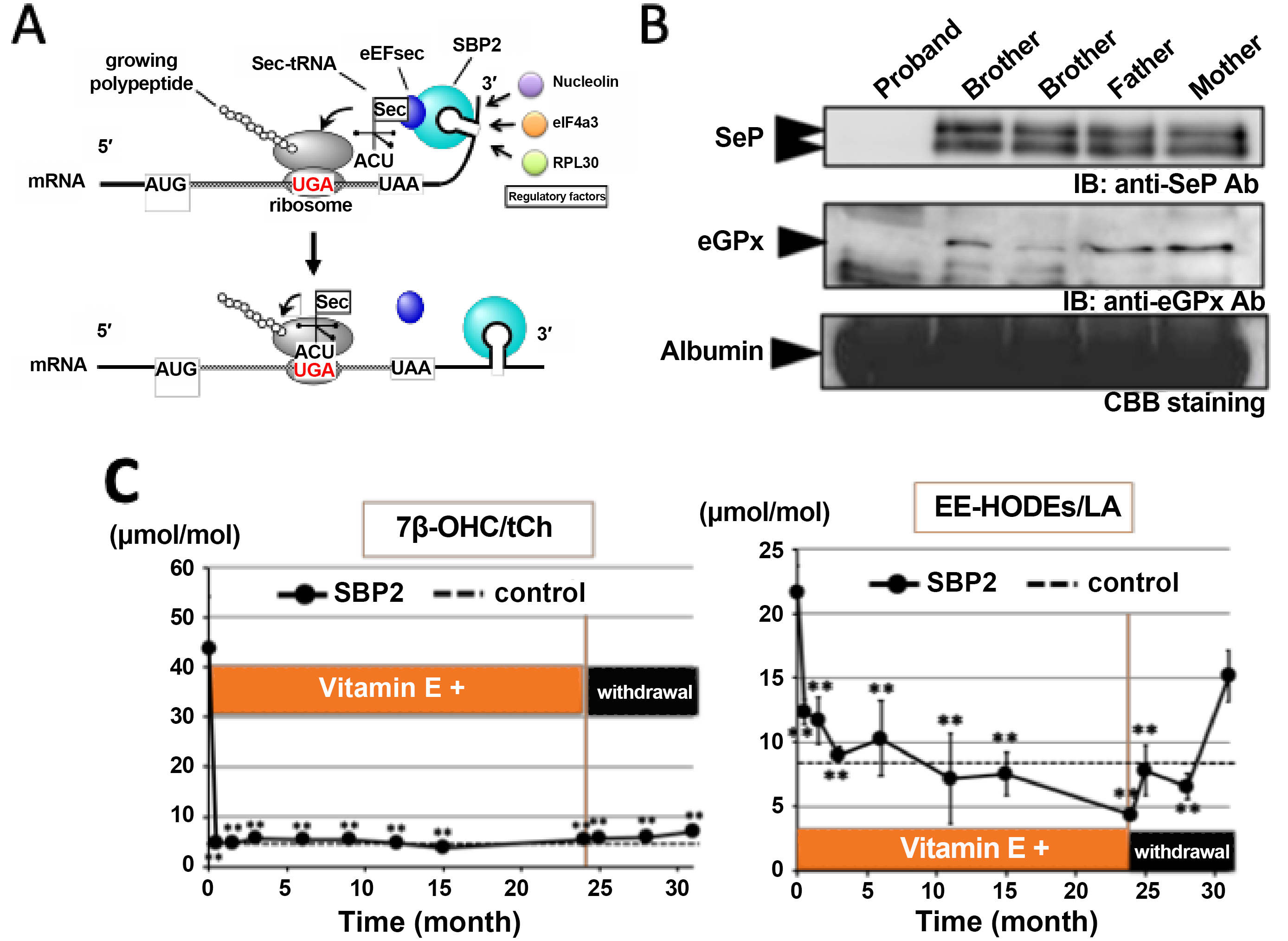
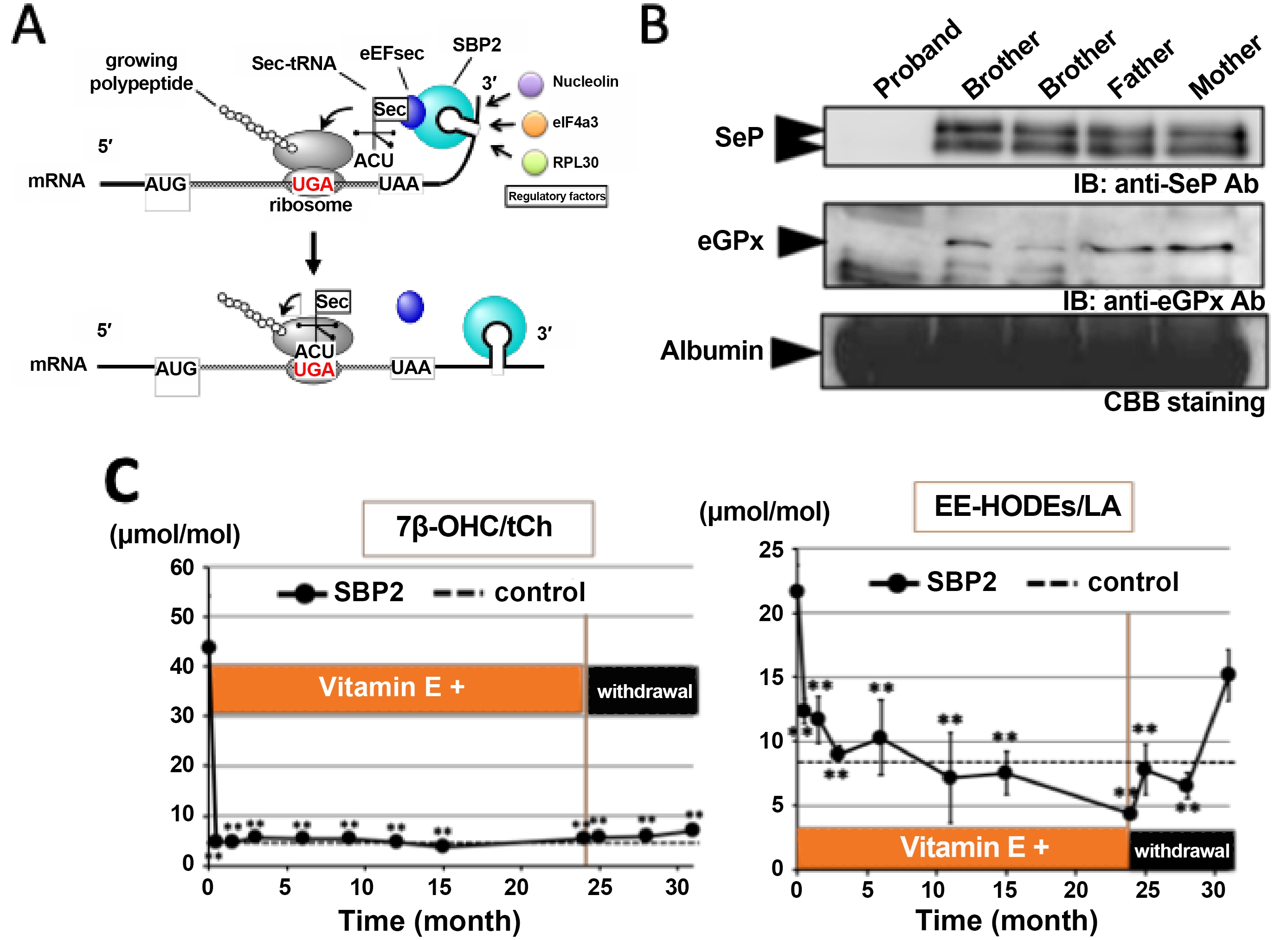
Figure 2. SBP2 deficiency promotes lipid peroxidation in vivo, and its oxidative phenotype is improved by vitamin E. (A) Schematic illustration of impaired SECIS-dependent selenoprotein synthesis; (B) Reduced expression of serum selenoproteins in samples from the SBP2-mutant patient. Lanes, from left to right, indicate the control, the proband, the proband’s brother, and the parents. Detailed clinical and experimental information is provided in reference[34]; (C) Effects of vitamin E supplementation on free radical-mediated lipid peroxidation products in the serum of the SBP2-mutant patient. Changes in 7β-OHCh/tCh and E,E-HODEs/LA during vitamin E treatment and after withdrawal are shown. The dashed control line indicates the fixed reference value obtained from the control sample and does not represent a longitudinal control measurement. Adapted from reference[34]. CC BY 4.0. SBP2: SECIS-binding protein 2; SECIS: selenocysteine insertion sequence; LA: linoleic acid; eGPx: extracellular glutathione peroxidase 3.
An important concept in selenium biology is the hierarchy of selenoprotein expression. Under conditions of limited selenium availability, individual selenoproteins are differentially affected, largely because of differences in the affinity of their SECIS elements for the translational machinery[6,10,33]. For example, GPx1 is highly sensitive to selenium status because of the relatively weak activity of its SECIS element, whereas thioredoxin reductase 1 is more resistant to selenium limitation and remains relatively stable even under selenium-deficient conditions. In contrast, impaired selenium utilization or biosynthesis, rather than simple selenium deficiency, causes broader defects in selenoprotein synthesis and leads to more severe selenoprotein deficiency and associated phenotypes.
In our previous studies, the SBP2-mutant patient provided a unique human model for this concept[34]. Because SBP2 is essential for SECIS-mediated selenocysteine incorporation, its mutation impairs the synthesis of multiple selenoproteins. We found that the patient with SBP2 mutations had undetectable levels of selenoprotein P and GPx3 in plasma and GPx1 in red blood cells, demonstrating profound selenoprotein deficiency (Figure 2B)[34,35]. Notably, impaired synthesis of iodothyronine deiodinases was associated with characteristic abnormalities in thyroid hormone profiles, including elevated thyroxine (T4), reduced triiodothyronine (T3), and normal or slightly elevated thyroid-stimulating hormone. These endocrine abnormalities were accompanied by characteristic clinical phenotypes, including short stature during childhood and delayed bone maturation[34-36]. In this SBP2 patient, we observed increased levels of free radical-mediated lipid peroxidation products, including E,E-HODEs and 7β-ChOH, indicating that impaired selenoprotein biosynthesis is associated with enhanced lipid oxidative damage in vivo[34]. These findings extend our observations in selenium-deficient cells and suggest that broad impairment of selenium utilization can promote lipid peroxidation not only in experimental models but also in human disease. The patient-derived data summarized in this review were derived from our previously published study[34], which was conducted with institutional ethical approval and informed consent as described in that report.
Next, we examined the effects of vitamin E treatment in the proband by monitoring serum α-T levels and oxidative biomarkers. Administration of α-T acetate (100 mg/day) for 2 years led to a time-dependent increase in serum α-T levels, whereas withdrawal of the treatment returned these levels to baseline within 7 months (Figure 2C)[34]. Vitamin E treatment effectively reduced lipid peroxidation products in the serum of the subject harboring SBP2 mutations. Notably, even a 2-week course of vitamin E treatment was sufficient to markedly lower 7β-OHCh levels, and this effect persisted for 2 years. In contrast, although E,E-HODEs were also reduced during the 2-year treatment period, they increased markedly after withdrawal of vitamin E treatment, whereas 7β-OHCh levels remained low. Collectively, these results indicate that vitamin E treatment effectively suppressed free radical-mediated lipid peroxidation in the subject with SBP2 mutations (Figure 2C)[34]. This finding is consistent with our earlier observations in selenium-deficient cells and reinforces the view that selenium-dependent antioxidant systems and vitamin E-dependent membrane protection function cooperatively to suppress oxidative membrane injury.
Taken together, our studies of SBP2-deficient patients provide human evidence that impaired selenoprotein biosynthesis promotes progressive lipid oxidation and increases oxidative vulnerability. The concurrent elevation of HODE and cholesterol oxidation products, together with their responsiveness to vitamin E, supports the idea that defective selenium metabolism generates a broader lipid oxidation phenotype than is often appreciated. These observations provide an important conceptual bridge between selenium metabolism and ferroptosis-related membrane damage.
4. Toxicological Targeting of Selenium Metabolism and Ferroptotic Vulnerability
Whereas selenium deficiency and SBP2 mutations illustrate nutritional and genetic impairment of selenium metabolism, toxic chemical exposure reveals a third context in which disruption of the selenoprotein biosynthetic system can increase ferroptosis sensitivity[37,38]. This perspective highlights selenium metabolism not merely as a nutritional factor, but as an active determinant of cellular vulnerability under toxic stress conditions.
Selenium used for selenoprotein synthesis is metabolized through multiple steps involving both organic and inorganic pathways. In the case of selenoprotein P as a selenium source, selenium present as selenocysteine within its polypeptide is taken up into cells via its receptors, such as ApoER2, and transported to lysosomes, where it is degraded to Sec[32,39]. Sec is further degraded to inorganic selenium, selenide, by Sec lyase and is then utilized by SEPHS2 to generate selenophosphate, which is used for Sec synthesis. For a long time, the metabolic steps linking Sec lyase and SEPHS2 remained unclear; however, recent studies have demonstrated that peroxiredoxin 6 (PRDX6) plays a critical role in selenium transfer, selenoprotein synthesis, and ferroptosis resistance[40-42]. Recent studies support a role for PRDX6 in cellular selenium utilization. PRDX6 has been proposed to receive selenium from selenocysteine lyase-derived selenide and from low-molecular-weight selenocompounds, such as selenodiglutathione and sodium selenite in the presence of glutathione. PRDX6 may therefore facilitate selenium delivery to SEPHS2 for selenophosphate production and subsequent Sec synthesis[40-42]. In addition, recent studies have shown that xCT/SLC7A11 contributes to selenium uptake from selenite-derived extracellular selenium species and supports selenium-dependent GPX4 expression, thereby linking selenium utilization to ferroptosis sensitivity[43-45].
This newly identified role of PRDX6 has attracted considerable attention and prompted new research directions, because PRDX6 is a target of diverse reactive species, including reactive oxygen species, reactive sulfur species, selenide, electrophiles, and environmental pollutants such as arsenite[37,46,47]. Notably, PRDX6 is a major target of hydrogen peroxide exposure, which induces irreversible overoxidation of its catalytic cysteine to sulfonic acid (Cys-SO3H)[48]. This overoxidation alters the isoelectric point of PRDX6 and can be detected by two-dimensional gel electrophoresis. We previously reported that overoxidized PRDX6 is increased in the red blood cells of patients with Alzheimer’s disease, raising the possibility that this modification reflects not only increased oxidative stress but also disturbance of selenium metabolism[47]. Among environmental toxicants, arsenite provides a particularly informative example. Our recent studies suggest that arsenite disrupts selenium metabolism and inhibits selenium-induced GPx expression, primarily by interfering with selenium utilization[37,49]. Notably, arsenite exposure reduced selenium incorporation into RNA, suggesting inhibition of Sec-tRNASec synthesis, a critical step in selenoprotein biosynthesis[49]. Furthermore, arsenite increased cellular susceptibility to ferroptosis, whereas genetic deletion of PRDX6 suppressed selenoprotein expression and exacerbated arsenite-induced ferroptosis. These findings provide new insight into the toxicological actions of arsenite, highlighting its ability to disrupt selenium metabolism and thereby promote lipid peroxidation and ferroptotic vulnerability. In addition to arsenite, other environmental toxicants may also perturb selenium-dependent redox defense and thereby influence ferroptosis susceptibility[50-53]. Mercury is a particularly relevant example, because mercury compounds have long been known to interact strongly with selenium and to impair the function of selenium-dependent antioxidant systems, including thioredoxin reductases and glutathione peroxidases[50]. More recent studies further suggest that both inorganic mercury and methylmercury can trigger ferroptotic injury through suppression of GPx4-associated defense and enhanced lipid peroxidation[51,52]. Lead exposure has likewise been linked to altered selenium status and oxidative stress[53], although its mechanistic connection to selenium metabolism-dependent ferroptosis remains less clearly defined. These observations suggest that disruption of selenium utilization may represent a broader toxicological mechanism by which multiple environmental pollutants enhance oxidative membrane vulnerability.
Recent studies have broadened the current view of selenium-mediated ferroptosis regulation beyond selenoprotein biosynthesis alone[54]. In addition to supporting the synthesis of GPx4 and other selenoproteins, selenium has been shown to drive a transcriptional adaptive program involving selenoproteome-related transcripts that enhances cellular resistance to ferroptosis. Moreover, selenium can also suppress ferroptosis through a rapid mechanism involving sulfide:quinone oxidoreductase-dependent ubiquinone reduction, thereby linking selenium metabolism to coenzyme Q10-mediated antioxidant defense[55]. These findings further support the concept that selenium regulates ferroptosis sensitivity at multiple levels, including transcriptional control, selenoprotein biosynthesis, and noncanonical redox metabolism.
5. Conclusion
In this review, we revisited selenium deficiency and impaired selenoprotein biosynthesis in the context of ferroptosis, with particular emphasis on insights gained from our previous studies. Our findings in selenium-deficient cells demonstrated a non-apoptotic form of cell death associated with lipid peroxidation, sensitivity to vitamin E and deferoxamine, and the accumulation of not only phospholipid oxidation products but also cholesterol oxidation products. These features suggest that impaired selenium metabolism can induce a ferroptosis-like mode of oxidative membrane injury (Figure 3).
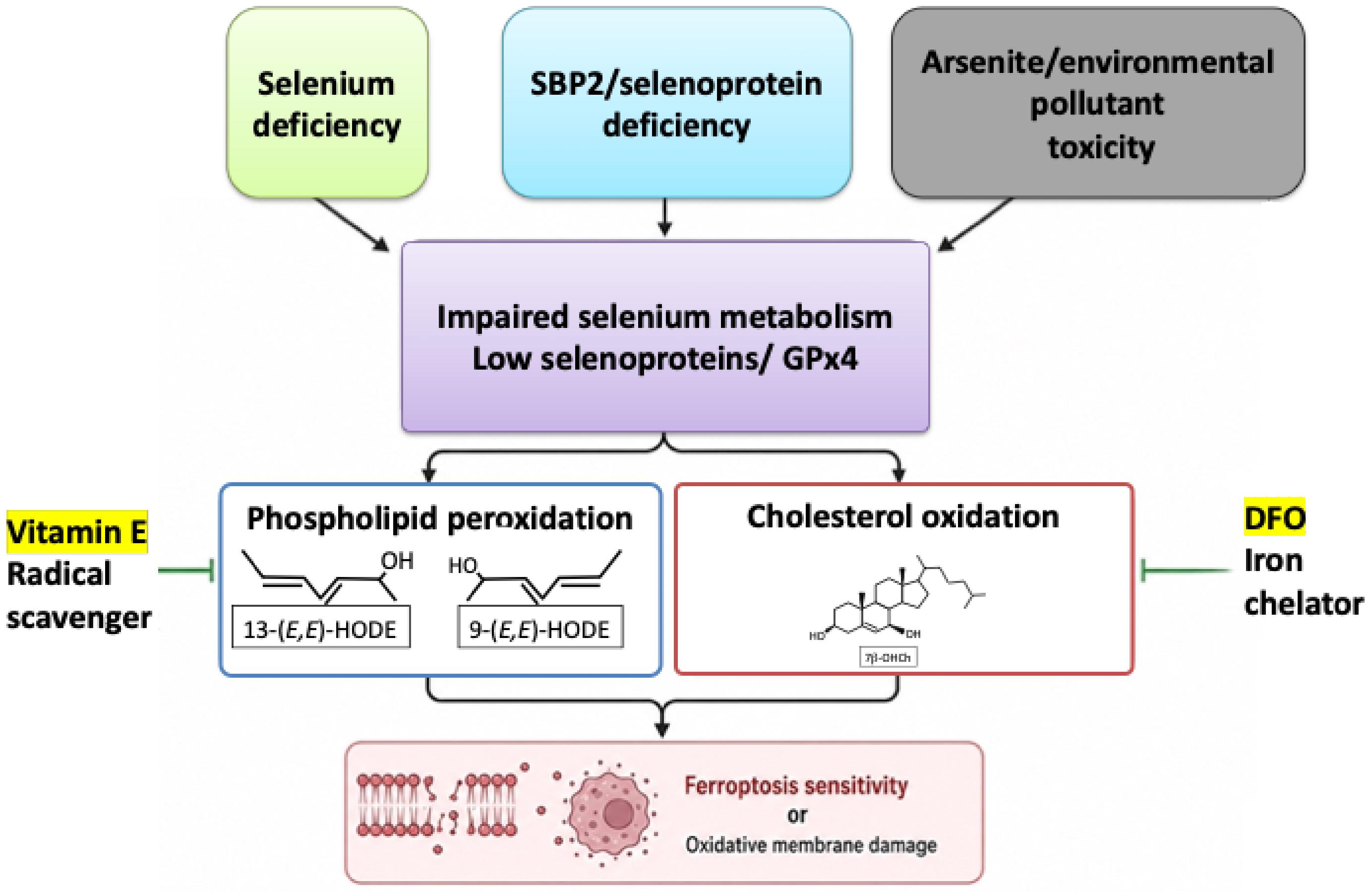
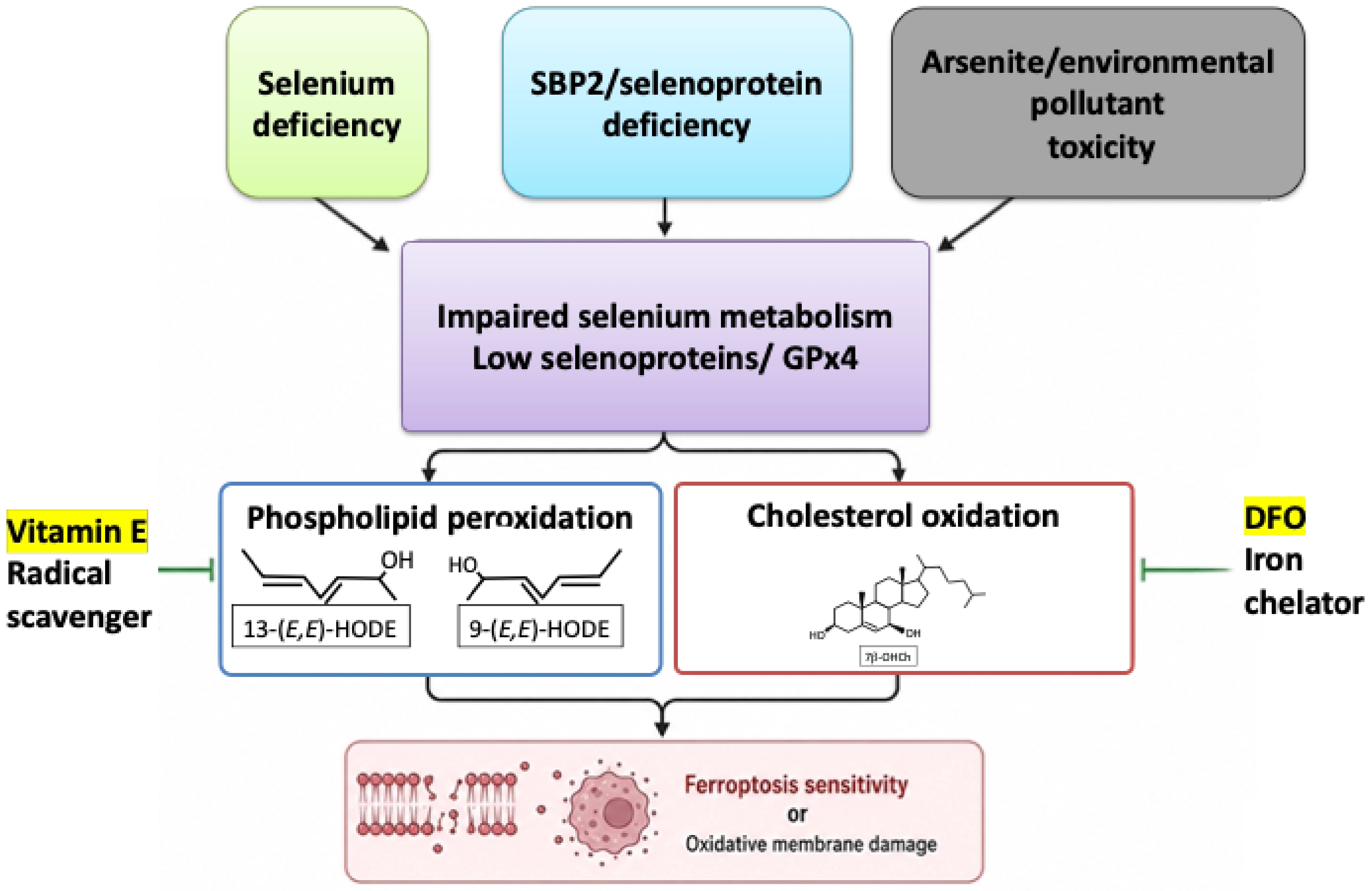
Figure 3. Connection between impaired selenium metabolism and ferroptosis-related membrane damage. Selenium deficiency, SBP2/selenoprotein deficiency, and arsenite/environmental pollutant toxicity converge on impaired selenium metabolism and decreased selenoprotein/GPx4-dependent defense. This state promotes both phospholipid peroxidation and cholesterol oxidation, thereby enhancing oxidative membrane damage and ferroptosis sensitivity. Representative oxidation products and protective factors are indicated. GPx4: glutathione peroxidase 4; SBP2: SECIS-binding protein 2; DFO: deferoxamine; HODE: hydroxyoctadecadienoic acid.
Our studies of SBP2-deficient patients further extended this concept to human disease. The concurrent increase in HODE and cholesterol oxidation products, together with their responsiveness to vitamin E, supports the idea that defective selenoprotein biosynthesis promotes progressive lipid oxidative damage in vivo.
Recent advances in selenium metabolism have further revealed that the selenoprotein biosynthetic system is vulnerable not only to nutritional deficiency and genetic defects but also to toxicological disruption. In particular, recent findings on PRDX6 and arsenite suggest that toxicants can interfere with selenium utilization, suppress selenoprotein expression, and increase ferroptosis sensitivity. These observations broaden the significance of selenium metabolism beyond a passive nutritional pathway and position it as an active and vulnerable regulator of lipid peroxide defense.
Taken together, these studies support the view that selenium metabolism should be regarded as a dynamic system that shapes lipid peroxidation, oxidative membrane damage, and ferroptotic vulnerability. Further studies on the interplay between selenium metabolism, membrane lipid oxidation, and ferroptosis will deepen our understanding of redox pathology and may provide new opportunities for the prevention and treatment of oxidative stress-related disorders.
Authors contribution
The author contributed solely to the article.
Conflicts of interest
Not applicable.
Ethical approval
Not applicable.
Consent to participate
Not applicable.
Consent for publication
Not applicable.
Availability of data and materials
Not applicable.
Funding
This work was supported in part by JSPS KAKENHI (Grant Nos. 21H05270, 21K19321, 24K22009 and 24H00590).
Copyright
© The Author(s) 2026.
References
-
1. Berndt C, Alborzinia H, Amen VS, Barayeu U, Bartelt A, Bayir H, et al. Ferroptosis in health and disease. Redox Biol. 2024;75:103211.[DOI]
-
2. Dixon SJ, Olzmann JA. The cell biology of ferroptosis. Nat Rev Mol Cell Biol. 2024;25(6):424-442.[DOI]
-
3. Stockwell BR, Angeli JPF, Bayir H, Bush AI, Conrad M, Dixon SJ, et al. Ferroptosis: A regulated cell death nexus linking metabolism, redox biology, and disease. Cell. 2017;171(2):273-285.[DOI]
-
4. Ingold I, Berndt C, Schmitt S, Doll S, Poschmann G, Buday K, et al. Selenium utilization by GPX4 is required to prevent hydroperoxide-induced ferroptosis. Cell. 2018;172(3):409-422.e21.[DOI]
-
5. Friedmann Angeli JP, Schneider M, Proneth B, Tyurina YY, Tyurin VA, Hammond VJ, et al. Inactivation of the ferroptosis regulator Gpx4 triggers acute renal failure in mice. Nat Cell Biol. 2024;16(12):1180-1191.[DOI]
-
6. Labunskyy VM, Hatfield DL, Gladyshev VN. Selenoproteins: Molecular pathways and physiological roles. Physiol Rev. 2014;94(3):739-777.[DOI]
-
7. Saito Y. Essential trace element selenium and redox regulation: Its metabolism, physiological function, and related diseases. Redox Exp Med. 2022;2022(1):R149-R158.[DOI]
-
9. Saito Y, Hayashi T, Tanaka A, Watanabe Y, Suzuki M, Saito E, et al. Selenoprotein P in human plasma as an extracellular phospholipid hydroperoxide glutathione peroxidase. J Biol Chem. 1999;274(5):2866-2871.[DOI]
-
10. Rayman MP. Selenium and human health. Lancet. 2012;379(9822):1256-1268.[DOI]
-
11. Saito Y. Diverse cytoprotective actions of vitamin E isoforms-role as peroxyl radical scavengers and complementary functions with selenoproteins. Free Radic Biol Med. 2021;175:121-129.[DOI]
-
12. Saito Y, Yoshida Y, Akazawa T, Takahashi K, Niki E. Cell death caused by selenium deficiency and protective effect of antioxidants. J Biol Chem. 2003;278(41):39428-39434.[DOI]
-
13. Saito Y, Yoshida Y, Niki E. Cholesterol is more susceptible to oxidation than linoleates in cultured cells under oxidative stress induced by selenium deficiency and free radicals. FEBS Lett. 2007;581(22):4349-4354.[DOI]
-
14. Saito Y, Noguchi N. 7-Hydroxycholestrol as a possible biomarker of cellular lipid peroxidation: Difference between cellular and plasma lipid peroxidation. Biochem Biophys Res Commun. 2014;446(3):741-744.[DOI]
-
15. Zheng J, Conrad M. Ferroptosis: When metabolism meets cell death. Physiol Rev. 2025;105(2):651-706.[DOI]
-
16. McKeehan WL, Hamilton WG, Ham RG. Selenium is an essential trace nutrient for growth of WI-38 diploid human fibroblasts. Proc Natl Acad Sci U S A. 1976;73(6):2023-2027.[DOI]
-
17. Hajeyah AA, Griffiths WJ, Wang Y, Finch AJ, O’Donnell VB. The biosynthesis of enzymatically oxidized lipids. Front Endocrinol. 2020;11:591819.[DOI]
-
19. Saito Y. Lipid peroxidation products as a mediator of toxicity and adaptive response—the regulatory role of selenoprotein and vitamin E. Arch Biochem Biophys. 2021;703:108840.[DOI]
-
22. Milne GL, Yin H, Hardy KD, Davies SS, Roberts LJ. Isoprostane generation and function. Chem Rev. 2011;111(10):5973-5996.[DOI]
-
23. Saito Y, Noguchi N, Niki E. Cholesterol is more readily oxidized than phospholipid linoleates in cell membranes to produce cholesterol hydroperoxides. Free Radic Biol Med. 2024;211:89-95.[DOI]
-
24. Noguchi N, Numano R, Kaneda H, Niki E. Oxidation of lipids in low density lipoprotein particles. Free Radic Res. 1998;29(1):43-52.[DOI]
-
25. Yoshida Y, Saito Y, Hayakawa M, Habuchi Y, Imai Y, Sawai Y, et al. Levels of lipid peroxidation in human plasma and erythrocytes: Comparison between fatty acids and cholesterol. Lipids. 2007;42(5):439-449.[DOI]
-
26. Royer MC, Lemaire-Ewing S, Desrumaux C, Monier S, de Barros JPP, Athias A, et al. 7-ketocholesterol incorporation into sphingolipid/cholesterol-enriched (lipid raft) domains is impaired by vitamin E. J Biol Chem. 2009;284(23):15826-15834.[DOI]
-
27. Schieffer D, Naware S, Bakun W, Bamezai AK. Lipid raft-based membrane order is important for antigen-specific clonal expansion of CD4+ T lymphocytes. BMC Immunol. 2014;15(1):58.[DOI]
-
28. Levitan I, Shentu TP. Impact of oxLDL on cholesterol-rich membrane rafts. J Lipids. 2011;2011:730209.[DOI]
-
29. Iuliano L. Pathways of cholesterol oxidation via non-enzymatic mechanisms. Chem Phys Lipids. 2011;164(6):457-468.[DOI]
-
30. Schweizer U, Fabiano M. Genetics of selenoproteins and selenoprotein metabolism—An overview of current concepts and emerging aspects. Redox Biol. 2026;91:104083.[DOI]
-
31. Copeland PR, Fletcher JE, Carlson BA, Hatfield DL, Driscoll DM. A novel RNA binding protein, SBP2, is required for the translation of mammalian selenoprotein mRNAs. EMBO J. 2000;19(2):306-314.[DOI]
-
32. Saito Y. Selenium transport mechanism via selenoprotein P: Its physiological role and related diseases. Front Nutr. 2021;8:685517.[DOI]
-
33. Schomburg L, Schweizer U. Hierarchical regulation of selenoprotein expression and sex-specific effects of selenium. Biochim Biophys Acta Gen Subj. 2009;1790(11):1453-1462.[DOI]
-
34. Saito Y, Shichiri M, Hamajima T, Ishida N, Mita Y, Nakao S, et al. Enhancement of lipid peroxidation and its amelioration by vitamin E in a subject with mutations in the SBP2 gene. J Lipid Res. 2015;56(11):2172-2182.[DOI]
-
35. Hamajima T, Mushimoto Y, Kobayashi H, Saito Y, Onigata K. Novel compound heterozygous mutations in the SBP2 gene: Characteristic clinical manifestations and the implications of GH and triiodothyronine in longitudinal bone growth and maturation. Eur J Endocrinol. 2012;166(4):757-764.
-
36. Dumitrescu AM, Liao XH, Abdullah MSY, Lado-Abeal J, Majed FA, Moeller LC, et al. Mutations in SECISBP2 result in abnormal thyroid hormone metabolism. Nat Genet. 2005;37(11):1247-1252.[DOI]
-
37. Takashima H, Makino R, Taguchi H, Ito J, Mishima E, Takenaka Y, et al. Arsenite sensitizes to ferroptosis by disrupting selenium metabolism and reducing GPx4 expression. Toxicology. 2026;522:154409.[DOI]
-
38. Yang L, Cai X, Li R. Ferroptosis induced by pollutants: An emerging mechanism in environmental toxicology. Environ Sci Technol. 2024;58(5):2166-2184.[DOI]
-
39. Mizuno A, Toyama T, Ichikawa A, Sakai N, Yoshioka Y, Nishito Y, et al. An efficient selenium transport pathway of selenoprotein P utilizing a high-affinity ApoER2 receptor variant and being independent of selenocysteine lyase. J Biol Chem. 2023;299(8):105009.[DOI]
-
40. Ito J, Nakamura T, Toyama T, Chen D, Berndt C, Poschmann G, et al. PRDX6 dictates ferroptosis sensitivity by directing cellular selenium utilization. Mol Cell. 2024;84(23):4629-4644.e9.[DOI]
-
41. Fujita H, Tanaka YK, Ogata S, Suzuki N, Kuno S, Barayeu U, et al. PRDX6 augments selenium utilization to limit iron toxicity and ferroptosis. Nat Struct Mol Biol. 2024;31(8):1277-1285.[DOI]
-
42. Chen Z, Inague A, Kaushal K, Fazeli G, Schilling D, da Silva TNX, et al. PRDX6 contributes to selenocysteine metabolism and ferroptosis resistance. Mol Cell. 2024;84(23):4645-4659.e9.[DOI]
-
43. Carlisle AE, Lee N, Matthew-Onabanjo AN, Spears ME, Park SJ, Youkana D, et al. Selenium detoxification is required for cancer-cell survival. Nat Metab. 2020;2(7):603-611.[DOI]
-
44. Lee N, Carlisle AE, Peppers A, Park SJ, Doshi MB, Spears ME, et al. xCT-driven expression of GPX4 determines sensitivity of breast cancer cells to ferroptosis inducers. Antioxidants. 2021;10(2):317.[DOI]
-
46. Fisher AB. Peroxiredoxin 6: A bifunctional enzyme with glutathione peroxidase and phospholipase A2Activities. Antioxid Redox Signal. 2011;15(3):831-844.[DOI]
-
47. Yoshida Y, Yoshikawa A, Kinumi T, Ogawa Y, Saito Y, Ohara K, et al. Hydroxyoctadecadienoic acid and oxidatively modified peroxiredoxins in the blood of Alzheimer’s disease patients and their potential as biomarkers. Neurobiol Aging. 2009;30(2):174-185.[DOI]
-
48. Saito Y, Nishio K, Ogawa Y, Kimata J, Kinumi T, Yoshida Y, et al. Turning point in apoptosis/necrosis induced by hydrogen peroxide. Free Radic Res. 2006;40(6):619-630.[DOI]
-
49. Takashima H, Makino R, Taguchi H, Takenaka Y, Akiyama Y, Tomikoka Y, et al. A novel ICP-MS strategy identifies arsenite as an inhibitor of selenocysteine-tRNASec charging. J Toxicol Sci. 2026;51(6):331-338.[DOI]
-
50. Spiller HA. Rethinking mercury: The role of selenium in the pathophysiology of mercury toxicity. Clin Toxicol. 2018;56(5):313-326.[DOI]
-
52. Xu X, Wang SS, Zhang L, Lu AX, Lin Y, Liu JX, et al. Methylmercury induced ferroptosis by interference of iron homeostasis and glutathione metabolism in CTX cells. Environ Poll. 2023;335:122278.[DOI]
-
54. Alim I, Caulfield JT, Chen Y, Swarup V, Geschwind DH, Ivanova E, et al. Selenium drives a transcriptional adaptive program to block ferroptosis and treat stroke. Cell. 2019;177(5):1262-1279.e25.[DOI]
-
55. Lee N, Park SJ, Lange M, Tseyang T, Doshi MB, Kim TY, et al. Selenium reduction of ubiquinone via SQOR suppresses ferroptosis. Nat Metab. 2024;6(2):343-358.[DOI]
Copyright
© The Author(s) 2026. This is an Open Access article licensed under a Creative Commons Attribution 4.0 International License (https://creativecommons.org/licenses/by/4.0/), which permits unrestricted use, sharing, adaptation, distribution and reproduction in any medium or format, for any purpose, even commercially, as long as you give appropriate credit to the original author(s) and the source, provide a link to the Creative Commons license, and indicate if changes were made.
Publisher’s Note
Science Exploration remains a neutral stance on jurisdictional claims in published
maps
and institutional affiliations. The views expressed in this article are solely those
of
the author(s) and do not reflect the opinions of the Editors or the publisher.
Share And Cite


Science Exploration Style
Saito Y. Selenium metabolism, lipid peroxidation, and ferroptosis sensitivity: Lessons from selenium deficiency. Ferroptosis Oxid Stress. 2026;2:202627. https://doi.org/10.70401/fos.2026.0036
Tips
Copy completed.
Submit a Manuscript
Author Instructions
Cite this Article
Article Metrics
0
View
0
Download
Cited
Article Updates

- Abstract
- Keywords
- 1. Introduction
- 2. Past Evidence from Our Studies: Selenium Deficiency-Induced Non-Apoptotic Cell Death and Lipid Peroxidation Products
- 3. Impaired Selenoprotein Biosynthesis and Lipid Peroxidation: Lessons from Selenocysteine Insertion Sequence (SECIS)-Binding Protein 2 (SBP2) Deficiency
- 4. Toxicological Targeting of Selenium Metabolism and Ferroptotic Vulnerability
- 5. Conclusion
- Authors contribution
- Conflicts of interest
- Ethical approval
- Consent to participate
- Consent for publication
- Availability of data and materials
- Funding
- References
- Copyright
 Get citation
Science Exploration Style

Saito Y. Selenium metabolism, lipid peroxidation, and ferroptosis sensitivity: Lessons from selenium deficiency. Ferroptosis Oxid Stress. 2026;2:202627. https://doi.org/10.70401/fos.2026.0036
copy
Share Link
copy
Subscribe