Targeting FSP1 to induce ferroptosis in cancer: From mechanisms to therapeutic strategies

Peng Lin
1,3

,
Jin-Dong Ma
4
,
Zhi-Nan Chen
1,2,*
,
Jiao Wu
1,2,*
*Correspondence to:
Jiao Wu, Department of Cell Biology and National Translational Science Center for Molecular Medicine, Fourth Military Medical University, Xi’an 710032, Shaanxi, China.
E-mail: jiaowubio@hotmail.com
Zhi-Nan Chen, Department of Cell Biology and National Translational Science Center for Molecular Medicine, Fourth Military Medical University, Xi’an 710032, Shaanxi, China. E-mail: znchen@fmmu.edu.cn
Zhi-Nan Chen, Department of Cell Biology and National Translational Science Center for Molecular Medicine, Fourth Military Medical University, Xi’an 710032, Shaanxi, China. E-mail: znchen@fmmu.edu.cn
Ferroptosis Oxid Stress. 2026;2:202625. 10.70401/fos.2026.0037
Received: May 28, 2026Accepted: July 27, 2026Published: July 27, 2026
Abstract
Ferroptosis is a form of programmed cell death driven by iron-dependent lipid peroxidation. Inducing ferroptosis in tumor cells has emerged as a crucial strategy for cancer treatment and overcoming therapeutic resistance. Ferroptosis suppressor protein 1 (FSP1), identified in recent years, is a key ferroptosis-inhibitory factor that operates independently of glutathione peroxidase 4 (GPX4). FSP1 is highly expressed in multiple malignant tumors, and its expression levels are tightly associated with unfavorable patient prognosis, treatment resistance, and tumor progression. Specific stresses within the tumor microenvironment, including hypoxia, metabolic stress, and the development of drug resistance, can drive a shift in ferroptosis resistance mechanisms from GPX4-dependent to FSP1-dependent pathways. Such plasticity renders FSP1 a druggable target in specific cancer genotypes or at particular stages of tumor progression. Indeed, genetic or pharmacological inhibition of FSP1 has yielded potent antitumor effects across diverse preclinical models. This review systematically summarizes the structural characteristics of FSP1, the molecular mechanisms through which it suppresses ferroptosis, the multilayered regulatory networks controlling its activity, and its roles in different malignancies. We deeply analyze the plasticity of FSP1 dependency in tumor cells in vivo and comprehensively review the latest preclinical progress on small-molecule FSP1 inhibitors and related combinatorial therapeutic strategies. This review aims to establish a theoretical framework to support the clinical translation of FSP1-targeted antitumor strategies.
Keywords
FSP1, ferroptosis, cancer therapy, lipid peroxidation, drug resistance reversal
1. Introduction
Evasion of cell death is a hallmark of cancer, and inducing tumor cell death remains a core therapeutic strategy in oncology[1]. Ferroptosis, a form of regulated cell death (RCD) driven by iron-dependent phospholipid peroxidation chain reactions, has attracted intensive research interest in recent years due to its unique metabolic characteristics and potential to overcome drug resistance[2]. Distinct from apoptosis, pyroptosis, or necroptosis, ferroptosis does not depend on classical upstream activation signals. Instead, it is determined by the dynamic crosstalk among intracellular iron metabolism, redox balance, and lipid composition. Therefore, ferroptosis is defined as a metabolic form of RCD[3]. The core driver of ferroptosis is peroxidative damage to phospholipids containing polyunsaturated fatty acids (PUFA-PLs). Lipid peroxidation proceeds via two distinct routes: the enzymatic pathway mainly mediated by lipoxygenases (LOXs) and non-enzymatic mechanisms[4]. Cellular iron homeostasis profoundly influences ferroptosis via non-enzymatic lipid peroxidation. The generation of free radicals by labile ferrous iron via Fenton and Fenton-like reactions initiates self-amplifying chain reactions, which in turn accelerate the peroxidation of PUFA-PLs and ultimately lead to ferroptosis[5].
Cells deploy multiple surveillance systems to continuously restrain excessive lipid peroxidation and actively prevent ferroptosis. The principal anti-ferroptosis systems include the glutathione (GSH)/glutathione peroxidase 4 (GPX4) pathway, the ferroptosis suppressor protein 1 (FSP1)/coenzyme Q10 (CoQ10) pathway, the GCH1/BH4/DHFR system, the DHODH/CoQH2 system, and the VKORC1L1 pathway[6-10]. In addition, endogenous radical-trapping antioxidants (RTAs), such as vitamin E, squalene, 7-dehydrocholesterol, hydropersulfides, and various tryptophan metabolites, participate in ferroptosis suppression[11-15]. Pathological insults can impair these protective mechanisms through restricted cystine uptake, GSH depletion, GPX4 inhibition, or inactivation of alternative antioxidant pathways such as the FSP1 pathway. When this occurs, cellular redox homeostasis surpasses a lethal threshold, leading to catastrophic lipid peroxidative damage and ultimately ferroptosis[16]. Ample evidence demonstrates that cellular sensitivity to ferroptosis is not a binary phenotype, but rather a tunable biological process regulated by the interplay among lipid metabolism, iron metabolism, and multiple lipid peroxidation defense systems[3].
To meet the metabolic demands of sustained proliferation and to cope with microenvironmental stresses such as hypoxia and nutrient deprivation, cancer cells remodel their lipid metabolism, iron homeostasis, and redox balance through metabolic reprogramming[17]. Although this adaptation grants them capabilities for survival, proliferation, metastasis, and drug resistance, it also introduces new metabolic vulnerabilities, most notably an enhanced sensitivity to ferroptosis. Such cells often exhibit ferroptosis-prone metabolic features, including high PUFA content, an elevated labile iron pool (LIP), and strong reliance on ferroptosis defense systems[2,18]. Several therapy-resistant cancer cell subpopulations display inherent susceptibility to ferroptosis. For instance, mesenchymal cancer cells that have undergone epithelial-mesenchymal transition (EMT) acquire high invasive capacity and significantly increase PUFA-phospholipid (PUFA-PL) content in their plasma membranes, making them heavily dependent on GPX4 to evade ferroptosis[19,20]. In addition, drug-tolerant persister (DTP) cells that escape apoptosis during targeted therapy or chemotherapy activate activating transcription factor 4 (ATF4)-related stress pathways and reshape lipid metabolism, ingeniously relying on GPX4 for survival[21,22]. Tumor cells harboring oncogenic KRAS mutations exhibit metabolic hyperactivity and elevated oxidative stress, yet they simultaneously activate nuclear factor erythroid 2-related factor 2 (NRF2) signaling and upregulate FSP1 expression to counteract ferroptotic stress[23,24]. Moreover, cancer stem cells (CSCs) with self-renewal capacity show increased iron uptake and dysregulated iron homeostasis, rendering them more sensitive to ferroptosis inducers[25]. Consequently, inducing ferroptosis is recognized as a key strategy for overcoming multimodal cell death resistance in tumors, with confirmed synergistic effects when combined with conventional therapies such as radiotherapy, chemotherapy, and immunotherapy[26].
In recent years, genetic or pharmacological ablation of ferroptosis defense pathways in tumor cells to trigger ferroptosis has emerged as a promising anticancer strategy. As the core intracellular ferroptosis suppressor, GPX4 has long been the primary target for drug development in this paradigm. However, GPX4 is ubiquitously expressed in normal tissues and is essential for basal lipid peroxidation detoxification[27]. Gpx4 knockout in mice leads to embryonic lethality, and patients with hereditary GPX4 deficiency exhibit severe pathologies such as early-onset dementia, severely narrowing the therapeutic window for systemic GPX4 inhibition[28]. More critically, in certain tumor cell lines, GPX4 inhibition fails to effectively induce ferroptosis, revealing the existence of compensatory GPX4-independent antioxidant systems[7,29]. FSP1 is the principal executor within this compensatory network. Targeting FSP1 offers distinct advantages over GPX4: Fsp1 knockout mice are viable and fertile, develop normally, and display no obvious pathological lesions in adulthood. Moreover, FSP1 is highly expressed in various tumor tissues, suggesting a tumor-selective therapeutic window. Most recently, seminal studies have demonstrated that in models of lung adenocarcinoma and melanoma lymph node metastasis in vivo, knockout or pharmacological inhibition of FSP1 alone exerts robust tumor-suppressive effects, a stark contrast to its largely dispensable role under standard in vitro culture conditions[30,31]. These findings provide a preclinical proof-of-concept that FSP1 inhibitors can be effective as monotherapy or as key components of combinatorial therapeutic regimens in vivo.
This review will systematically summarize the mechanisms whereby FSP1 suppresses ferroptosis, its regulatory networks, microenvironment-dependent plasticity, and preclinical progress in FSP1-targeted anticancer therapy. Finally, challenges and future directions will be discussed.
2. Functions and Molecular Mechanisms of FSP1
2.1 Molecular characteristics of FSP1
2.1.1 FSP1 protein structure and membrane localization
The human FSP1 gene is located on chromosome 10q21.3-q22.1 and encodes a polypeptide of 373 amino acids. FSP1 belongs to the type II NADH:quinone oxidoreductase family. It was initially named apoptosis-inducing factor mitochondrial 2 (AIFM2) due to its structural homology with apoptosis-inducing factor (AIF) and was subsequently renamed FSP1 following the discovery of its function in inhibiting ferroptosis in a GSH-independent manner[7,29]. Unlike AIF, FSP1 lacks a canonical N-terminal mitochondrial targeting sequence and is therefore not primarily localized to mitochondria. Instead, it is widely distributed across various cellular membrane compartments, including the plasma membrane, endoplasmic reticulum (ER), Golgi apparatus, lysosomes, perinuclear membranes, and lipid droplets[31,32]. This broad membrane distribution provides FSP1 with the basis to execute ferroptosis surveillance at multiple subcellular sites (Figure 1B).
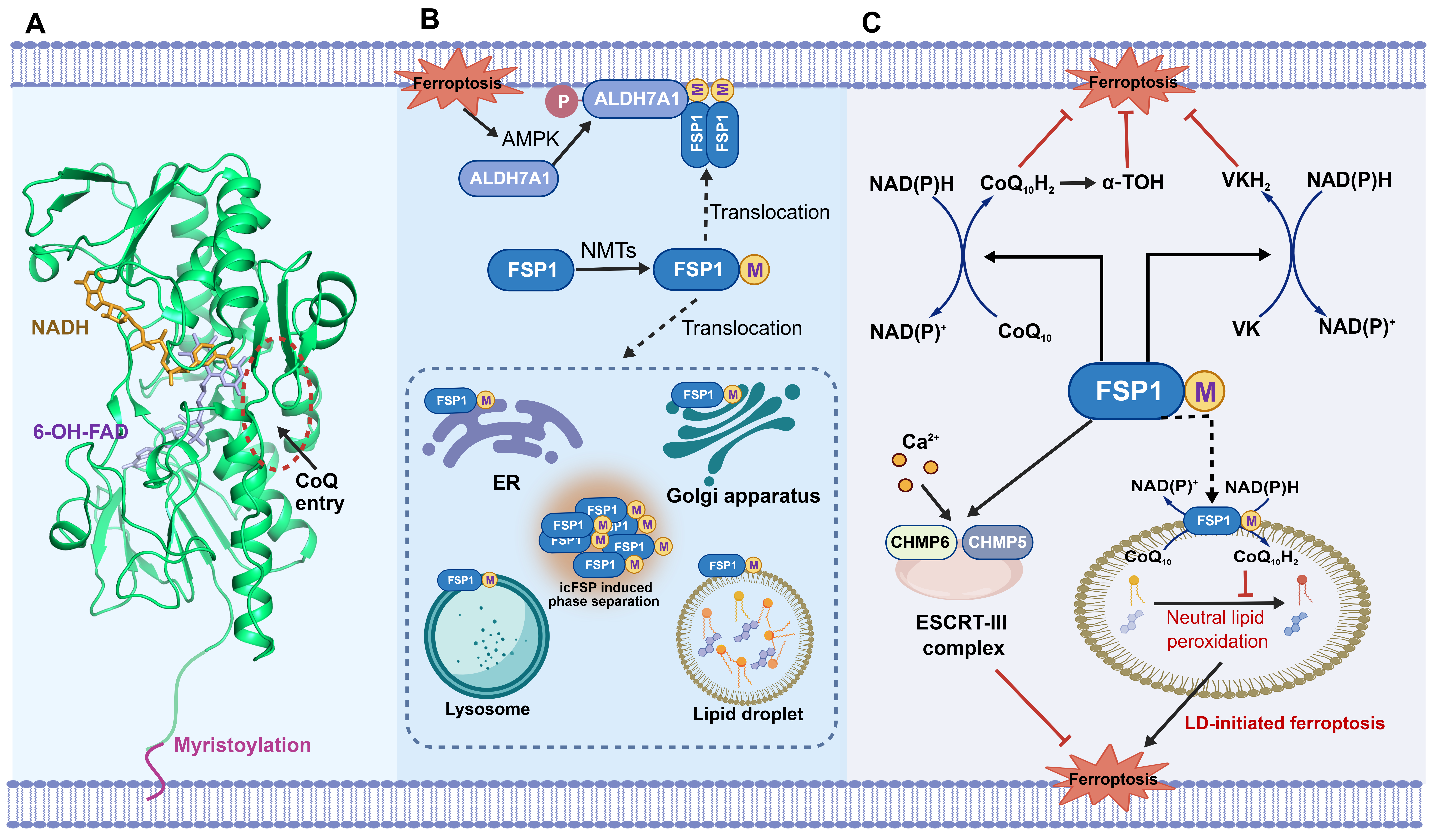
Figure 1. FSP1 structure, localization, and mechanisms of ferroptosis inhibition in cancer. (A) Structure of human FSP1 (PDB ID: 8WIK). N-terminal myristoylation motif (purple) mediates the membrane localization of FSP1. NADH (orange) and 6-OH-FAD (purple) are displayed as sticks. The CoQ substrate entry of FSP1 is indicated by a red dashed circle; (B) Subcellular localization of FSP1. FSP1 localizes to the plasma membrane and various organelle membranes, including the ER, Golgi apparatus, lysosomes, and lipid droplets, through its N-terminal myristoylation and interaction with phosphorylated ALDH7A1 under ferroptotic stress. FSP1 can also form biomolecular condensates through phase separation under specific conditions, including icFSP1 treatment and TMEM87A ablation-induced excessive acidification of the Golgi apparatus; (C) Molecular mechanisms of ferroptosis inhibition in cancer, including the FSP1/CoQ10/NAD(P)H axis, the FSP1/vitamin K/NAD(P)H axis, and the FSP1/ESCRT-III membrane repair pathway. In addition, FSP1 localizes to the surface of lipid droplets and inhibits the peroxidation of neutral lipids by reducing CoQ10 to ubiquinol, thereby protecting against ferroptosis triggered by lipid peroxidation in lipid droplets. Created with BioGDP (https://BioGDP.com)[154]. FSP1: ferroptosis suppressor protein 1; NADH: reduced nicotinamide adenine dinucleotide; CoQ: coenzyme Q; 6-OH-FAD: 6-hydroxy-flavin adenine dinucleotide; AMPK: AMP-activated protein kinase; ALDH7A1: aldehyde dehydrogenase 7 family member A1; TMEM87A: transmembrane protein 87A; NMTs: N-myristoyltransferases; ER: endoplasmic reticulum; α-TOH: alpha-tocopherol; VK: vitamin K; CHMP6: charged multivesicular body protein 6; CHMP5: charged multivesicular body protein 5; LD: lipid droplet; ESCRT-III: endosomal sorting complex required for transport-III.
Structurally, FSP1 contains three domains: an N-terminal hydrophobic membrane-binding domain (residues 1-27), an NADH oxidoreductase domain (residues 81-285), and a flavin adenine dinucleotide (FAD)-binding domain (residues 286-308). Its catalytic reductase activity is modulated by cofactors and substrates such as NAD(P)H, FAD, CoQ10, and vitamin K[33,34]. The N-terminal myristoylation motif constitutes a critical structural determinant for the anti-ferroptotic function of FSP1. This fatty acid modification, catalyzed by N-myristoyltransferases (NMTs), drives FSP1 recruitment to diverse subcellular membranes. When the FSP1 N-terminus undergoes a G2A mutation or when cells are treated with the NMT inhibitor IMP-1088, FSP1 loses its membrane-targeting capacity, and its anti-ferroptotic function is consequently abolished (Figure 1A)[7]. Notably, while myristoylation is crucial for membrane targeting, myristoylation alone is insufficient to ensure stable membrane anchorage of FSP1. Evidence indicates that the dimerization of FSP1, likely through a dual myristoyl anchor structure, may enhance its binding stability on cellular membranes[35]. At a more refined regulatory level, upon elevated ferroptotic stress, the membrane recruitment of FSP1 is promoted via its interaction with the phosphorylated form of aldehyde dehydrogenase 7A1 (ALDH7A1) in cancer cells. Ferroptotic stress activates the AMPK pathway, which facilitates ALDH7A1 translocation to cellular membranes, thereby providing a platform for FSP1 membrane residence (Figure 1B). In certain contexts, this mechanism not only couples FSP1 activity with upstream metabolic signals but also offers new insights into the regulatory dynamics of FSP1 distribution on specific membrane compartments, including the plasma membrane, the Golgi complex, and lipid droplets[36].
2.1.2 FSP1 phase separation under specific conditions
FSP1 contains intrinsically disordered regions that drive liquid-liquid phase separation (LLPS), enabling the formation of condensates under specific conditions[37]. Recent studies have shown that Golgi membrane lipid peroxidation during early ferroptosis disturbs Golgi luminal acid-base homeostasis. The Golgi-resident transmembrane protein TMEM87A maintains FSP1 function by buffering Golgi luminal pH; knockdown of TMEM87A causes Golgi hyperacidification, driving phase-separated FSP1 condensates formation and concomitant loss of its enzymatic activity[38]. Similarly, the human FSP1-specific inhibitor icFSP1 induces FSP1 dissociation from lipid membranes and triggers its condensation, altering subcellular localization and abrogating its ability to scavenge phospholipid-derived free radicals[37]. This provides a novel pharmacological strategy for FSP1 targeting, as phase separation-mediated loss of function occurs independently of direct enzymatic inhibition and may confer improved drug selectivity. Notably, although FSP1 has been observed to undergo phase separation and form condensates under specific conditions, such as treatment with icFSP1 or excessive acidification of the Golgi apparatus, little evidence supports the notion that endogenous FSP1 engages in phase separation as a regulatory mechanism under physiological conditions. Physicochemical factors such as pH and temperature are well-established modulators of protein phase separation. Whether FSP1 undergoes phase separation under other conditions and whether FSP1 phase separation acts as a mechanism to modulate FSP1 function and ferroptosis sensitivity via spatial distribution warrants further investigation.
In summary, FSP1 achieves spatial distribution plasticity through multiple mechanisms, including myristoylation, protein-protein interactions, and inhibitor-induced phase separation, thereby establishing a ferroptosis surveillance network with broad spatial coverage and localized response capacity across distinct cellular membrane compartments.
2.2 Molecular mechanisms of FSP1 in inhibiting ferroptosis
Beyond its canonical role in suppressing ferroptosis, FSP1 exerts diverse physiological functions. FSP1 is highly expressed in brown adipocytes, where, upon cold or β-adrenergic stimulation, it anchors to the outer leaflet of the inner mitochondrial membrane to promote oxygen consumption, uncoupled respiration, and thermogenesis[39]. Moreover, FSP1 can regulate caspase-independent apoptosis[40]. However, its ferroptosis-inhibitory activity is undoubtedly the central focus of current cancer research.
2.2.1 The FSP1/CoQ10/NAD(P)H antioxidant axis
FSP1 suppresses ferroptosis primarily through a three-pronged mechanism, the core of which is the FSP1/CoQ10/NAD(P)H antioxidant axis. Upon membrane recruitment, FSP1 binds NAD(P)H via its NAD(P)H-binding domain and transfers two electrons to its FAD-binding domain, generating FADH2. These electrons are subsequently passed to oxidized ubiquinone (CoQ10), reducing it to ubiquinol (CoQ10H2)[35,41]. As a highly potent lipophilic RTA, CoQ10H2 directly scavenges lipid peroxyl radicals, thereby halting the lipid peroxidation chain reaction and arresting ferroptotic progression. In addition, CoQ10H2 can indirectly regenerate α-tocopherol (vitamin E), another key membrane-resident lipophilic radical-trapping antioxidant, further reinforcing the membrane antioxidant defense network (Figure 1C)[42].
Notably, a recent study has extended the anti-ferroptotic function of FSP1 to lipid droplets (LDs), which are dynamic organelles that store neutral lipids. FSP1 localizes to the LD surface and suppresses peroxidation of neutral lipids, including triacylglycerols and cholesterol esters, by reducing CoQ10 to ubiquinol[43]. When FSP1 function is compromised, LDs enriched with PUFAs can initiate a LD-mediated ferroptosis pathway (Figure 1C). This finding expands the anti-ferroptotic role of FSP1 from membrane phospholipid bilayers to stored lipids, adding a new dimension to its regulation of global cellular lipid redox homeostasis.
2.2.2 The FSP1/vitamin K/NAD(P)H pathway
Beyond CoQ10, FSP1 also possesses vitamin K reductase catalytic activity. Mishima et al. demonstrated that FSP1 utilizes NAD(P)H to reduce vitamin K forms, including phylloquinone, menaquinone-4, and menadione, to vitamin K hydroquinone (VKH2). VKH2 serves as a potent RTA that effectively blocks lipid peroxidation (Figure 1C). In both Gpx4-deficient mouse embryonic fibroblasts and GPX4-null human cancer cell lines, all three forms of vitamin K can suppress ferroptosis, and this protective effect is diminished by FSP1 inhibition[44].
The FSP1/CoQ10 axis and the FSP1/VK axis collectively constitute the functional landscape of the multi-substrate, multi-pathway anti-ferroptosis activity of FSP1. Both pathways use NAD(P)H as the source of reducing equivalents and generate lipophilic RTAs as end products. These two axes may exert redundant yet complementary protection for cell membranes against lipid peroxidation across distinct tissue microenvironments and metabolic contexts.
2.2.3 The FSP1/ESCRT-III membrane repair pathway
Beyond regenerating antioxidants to chemically neutralize lipid peroxidation, FSP1 also counteracts ferroptosis by promoting physical membrane repair. During ferroptosis progression, nanoscale pores form in the plasma membrane, leading to ion imbalance and water influx[45]. The ESCRT-III-dependent endosomal sorting complex, an evolutionarily conserved protein complex with membrane scission machinery, plays a critical role in repairing plasma membrane lesions triggered by multiple forms of regulated cell death, including ferroptosis, necroptosis, and pyroptosis[46-48].
Studies have revealed that FSP1 facilitates ESCRT-III recruitment to sites of the damaged plasma membrane in a CoQ10-independent manner, thereby enhancing cellular membrane reparative capacity[49]. Ferroptosis inducers such as RSL3 and erastin drive the accumulation of core ESCRT-III subunits CHMP5 and CHMP6 at the plasma membrane through ER stress signaling and calcium influx-mediated cascades (Figure 1C)[50]. FSP1 knockdown suppresses this process, whereas overexpression of CHMP5 rescues the ferroptosis-sensitive phenotype in FSP1-silenced cells. Genetic suppression of ESCRT-III has been shown to sensitize tumors to ferroptosis in immunodeficient mice and to enhance the antitumor activity of RSL3.
These findings indicate that FSP1 provides an additional layer of ferroptosis protection via the ESCRT-III membrane repair mechanism. Importantly, the FSP1/ESCRT-III pathway also operates in normal somatic cells and immune cells[51]. Therefore, when FSP1 is pharmacologically targeted, rigorous assessment is needed to prevent off-target disruption of homeostatic membrane repair in non-malignant cells.
2.2.4 Interplay between the FSP1 pathway and other ferroptosis defense systems
As previously described, multiple anti-ferroptosis systems operate in concert within cells, and functional interactions exist among them. FSP1 and GPX4 constitute the most important parallel and complementary anti-ferroptosis systems. When GPX4 is inhibited by RSL3, FSP1 expression is compensatorily upregulated, conferring adaptive resistance to GPX4 inhibitors in tumor cells[52]. Conversely, the FSP1 inhibitor iFSP1 enhances the ferroptosis-inducing effect of GPX4 inhibitors across various cancer cell lines[29]. This negative feedback crosstalk suggests a tight interplay between the two systems in maintaining redox homeostasis. Further elucidation of the coordination mechanisms between FSP1 and GPX4 may help reverse tumor cell resistance to ferroptosis by disrupting this negative feedback crosstalk.
Dihydroorotate dehydrogenase (DHODH) is an enzyme located on the mitochondrial inner membrane. It reduces CoQ to CoQH2, which functions as an RTA that neutralizes phospholipid peroxyl radicals (PLOO•), thereby inhibiting ferroptosis[53]. FSP1 and DHODH share the substrate CoQ10 but operate at the plasma membrane and mitochondrial inner membrane, respectively. Studies have shown that in FSP1-knockout cells, the DHODH inhibitor brequinar fails to induce ferroptosis despite normal DHODH expression, indicating that the ferroptosis-inducing effect of brequinar is primarily achieved through FSP1 inhibition rather than DHODH inhibition[53].
VKORC1L1 is an endoplasmic reticulum-resident transmembrane protein that exerts its anti-ferroptotic function by reducing vitamin K to VKH2[54]. Both FSP1 and VKORC1L1 can utilize vitamin K as a substrate but are localized to the plasma membrane and the endoplasmic reticulum, respectively[44]. They may act synergistically to resist lipid peroxidation within distinct subcellular compartments. Whether a substrate competition relationship exists between FSP1 and VKORC1L1 warrants further investigation.
2.3 Mechanisms of FSP1 regulation
The expression and activity of FSP1 are under the precise control of an intricate regulatory network that operates at multiple levels, including transcriptional, post-transcriptional, post-translational, and metabolic regulation. These regulatory layers not only maintain the basal expression of FSP1 under physiological conditions but also dynamically remodel FSP1 function during tumor progression, microenvironmental stress, and the emergence of drug resistance.
2.3.1 Transcriptional regulation
The transcriptional regulation of FSP1 centers on the NRF2-KEAP1 signaling axis. Under unstressed conditions, NRF2 is constitutively ubiquitinated and degraded via binding to KEAP1, thereby maintaining low basal NRF2 levels. When cells encounter oxidative stress or when KEAP1 undergoes loss-of-function mutations, which are particularly prevalent in lung adenocarcinoma, NRF2 escapes degradation and translocates to the nucleus. There, it binds to antioxidant response elements (AREs) to activate the coordinated transcription of a battery of antioxidant genes, including FSP1[55]. In KEAP1-deficient lung cancer cells, FSP1 is among the most highly upregulated genes driven by NRF2. Knocking out NRF2 significantly reduces FSP1 expression and enhances ferroptosis sensitivity[56]. Beyond oxidative stress, oncogenic KRAS mutations co-upregulate FSP1 transcription through their downstream mitogen-activated protein kinase (MAPK) signaling pathway and the concomitant activation of NRF2, directly linking oncogenic drive to redox defense (Figure 2A)[24].
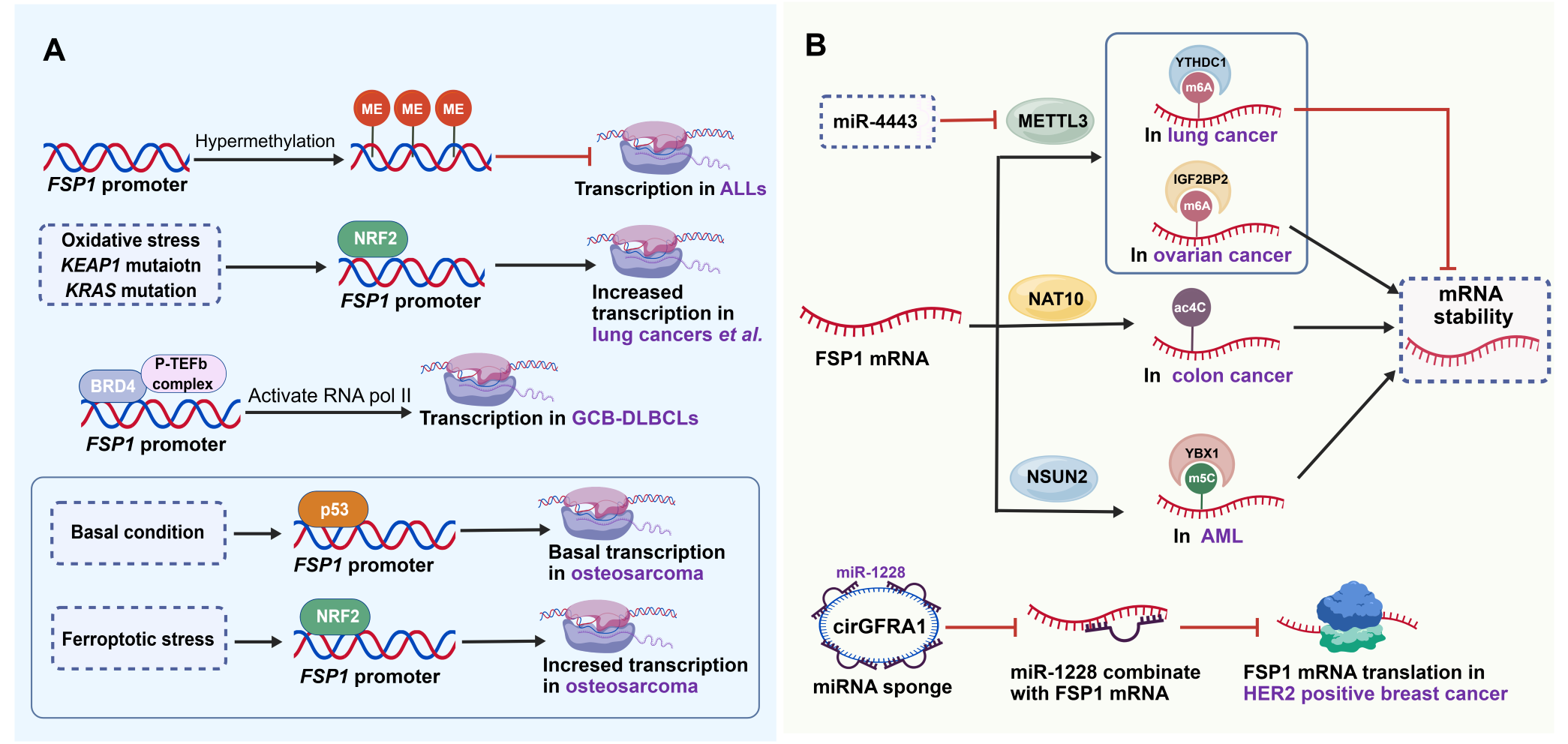
Figure 2. Transcriptional and post-transcriptional regulation of FSP1 in cancer. (A) Transcriptional regulation of FSP1 in cancer. FSP1 transcription is regulated by multiple mechanisms. DNA methylation within the FSP1 promoter suppresses its transcription. Under specific conditions, transcription factors such as NRF2, BRD4, and p53 initiate FSP1 transcription; (B) METTL3-catalyzed m⁶A modification, NAT10-catalyzed ac4C modification, and NSUN2-mediated RNA m⁵C methylation all modulate FSP1 mRNA stability, thereby modulating the sensitivity of cancer cells to ferroptosis. In addition, non-coding RNAs (miRNAs and circRNAs) also regulate FSP1 mRNA abundance via multiple mechanisms. Created with BioGDP (https://BioGDP.com)[154]. KEAP1: kelch-like ECH-associated protein 1; KRAS: Kirsten rat sarcoma viral oncogene homolog; ALL: acute lymphoblastic leukemia; BRD4: bromodomain-containing protein 4; P-TEFb: positive transcription elongation factor b; RNA pol II: RNA polymerase II; GCB-DLBCLs: germinal center B-cell-like diffuse large B-cell lymphoma; p53: tumor protein p53; NRF2: nuclear factor erythroid 2-related factor 2; METTL3: methyltransferase-like 3; YTHDC1: YTH domain-containing protein 1; IGF2BP2: insulin-like growth factor 2 mRNA-binding protein 2; NAT10: N-acetyltransferase 10; m6A: N6-methyladenosine; ac4C: N4-acetylcytidine; NSUN2: NOP2/Sun RNA methyltransferase 2; m5C: 5-methylcytidine; YBX1: Y-box binding protein 1; AML: acute myeloid leukemia; HER2: human epidermal growth factor receptor 2.
The classical tumor suppressor p53 also participates in the transcriptional regulation of FSP1 in a context-dependent manner. In HEK293 cells, either by overexpression of p53 or by treatment with the DNA-damaging agent 5-fluorouracil (which induces endogenous p53), p53 can bind to the FSP1 (also known as AMID) promoter to activate its transcription[57]. The activation of p53 in response to radiation-induced DNA damage can enhance the expression of FSP1 in human lymphoblast cell lines[58]. A study found that ATF6 regulates severe acute pancreatitis and concurrent multiple organ injury via p53/AIFM2 (FSP1)-dependent apoptosis[59]. In osteosarcoma cells, p53 governs basal FSP1 expression levels, while NRF2 mediates FSP1 upregulation under pro-oxidative or pro-ferroptotic conditions (Figure 2A)[60]. Notably, nutlin-mediated activation of p53 failed to increase FSP1 expression in osteosarcoma cell lines[7]. These seemingly contradictory observations indicate that the regulation of FSP1 by p53 is likely to be context-dependent. Whether p53 can affect ferroptosis through transcriptional regulation of FSP1 expression needs further investigation. This exemplifies the complex, context-dependent dual role of p53 in ferroptosis regulation.
Epigenetic modifications act as a “switch” in the transcriptional regulation of FSP1. In acute lymphoblastic leukemia (ALL) cells, the CpG island in the FSP1 promoter is hypermethylated, leading to near-complete suppression of FSP1 transcription. Studies have shown that ubiquitin-like with PHD and RING finger domains 1 (UHRF1) recognizes and recruits DNA methyltransferase 1 (DNMT1) to the FSP1 promoter, resulting in DNA hypermethylation and epigenetic silencing of FSP1, which promotes ferroptosis in alveolar type 2 epithelial cells[61]. In contrast to the silencing mechanism, the transcriptional activator BRD4 in germinal center B-cell-like diffuse large B-cell lymphoma (GCB-DLBCL) directly binds to the FSP1 promoter and upregulates FSP1 mRNA levels by recruiting the positive transcription elongation factor b (P-TEFb) complex to enhance RNA polymerase II activity (Figure 2A)[62,63].
2.3.2 Post-transcriptional regulation
The processing, modification, and stability of FSP1 mRNA are intricately regulated by diverse post-transcriptional mechanisms. RNA modifications play a key role in determining FSP1 mRNA fate. METTL3-catalyzed N6-methyladenosine (m6A) modification regulates FSP1 expression by affecting mRNA stability, yet its effect exhibits dual specificity depending on cell type and context. In aortic smooth muscle cells, METTL3 promotes FSP1 mRNA degradation, thereby facilitating ferroptosis[64]. Conversely, in glioma, METTL3-mediated m6A modification stabilizes FSP1 mRNA, inhibiting ferroptosis and promoting tumor progression[65]. This discrepancy may arise from differential recognition of m6A sites on FSP1 mRNA by distinct reader proteins, such as YTHDF2 and YTHDC1. YTHDC1 is considered a negative post-transcriptional regulator of FSP1. In lung cancer cells, YTHDC1 recognizes m6A sites in the 3′ untranslated region of FSP1 mRNA, promoting the formation of a truncated, unstable mRNA isoform and thereby reducing FSP1 protein levels[66]. As an m6A reader protein, insulin-like growth factor 2 mRNA-binding protein 2 (IGF2BP2) recognizes the m6A modification on FSP1 mRNA, thereby enhancing its stability. The IGF2BP2–FSP1 axis ultimately mediates cisplatin resistance in ovarian cancer and hepatocellular carcinoma (HCC) progression (Figure 2B)[67,68].
In contrast, N4-acetylcytidine (ac4C) modification, catalyzed by N-acetyltransferase 10 (NAT10), positively regulates FSP1 expression. In colorectal cancer, NAT10 enhances the stability and translation efficiency of FSP1 mRNA through ac4C modification, thereby increasing ferroptosis resistance and tumor metastatic capacity[69,70]. Similarly, NSUN2 mediates RNA 5-methylcytosine (m5C) methylation on FSP1 mRNA, which subsequently enhances the recognition and stability of FSP1 mRNA through the m5C reader protein YBX1. Consequently, the NSUN2-YBX1-FSP1 signaling axis confers ferroptosis resistance in acute myeloid leukemia (AML) cells (Figure 2B)[71].
Non-coding RNAs (ncRNAs) play a critical role in the fine-tuning of FSP1 expression. In HCC, the long non-coding RNA (lncRNA) lncFAL directly binds to the FSP1 protein and competitively inhibits TRIM69-dependent ubiquitination and degradation of FSP1, thereby enhancing FSP1 protein stability and ferroptosis resistance[72]. In breast cancer, circular RNA circGFRA1 acts as a molecular sponge for miR-1228, relieving miR-1228-mediated translational inhibition of FSP1 mRNA and promoting FSP1 expression and malignant progression of HER2-positive breast cancer[73]. Exosome-mediated intercellular communication extends the regulatory role of ncRNAs from the intracellular space to the tumor microenvironment. Exosomal miR-4443, derived from cisplatin-resistant non-small cell lung cancer (NSCLC) cells, can be delivered to cisplatin-sensitive cells. It upregulates FSP1 protein expression by inhibiting METTL3, thereby reducing the m6A modification level of FSP1 mRNA and ultimately conferring cisplatin resistance to the recipient cells (Figure 2B)[74]. Similarly, exosomal miR-130a-3p derived from drug-resistant esophageal cancer cells targets METTL14, thereby inhibiting the m6A methylation of FSP1 in recipient cells and consequently mediating cisplatin resistance[75]. These findings provide important clues for understanding the intercellular transmission of chemotherapy resistance within the tumor microenvironment.
2.3.3 Post-translational modification regulation
Tumor cells employ a combination of post-translational modifications (PTMs) to finely tune FSP1 protein abundance across different cancer types or stages of tumor progression. The ubiquitin-proteasome system constitutes a core PTM governing FSP1 protein stability, with multiple E3 ubiquitin ligases and deubiquitinases forming a sophisticated dynamic equilibrium network.
In HCC, the E3 ligase TRIM54 mediates K48-linked polyubiquitination and proteasomal degradation of FSP1. Sorafenib accelerates the interaction between TRIM54 and FSP1 through the ERK pathway, thereby promoting FSP1 degradation and enhancing chemosensitivity[76]. TRIM69 similarly catalyzes the ubiquitination and degradation of FSP1, while lncFAL competitively blocks this process by directly binding to FSP1, forming a convergence point of post-translational and post-transcriptional regulation[72]. In contrast, the E3 ligase TRIM21 catalyzes K63-linked ubiquitination of FSP1, facilitating its transport to the plasma membrane and enhancing its ferroptosis-protective function[77]. In gastric cancer, the E3 ubiquitin ligase SMURF1 has been identified as another FSP1-ubiquitinating enzyme, with USP29 acting as a counteracting deubiquitinase; they jointly regulate FSP1 ubiquitination and chemoresistance[78]. In intrahepatic cholangiocarcinoma, MUC1 recruits Src kinase to phosphorylate the deubiquitinase USP10, enhancing its deubiquitination of FSP1 and thus stabilizing the protein. Simultaneously, Src-mediated phosphorylation of NMT1 promotes FSP1 myristoylation and membrane localization, synergistically activating the FSP1/CoQ10 pathway through theses dual mechanisms (Figure 3)[79].
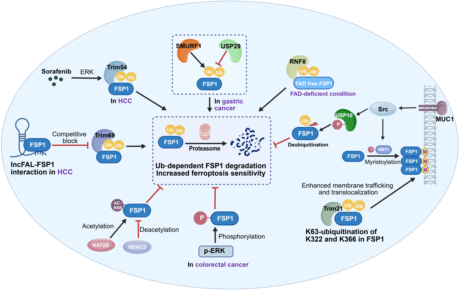
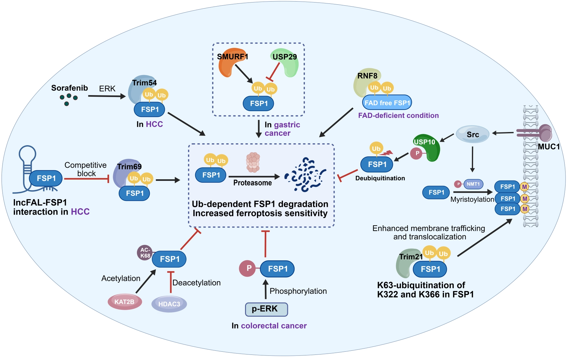
Figure 3. Post-translational modification regulation of FSP1 in cancer. The ubiquitin-proteasome system represents a core PTM mechanism that governs FSP1 protein stability. The E3 ubiquitin ligases TRIM54, TRIM69, TRIM21, SMURF1, and RNF8 each mediate the ubiquitination of FSP1, promoting FSP1 degradation and increasing the sensitivity of tumor cells to ferroptosis. In contrast, USP29 and USP10 catalyze FSP1 deubiquitination, thereby stabilizing FSP1 protein. Furthermore, acetylation and phosphorylation of FSP1 suppress its ubiquitination and subsequent degradation, conferring ferroptosis resistance in tumors. Created with BioGDP (https://BioGDP.com)[154]. HCC: hepatocellular carcinoma; ERK: extracellular signal-regulated kinase; TRIM54: tripartite motif containing 54; Ub: ubiquitin; SMURF1: SMAD specific E3 ubiquitin protein ligase 1; USP29: ubiquitin specific peptidase 29; RNF8: ring finger protein 8; FAD: flavin adenine dinucleotide; Src: SRC proto-oncogene, non-receptor tyrosine kinase; MUC1: mucin 1, cell surface associated; USP10: ubiquitin specific peptidase 10; NMT1: N-myristoyltransferase 1; TRIM21: tripartite motif containing 21; LncFAL: long non-coding RNA ferroptosis associated lncRNA; TRIM69: tripartite motif containing 69; KAT2B: lysine acetyltransferase 2B; PTM: post-translational modification; FSP1: ferroptosis suppressor protein 1; HDAC3: histone deacetylase 3; p-ERK: phosphorylated extracellular signal-regulated kinase.
A recent study identified the E3 ubiquitin ligase RNF8 as a quality-control factor that specifically recognizes the “empty” FSP1 protein lacking the FAD cofactor and mediates its ubiquitination and degradation. When intracellular riboflavin is deficient or FAD synthesis is impaired, the pool of cofactor-unbound FSP1 increases, and its degradation is accelerated via RNF8-mediated ubiquitination, leading to a subsequent decrease in FSP1 protein levels (Figure 3)[80,81]. Thus, RNF8 serves as a bridge between “metabolic sensing” and the “protein quality control system”. This finding directly couples FSP1 protein stability with upstream riboflavin/FAD metabolic status.
Acetylation also regulates FSP1 stability. Cytosolic acetyl-coenzyme A (acetyl-CoA), generated through the ATP-citrate lyase (ACLY)-catalyzed citrate metabolism pathway, sustains FSP1 acetylation at lysine 168. This modification inhibits its recognition by β-TrCP and subsequent degradation via the ubiquitin-proteasome pathway. KAT2B and HDAC3 mediate the acetylation and deacetylation of this site, respectively, forming a “writer-eraser” pair for acetylation regulation. Knockdown of the mitochondrial citrate carrier SLC25A1, an upstream component of the acetyl-CoA metabolic pathway, depletes the cytosolic acetyl-CoA pool, reduces FSP1 acetylation, and enhances its degradation and ferroptosis sensitivity (Figure 3)[82]. This complete molecular chain, from mitochondrial carbon metabolism to FSP1 acetylation and finally to ferroptosis susceptibility, directly links cellular carbon metabolism with antioxidant defense.
Recently, phosphorylation has also been found to participate in FSP1 regulation. In colorectal cancer, downregulation of the creatine transporter SLC6A8 leads to insufficient cellular creatine uptake, which activates ERK2 to phosphorylate FSP1 at threonine 109. This phosphorylation stabilizes the FSP1 protein and enhances ferroptosis resistance[83]. Creatine supplementation inhibits downstream FSP1 phosphorylation, promotes ferroptosis, and potentiates the efficacy of immune checkpoint inhibitors (Figure 3).
2.3.4 Regulation of FSP1 subcellular localization
The intracellular propagation of ferroptosis follows a highly compartmentalized pattern. Lipid oxidative damage typically originates in membranous organelles such as the endoplasmic reticulum (ER), lysosomes, or lipid droplets, and then spreads directionally to other organelles and the plasma membrane (Figure 1B). Lipid peroxidation within lysosomes increases lysosomal membrane permeability, leading to iron release and the initiation of widespread intracellular lipid peroxidation. The ER serves as a central hub for the accumulation and signal amplification of peroxidized lipids, while mitochondria receive oxidative signals through mitochondria–ER contact sites (MERCs) and act as “amplifiers”[2]. In this propagation cascade, the subcellular localization and phase separation state of FSP1 exhibit dynamically adaptive features under specific conditions, determining the spatial precision with which it executes local antioxidant protection across different membranes.
As discussed above, the subcellular localization of FSP1 is coordinately regulated by multiple mechanisms. Myristoylation serves as the fundamental prerequisite for membrane anchoring, and its efficiency is dually controlled by NMT activity and the availability of the substrate myristoyl-CoA[7]. K63-linked ubiquitination catalyzed by TRIM21 facilitates FSP1 transport to the plasma membrane[77]. Under ferroptotic stress, stable residence of FSP1 on specific membranes is reinforced through its interaction with phosphorylated ALDH7A1. ALDH7A1 can catalyze the production of membrane-bound NADH, providing a localized cofactor supply for FSP1 (Figure 4)[36]. This spatial coupling of cofactor metabolism and antioxidant execution substantially enhances the efficiency of FSP1.
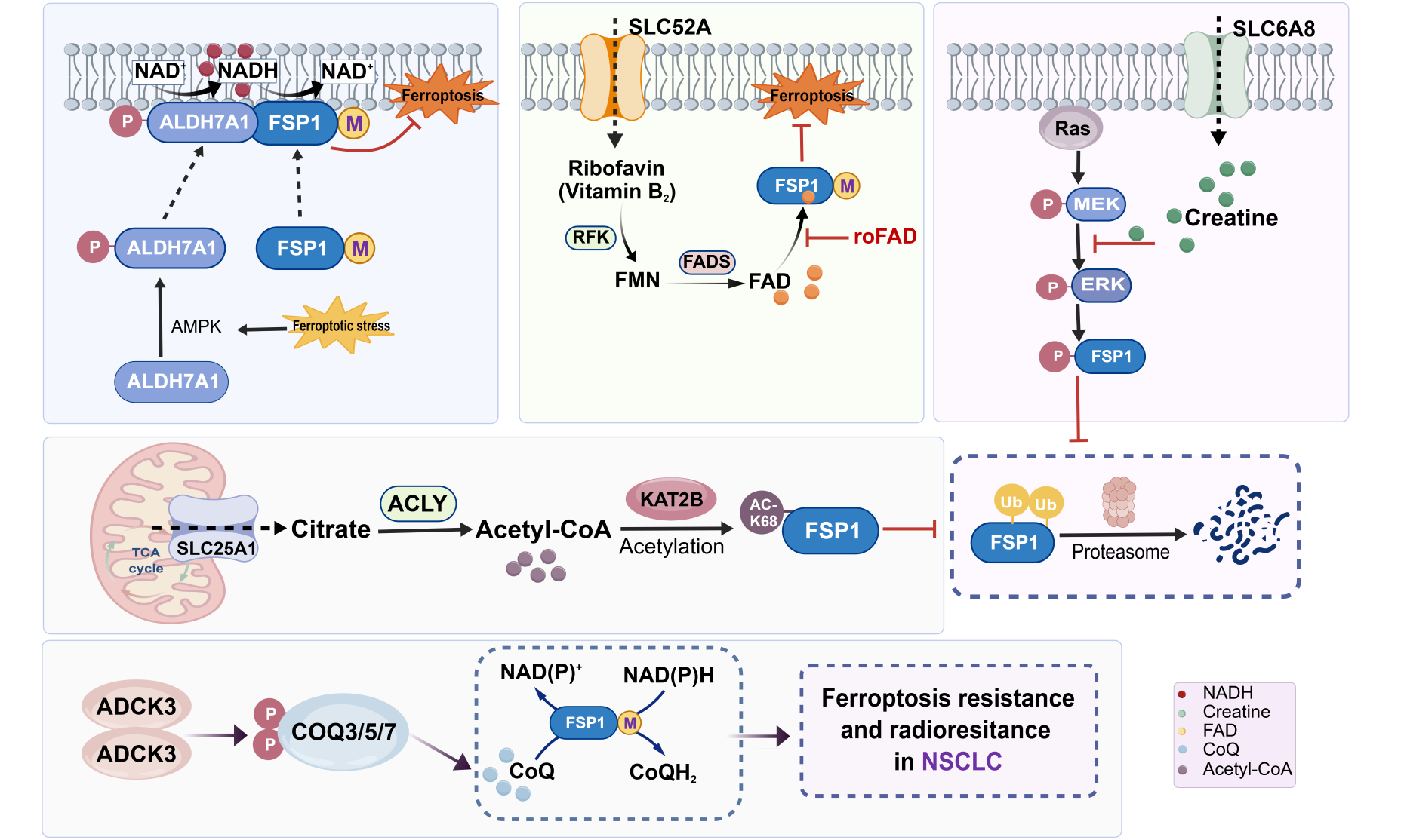
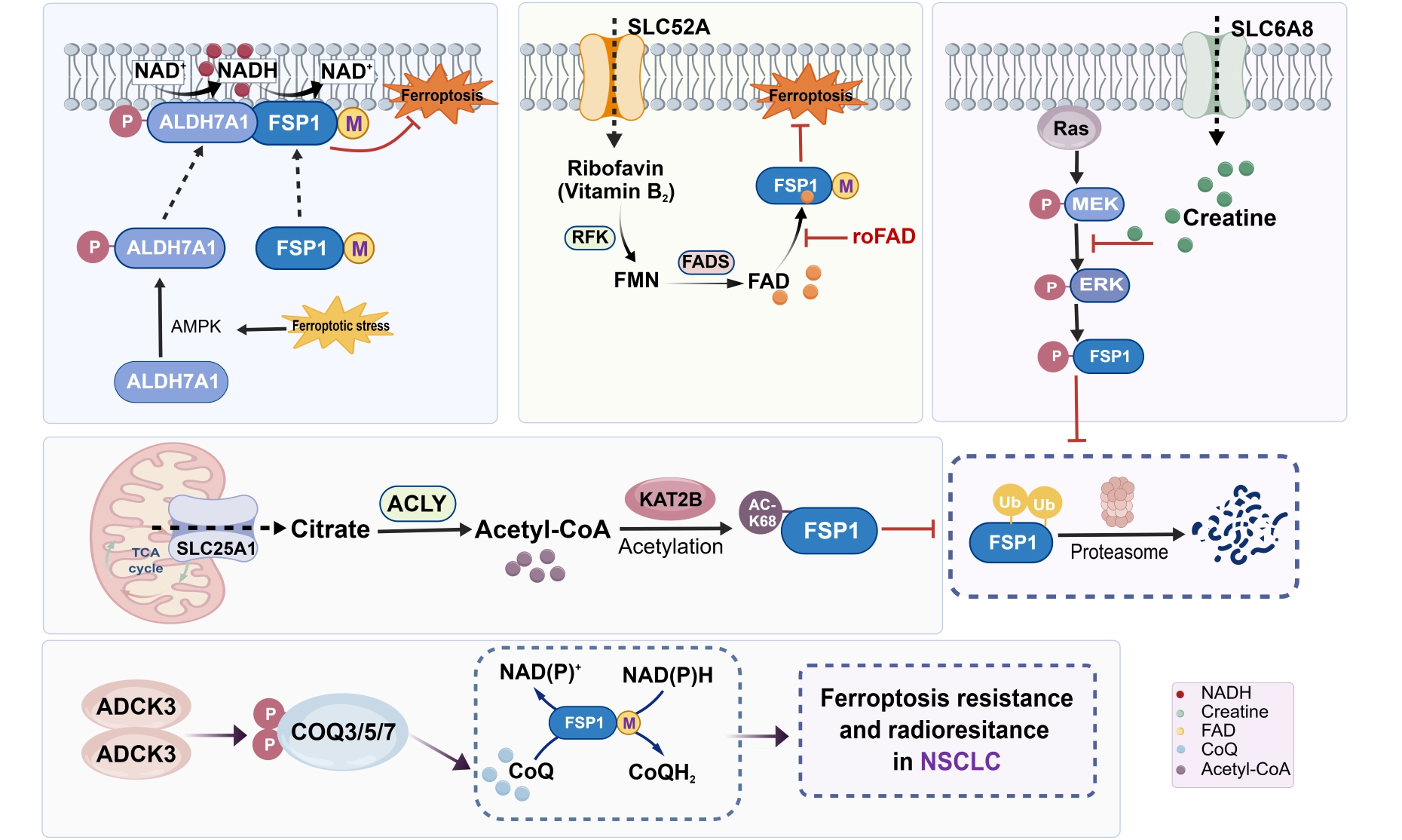
Figure 4. Regulation of FSP1 by metabolic molecules in cancer. Metabolic molecules, including NADH, FAD, CoQ, creatine, and acetyl-CoA, precisely modulate the subcellular distribution, stability, and enzymatic activity of FSP1 through multiple mechanisms, including cofactor incorporation, cofactor supply, substrate availability, and PTMs. Created with BioGDP (https://BioGDP.com)[154]. FSP1: ferroptosis suppressor protein 1; NADH: reduced nicotinamide adenine dinucleotide; PTM: post-translational modifications; ALDH7A1: aldehyde dehydrogenase 7 family member A1; AMPK: AMP-activated protein kinase; SLC52A: solute carrier family 52 member; RFK: riboflavin kinase; FMN: flavin mononucleotide; FADS: flavin adenine dinucleotide synthetase; FAD: flavin adenine dinucleotide; roFAD: roseoflavin adenine dinucleotide; SLC6A8: solute carrier family 6 member 8; Ras: rat sarcoma viral oncogene homolog; MEK: mitogen-activated protein kinase kinase; ERK: extracellular signal-regulated kinase; SLC25A1: solute carrier family 25 member 1; TCA cycle: tricarboxylic acid cycle; ACLY: ATP-citrate lyase; Acetyl-CoA: acetyl coenzyme A; KAT2B: lysine acetyltransferase 2B; AC-K68: acetylated lysine 68; ADCK3: aarF domain containing kinase 3; COQ3/5/7: coenzyme Q3/5/7; NSCLC: non-small cell lung cancer.
Under physiological conditions, FSP1 at sites of membrane lipid peroxidation efficiently eliminates local lipid peroxyl radicals and promotes membrane repair, thereby inhibiting ferroptosis. However, when phase separation is induced by drugs or abnormal pH, it can instead lead to FSP1 inactivation and thus promote ferroptosis. The FSP1 inhibitor icFSP1 triggers FSP1 dissociation from membranes and induces its phase separation. This non-physiological condensation likely alters FSP1 conformation and spatial distribution, impeding its function in scavenging phospholipid radicals[37]. Regarding the chemical environment, the transmembrane protein TMEM87A prevents aberrant aggregation and inactivation of FSP1 on the Golgi membrane by maintaining Golgi pH homeostasis, indicating that local intracellular pH may be a critical chemical factor regulating FSP1 phase separation[38]. Notably, early Golgi membrane lipid oxidation during ferroptosis can itself lead to elevated Golgi luminal pH. Whether the condensed state of FSP1 constitutes a component of the cellular machinery that senses ferroptotic stress remains to be elucidated.
The subcellular localization of FSP1 also exhibits significant plasticity in different tumor microenvironments. In lymph node-metastatic melanoma cells, hypoxia within the lymph node drives FSP1 redistribution from a diffuse distribution across multiple membranes to concentrated perinuclear lysosomal localization[31]. This suggests that microenvironment-dependent redistribution of FSP1 may represent a spatial adaptation strategy by which tumor cells optimize local antioxidant defense, although the molecular mechanism and functional significance remain to be explored.
Given the differential distribution of FSP1 across various cellular membranes and its functional state switch induced by phase separation, it is plausible to hypothesize that the dynamics of FSP1 subcellular localization may represent a key node controlling the direction and efficiency of ferroptosis propagation between different intracellular compartments. Future research should dissect the mechanisms governing FSP1 trafficking between distinct membrane structures and how variations in organelle membrane lipid composition affect the local catalytic activity of FSP1.
2.3.5 Regulation of FSP1 by metabolic molecules
As an oxidoreductase, FSP1 requires cofactors such as NAD(P)H, FAD, or 6-hydroxy-FAD to maintain its enzymatic activity, and its catalytic substrates include metabolic molecules such as CoQ10 and vitamin K. The metabolic flux and local enrichment of these cofactors and substrates precisely tune the distribution, stability, and enzymatic activity of FSP1.
The antioxidant function of FSP1 depends on an adequate supply of the NADH cofactor. A recent study discovered that ALDH7A1 catalyzes the generation of membrane-bound NADH and directly supplies this cofactor to membrane-localized FSP1. Membrane-bound NADH and cytosolic soluble NADH likely belong to independent molecular pools. This compartmentalization ensures that FSP1 receives a sustained and efficient electron supply within the membrane environment without being perturbed by fluctuations in cytosolic NADH[36]. This finding reveals the unique safeguarding role of cofactor compartmentalization for the anti-ferroptotic function of FSP1 (Figure 4).
Riboflavin (vitamin B2) and its metabolite FAD constitute the most fundamental metabolic support for the structural stability and function of FSP1. When cellular FAD is deficient or the FAD-binding site on FSP1 is mutated, FSP1 misfolds and undergoes degradation via the RNF8-mediated ubiquitin-proteasome pathway. Riboflavin kinase (RFK) and FAD synthetase (FADS) are the key enzymes that catalyze the conversion of riboflavin to FAD; loss of their expression specifically impairs FSP1 function and broadly affects cellular ferroptosis sensitivity (Figure 4). Under physiologically relevant riboflavin-deficient conditions, both FSP1 expression levels and cellular ferroptotic phenotypes are significantly altered, indicating that slight fluctuations in riboflavin content are sufficient to perturb cellular lipid redox homeostasis[80,81]. Importantly, the riboflavin concentration in standard cell culture media far exceeds that of physiological human plasma, which maximizes FSP1 stability and likely masks the functional vulnerability of FSP1 under the lower riboflavin availability in vivo. This observation also suggests that the riboflavin metabolic status of individual patients may need to be considered when designing clinical trials for FSP1 inhibitor-based anti-cancer therapies.
Furthermore, as a direct substrate of FSP1, the cellular synthesis of coenzyme Q also directly impacts the functional output of FSP1. Statins block HMG-CoA reductase in the mevalonate pathway, thereby reducing the supply of CoQ synthesis precursors and indirectly inhibiting the FSP1 axis[84] (Figure 4). This mechanism also explains the clinical observation that combining statins with radiotherapy can restore radiosensitivity in radioresistant patients.
Acetyl-CoA, a central metabolite, bridges carbon metabolism and ferroptosis resistance by modulating FSP1 acetylation levels. The mitochondrial citrate carrier SLC25A1 transports citrate to the cytoplasm, where it is converted to acetyl-CoA by ACLY. An abundant acetyl-CoA pool sustains KAT2B-catalyzed FSP1 acetylation, thereby inhibiting its ubiquitination-mediated degradation (Figure 4). Knockdown of SLC25A1 or ACLY depletes the cytosolic acetyl-CoA pool, reduces FSP1 stability, and enhances ferroptosis sensitivity[82].
The energy buffer molecule creatine regulates ferroptosis sensitivity through the SLC6A8-creatine-ERK2-FSP1 axis. Creatine directly binds to the substrate-binding domain of ERK2, hindering its activation by MEK1. When downregulation of SLC6A8 leads to insufficient cellular creatine uptake, ERK2 is activated and phosphorylates FSP1 at threonine 109, stabilizing the FSP1 protein and thereby conferring ferroptosis resistance (Figure 4)[83]. Of note, creatine has been reported to exert dual effects, either antioxidant or pro-ferroptotic, which may be attributable to dose dependency, tumor type specificity, and microenvironmental context.
In summary, metabolic molecules modulate the functional state of FSP1 through multiple dimensions, including cofactor incorporation, cofactor supply, substrate availability, and PTMs. The spatial heterogeneity and temporal dynamics of metabolic molecule concentrations result in marked differences in FSP1 activity across different microenvironments in vivo. This not only explains why in vitro results are difficult to translate in vivo in certain contexts, but also provides a metabolic basis for defining precise therapeutic windows during FSP1-targeted interventions.
3. The Role of FSP1 in Cancer
3.1 FSP1 expression profile in cancer and prognostic value
FSP1 is overexpressed in a wide spectrum of malignant tumors, and its high expression is typically associated with poor clinical prognosis[85-87]. In HCC, approximately 37% of tumor specimens exhibit elevated FSP1 expression, which correlates with shorter overall survival[88]. In lung cancer, NRF2-driven transcriptional upregulation of FSP1 is particularly prominent in KRAS-mutant and KEAP1-mutant NSCLC. Moreover, FSP1 levels increase with tumor progression in human lung adenocarcinoma samples, whereas GPX4 levels remain relatively stable and show no significant correlation with patient survival[30]. In colorectal cancer (CRC), FSP1 upregulation is significantly associated with advanced TNM stage, lymph node metastasis, distant metastasis, and poor prognosis[89]. In pancreatic ductal adenocarcinoma (PDAC), tumors harboring activating KRAS mutations drive high FSP1 expression via the MAPK/NRF2 pathway[24]. In triple-negative breast cancer (TNBC), FSP1 expression is significantly higher than that in normal breast tissue and is linked to shorter survival and a higher risk of metastasis[90]. In specific subtypes of AML, both GPX4 and FSP1 are highly expressed, and high FSP1 levels indicate poor prognosis[91].
Nevertheless, contradictory cases have been reported. A study by Takahara et al. in lung adenocarcinoma found that low FSP1 expression was significantly associated with poorer recurrence-free survival[92]. A plausible explanation is that tumors with intrinsically low FSP1 expression may compensate by upregulating GPX4, SLC7A11, or other NRF2-driven antioxidant pathways, allowing aggressive growth to proceed despite the presence of ferroptosis vulnerability. This counterexample suggests that when evaluating FSP1 as a prognostic biomarker, a comprehensive assessment must integrate the tumor molecular subtype, the activity status of non-FSP1 ferroptosis defense pathways, and the abundance of ferroptosis-related metabolites in the tumor microenvironment.
The malignant phenotype-driving function of FSP1 extends beyond ferroptosis resistance to driving tumor metastasis. High FSP1 expression can stabilize NRF2 by maintaining a low lipid-ROS microenvironment. Nuclear NRF2 then directly upregulates the EMT transcription factors Snail and Slug, leading to loss of E-cadherin and induced expression of vimentin and N-cadherin, thereby endowing tumor cells with greater migratory and invasive capacity[93]. In HCC, high FSP1 expression can also promote mitochondrial biogenesis by activating the SIRT1/PGC-1α axis, enhancing the metabolic activity and metastatic potential of tumor cells in a ferroptosis-independent manner[88].
FSP1-mediated therapeutic resistance is one of its most important clinical characteristics. In colorectal cancer, high levels of FSP1 and GPX4 synergistically maintain the EMT program and shield cells from 5-fluorouracil-induced cytotoxicity[94]. In ovarian cancer, the NRF2-JAM3-FSP1 axis mitigates lipid peroxidation and mediates cross-resistance to cisplatin and poly(ADP-ribose) polymerase (PARP) inhibitors[95]. Elevated FSP1 levels similarly attenuates the antitumor efficacy of sorafenib in HCC and abiraterone in prostate cancer[96]. FSP1-dependent ferroptosis evasion also contributes to tumorigenesis and lenvatinib resistance in HCC[97]. Notably, FSP1 also promotes DNA damage repair through a ferroptosis-independent mechanism. In ovarian cancer cells, FSP1 physically interacts with Ku70, promoting DNA-PKcs-mediated non-homologous end joining (NHEJ) repair efficiency, thereby directly contributing to PARP inhibitor resistance[98]. This non-canonical function broadens the biological scope of FSP1 in mediating drug resistance and suggests that FSP1-targeted therapy could achieve more potent antitumor effects by simultaneously blocking two independent processes: ferroptosis suppression and proficient DNA damage repair.
3.2 Plasticity of FSP1 dependency in cancer
Tumor cell reliance on the FSP1 anti-ferroptotic pathway displays dynamic plasticity shaped by intrinsic tumor alterations and microenvironmental conditions. This plasticity not only profoundly influences the scope of FSP1’s applicability as a therapeutic target but also determines the optimal time windows for clinical intervention.
3.2.1 Microenvironmental discrepancies between in vitro and in vivo remodel FSP1 dependency
Under standard in vitro culture conditions, FSP1 knockout or pharmacological inhibition is typically insufficient to induce robust ferroptosis, indicating that FSP1 is largely dispensable in most cancer cell lines. However, in an in vivo genetically engineered mouse model of primary lung cancer, tumor-specific knockout of FSP1 yielded tumor-suppressive effects comparable to GPX4 ablation, and this phenotype was rescued by the ferroptosis inhibitor liproxstatin-1 or vitamin E[30]. A similar phenomenon has been validated in a melanoma lymph node metastasis model: the hypoxic conditions of the lymphatic microenvironment induce GPX4 degradation via the ubiquitin-proteasome pathway, depriving metastatic cells of GPX4-mediated antioxidant defense and rendering them highly FSP1-dependent. While FSP1 monoinhibition barely impaired the viability of lymph node-metastatic cells in vitro, FSP1 inhibitors significantly suppressed intranodal tumor expansion in vivo[31]. Consistent observations have also been documented in chromophobe renal cell carcinoma[87]. The divergent FSP1 dependency between in vitro and in vivo contexts arises from multiple layered mechanisms: (1) The in vivo tumor microenvironment imposes a greater oxidative stress burden, leading to persistent lipid peroxidation and forcing cancer cells to rely on FSP1 for antioxidant buffering; (2) Metabolite concentrations (e.g., supraphysiologic riboflavin levels) and oxygen tension in standard in vitro culture media deviate substantially from the native in vivo milieus, masking the true extent of FSP1 dependency; (3) The three-dimensional architecture and mechanical stress generated by cell-matrix interactions in vivo may exacerbate membrane damage upon ferroptosis, thereby elevating cellular dependence on the FSP1/ESCRT-III membrane repair cascade.
3.2.2 Microenvironment-driven remodeling of FSP1 dependency in metastasis
Tumor colonization at distinct anatomical metastatic niches is accompanied by profound microenvironmental shifts, resulting in adaptive remodeling of FSP1 dependency. Further work by De Palma et al. demonstrated that hypoxia, a hallmark of the lymph node microenvironment, accelerates GPX4 protein degradation via the ubiquitin-proteasome pathway, markedly lowering GPX4 protein abundance in lymph node-metastatic cells. In these cells, FSP1 expression is compensatorily upregulated, and the protein relocalizes to perinuclear lysosomes to exert compartmentalized antioxidant defense. The growth of FSP1-knockout melanoma cells within lymph nodes was markedly suppressed, with some cases even exhibiting complete tumor regression. By contrast, subcutaneously implanted tumors from the same origin displayed negligible responses to FSP1 ablation[31,99]. These findings clearly demonstrate that tumor cell dependence on FSP1 can be reshaped by the specific conditions of the metastatic microenvironment.
3.2.3 Remodeling of FSP1 dependency in acquired therapy resistance
During the acquisition of radioresistance in repeatedly irradiated NSCLC cells, the CoQ synthesis kinase ADCK3 is activated to boost CoQ biosynthesis, while SLC7A11 expression is suppressed upon radiation exposure, leading to diminished GSH synthesis. These alterations drive a shift in the ferroptosis defense machinery of radioresistant tumor cells from the GSH/GPX4 axis to the FSP1/CoQ axis[84]. In DTP cells derived from TNBC, GPX4 downregulation is accompanied by marked compensatory upregulation of FSP1. Inhibiting FSP1 alone can restore chemosensitivity in drug-resistant cells, whereas dual inhibition of GPX4 and FSP1 selectively triggers ferroptosis in proliferative DTP populations and eliminates residual disease[52]. Notably, under high ROS stress induced by cisplatin, ovarian tumor cells appear to preferentially utilize the USP18-IGF2BP2-FSP1 bypass pathway rather than the canonical USP18-SLC7A11 pathway to evade ferroptosis[67]. These defense mode switches generate a “vulnerable therapeutic window” during which drug-tolerant lesions remain incompletely resistant. Targeting FSP1 within this window may suppress the outgrowth of resistant clones and eliminate residual disease.
3.2.4 FSP1 dependency in cancer stem cells
CSCs represent a core driver of tumor therapeutic resistance, recurrence, and metastasis. CSCs frequently display enhanced iron uptake driven by high TFRC expression and elevated LIP levels. This aberrant iron metabolism sustains stemness maintenance yet simultaneously renders CSCs more vulnerable to ferroptosis[25,100]. In A549 CD133+ populations (an in vitro cellular model of lung CSCs), dual inhibition of GPX4 and FSP1 potently triggers ferroptosis and impairs their self-renewal capacity[101]. TNBC exhibits the CD44+/CD24- phenotype of breast cancer stem cells (BCSCs) and harbors robust tumor-initiating capacity; its malignant progression is tightly correlated with breast cancer stemness features[102]. Elevated FSP1-dependent ubiquinone redox signaling confers ferroptosis resistance to TNBC; blockade of the FSP1/CoQ10 axis reverses this protective effect and restrains TNBC malignant progression[90]. Notably, NRF2 is highly expressed in cancer stem cells, and FSP1 functions as a direct downstream effector of NRF2[103]. Accordingly, it is reasonable to hypothesize that the FSP1/CoQ10 axis may also be transcriptionally upregulated in CSCs, which warrants further experimental validation.
In summary, FSP1 dependency is dynamically regulated across multiple regulatory layers, encompassing tumor-intrinsic traits (genetic alterations, disease stage, therapeutic resistance, stemness) and extrinsic microenvironmental cues (hypoxia, metabolite availability, lipid peroxidation load). For clinical translation, accurate stratification of FSP1 dependency across different tumor subtypes and progressive disease stages is crucial: this will facilitate the identification of patient cohorts most responsive to FSP1-targeted therapies and guide the selection of optimal therapeutic windows for FSP1 inhibitory intervention.
3.3 Impact of FSP1 inhibition on the tumor immune microenvironment
3.3.1 Bidirectional immunomodulation of ferroptosis in the TME
A complex bidirectional regulatory relationship exists between ferroptosis and the tumor immune microenvironment. Ferroptosis can either boost or suppress antitumor immune responses. The net outcome is highly dependent on the specific cell type undergoing ferroptosis, the progression stage of cell death, and the local microenvironment[104].
In terms of promoting antitumor immunity, ferroptosis has been confirmed to be immunogenic[2]. Prophylactic vaccination experiments by Efimova et al. demonstrated that early ferroptotic tumor cells treated with RSL3 promote the phenotypic maturation of bone marrow-derived dendritic cells, eliciting a vaccine-like effect in immunocompetent mice[105]. Damage-associated molecular patterns (DAMPs) released by ferroptotic cells, including high mobility group box 1 (HMGB1), ATP, and type I interferons, promote the maturation and recruitment of antigen-presenting cells[105-107]. Ferroptosis directly participates in T cell–mediated tumor killing: activated CD8+ T cells can trigger tumor cell ferroptosis through the IFN-γ pathway and ACSL4-coupled arachidonic acid metabolism, thereby coupling immune attack to ferroptotic death[108]. Activated CD8+ T cells also deplete intracellular GSH in tumor cells by inhibiting SLC7A11 mediated by IFN-γ to promote ferroptosis[109]. Common ferroptosis-inducing strategies, such as inhibiting GPX4 or FSP1, can enhance immune cell infiltration within the tumor microenvironment and exhibit synergistic effects with immune checkpoint inhibitors (ICIs)[110,111].
Despite the pro-antitumor immunogenic effects described above, ferroptosis can also have detrimental effects on antitumor immunity. Immune cells within the TME can themselves fall victim to ferroptosis. Under the same strain and genetic background, CD8+ T cells are markedly more sensitive to GPX4 inhibitors than B16 melanoma cells[112,113]. T cells scavenge oxidized lipids from the TME via CD36, which can induce lipid peroxidation and functional exhaustion[114,115]. T cells from mice with T-cell–specific GPX4 knockout rapidly accumulate abundant lipid peroxides upon activation and subsequently undergo ferroptosis[116]. Follicular helper T cells (Tfh) and regulatory T cells (Tregs) also exhibit pronounced sensitivity to GPX4 loss[117,118]. The differential sensitivity of various immune cell populations to ferroptosis creates distinct therapeutic opportunities in oncology. Selectively inducing ferroptosis in Tregs can mitigate their immunosuppressive activity and enhance antitumor responses[118]. Moreover, immunosuppressive M2-type tumor-associated macrophages (TAMs) are relatively sensitive to ferroptosis, and ferroptotic stimulation can reprogram them toward a pro-inflammatory, antitumor M1 phenotype[119-121]. These nuanced, cell-type-specific responses underscore the complexity of ferroptosis-mediated immune regulation and provide a theoretical basis for cell-subtype-targeted ferroptosis-inducing strategies.
The dual immune effects of ferroptosis reflect its multifaceted roles in the tumor immune microenvironment, highlighting that spatiotemporal precision (which cell type, when, and where) is critical for combining ferroptosis induction with immunotherapy.
3.3.2 Immunomodulatory advantages of FSP1 targeting
Compared with GPX4 targeting, targeting FSP1 offers unique advantages in preserving antitumor immunity. First, cell death induced by FSP1 deficiency is primarily a tumor cell-intrinsic response that does not induce significant alterations in major immune cell populations[122]. Second, recent studies have identified FSP1 as a key molecule enabling Tregs to resist ferroptosis and sustain immunosuppression. In mouse models, systemic deletion of FSP1 in T cells, or its specific deletion in Tregs, enhances tumor control by selectively impairing the immunosuppressive capacity of intratumoral Tregs without triggering autoimmune pathology[123]. Furthermore, FSP1 inhibition indirectly upregulates the expression of chemokines including CXCL9 and CX3CL1, thereby boosting the recruitment of dendritic cells, macrophages, and T cells into tumors[51]. Multiple studies have confirmed the synergistic therapeutic potential of combining FSP1 inhibitors with immune checkpoint blockade therapy[111,124,125].
4. Therapeutic Strategies Targeting FSP1 in Cancer
Targeting FSP1 to induce ferroptosis offers several theoretical advantages as an antitumor strategy. First, it presents a superior therapeutic safety window: FSP1 is highly expressed in many malignant tumors, whereas Fsp1 knockout mice are viable and fertile with no apparent pathological phenotype, suggesting that FSP1 targeting is far less toxic to normal tissues than GPX4 inhibition. Second, certain tumor cells exhibit context-specific dependency on FSP1. Under in vivo microenvironmental conditions such as hypoxia and elevated lipid peroxidation burden, this dependency is markedly enhanced, creating a favorable therapeutic window for targeting metastatic and therapy-resistant tumors[30,31]. Third, FSP1 inhibitors display a better immune safety profile compared with GPX4 inhibitors, making them particularly suitable for combination with immunotherapy. As of early 2026, several FSP1 inhibitors are in preclinical development for antitumor therapy.
4.1 Inhibitors targeting FSP1
4.1.1 Specific FSP1 inhibitors
iFSP1 is the first reported human FSP1-specific inhibitor[29]. It binds to the F360 residue of FSP1 via aromatic π-π stacking interactions, competitively inhibiting its enzymatic activity[126]. However, iFSP1 is active only against human FSP1 and not mouse FSP1, which limits its application in mouse models (Table 1)[126]. Recent studies have used iFSP1 as a scaffold to design and synthesize proteolysis-targeting chimeras (PROTAC) degraders targeting FSP1, such as compound 2307[127].
Table 1. Summary of specific small-molecule FSP1 inhibitors.
| Compound name | Molecular mechanism | Species specificity | Refs. |
| iFSP1 | Competitive inhibitor; binds to the quinone-binding pocket. | human FSP1 | [29,126] |
| viFSP1 | Non-competitive inhibitor; binds to the NAD(P)H-binding pocket. | human FSP1, mouse FSP1 | [126] |
| FSEN1 | Structural modeling indicates competitive binding to the CoQ-binding pocket, though initial kinetic assays classified it as a non-competitive inhibitor. | human FSP1 | [128,129] |
| icFSP1 | Induces FSP1 dissociation from lipid membranes and triggers phase separation into condensates without directly suppressing its enzymatic activity. | human FSP1 | [31,37] |
| Roseoflavin | Competitively interferes with FAD biosynthesis, deprives FSP1 of its essential cofactor, and triggers ubiquitin-proteasome-mediated degradation of FSP1. | human FSP1 | [80,81] |
| Temsirolimus | Directly binds to and inhibits FSP1 enzymatic activity. | human FSP1 | [130] |
| NPD4928 | Directly binds to and inhibits FSP1. | human FSP1 | [131] |
| Compound 39 | Directly binds to and inhibits FSP1. | human FSP1 | [132] |
| Salidroside | Directly inhibits FSP1 activity. | human FSP1 | [133] |
| Quinolinone alkaloids | Directly inhibit FSP1 activity. | human FSP1 | [134] |
FSP1: ferroptosis suppressor protein 1; NAD(P)H: nicotinamide adenine dinucleotide (phosphate); CoQ: coenzyme Q; FAD: flavin adenine dinucleotide.
viFSP1, a versatile FSP1 inhibitor, non-competitively binds to the NAD(P)H-binding pocket of FSP1 and effectively inhibits both human and mouse FSP1. In multiple human and mouse cancer cell lines, combining viFSP1 with the GPX4 inhibitor RSL3 synergistically enhances ferroptosis induction[126].
FSEN1 and its analogs were initially identified as non-competitive inhibitors of FSP1 via enzymatic kinetic assays. The corresponding work proposed that FSEN1 binds to FSP1 only after FSP1 has pre-bound NADH and CoQ, forming an ineffective complex and halting the catalytic cycle by locking the enzyme-substrate complex. Nevertheless, subsequent structural modeling of the FSP1-FSEN1 complex revealed that FSEN1 occupies the CoQ-binding pocket, indicating that it acts as a competitive inhibitor[128]. The reason for the discrepancy remains elusive. FSEN1 exhibits favorable pharmacokinetic profiles and metabolic stability in mice. The structurally diverse panel of FSP1 inhibitors (FSEN1-19) provides a rich toolbox of candidate molecules for FSP1 inhibition-based anticancer therapies[129].
icFSP1, a 3-phenylquinazolinone compound, represents a new strategy for FSP1 inhibition. Rather than directly blocking enzymatic activity, icFSP1 triggers FSP1 to dissociate from lipid membranes and undergo phase separation into condensates, thereby altering its subcellular localization. In vivo, icFSP1 significantly suppresses tumor growth and elevates 4-hydroxynonenal (4-HNE). Compared with iFSP1, it exhibits greater metabolic stability and enhanced in vivo efficacy[37]. However, icFSP1 shows no significant activity in a lymph node-metastatic melanoma model, possibly because FSP1, having relocalized to perinuclear lysosomes under this condition, acquires functional independence from condensate formation[31]. This also suggests that the applicability of different FSP1 inhibitors may require individualized selection based on the subcellular localization state of FSP1 within a given microenvironment.
As elaborated above, riboflavin metabolism is critical for maintaining FSP1 activity. Recent research has identified roseoflavin as an antimetabolite of riboflavin. By mimicking the structure of riboflavin, roseoflavin competitively interferes with FAD synthesis, depriving FSP1 of its essential cofactor and leading to its ubiquitin-proteasome-mediated degradation, thereby promoting ferroptosis[80,81]. A unique advantage of roseoflavin lies in its bioactivity at nanomolar concentrations. Moreover, because its activation relies on the conserved enzymatic machinery of the riboflavin metabolic pathway, tumor cells can hardly develop resistance to roseoflavin without compromising their own riboflavin metabolism.
The toolbox of FSP1 inhibitors has expanded considerably in recent years. The marketed drug temsirolimus directly inhibits the enzymatic activity of FSP1, sensitizing HCC cells to ferroptosis[130]. NPD4928 is a novel compound that directly binds and inhibits FSP1, sensitizing cancer cells to ferroptosis; in PANC-1 cells, NPD4928 induces ferroptosis more effectively than FSP1 knockout[131]. Recent studies have also generated a series of triazolothiadiazole derivatives with FSP1 inhibitory activity. Among them, compound 39 exhibits potent activity, effectively increasing intracellular lipid peroxide accumulation and enhancing sensitivity to ferroptosis in various tumor cells[132]. Additionally, several natural compounds, including salidroside and quinolinone alkaloids derived from Evodia rutaecarpa fruit extracts, have been reported to directly inhibit FSP1 activity and thereby suppress tumor progression[133,134].
4.1.2 Indirect FSP1 inhibitors
Statins indirectly suppress the FSP1/CoQ axis by inhibiting HMG-CoA reductase in the mevalonate pathway, thereby reducing the supply of CoQ synthesis precursors[90,135]. Sorafenib promotes the interaction between TRIM54 and FSP1 via the ERK pathway, accelerating FSP1 ubiquitination and degradation[76]. At high concentrations, the DHODH inhibitor brequinar induces ferroptosis by FSP1 suppression instead of DHODH inhibition[53]. Propofol, a commonly used intravenous anesthetic, enhances the sensitivity of cervical cancer cells to paclitaxel chemotherapy by inhibiting the FSP1 pathway[136]. TOMM40, a physical binding partner of FSP1, may inhibit FSP1 activity by altering its mitochondrial localization, representing a potential novel target for indirect intervention[137,138].
A variety of natural products also inhibit FSP1 through distinct mechanisms. Astragaloside IV downregulates FSP1 expression via the FTO/YTHDF2/m⁶A signaling axis to induce ferroptosis in gastric cancer cells[139]. The oridonin derivative XD suppresses HCC progression in mice by inhibiting the G6PD/PGD/FSP1/DHODH axis[140]. Curcumin induces ferroptosis in colorectal cancer and lung cancer stem cells through dual inhibition of the GSH/GPX4 and FSP1/CoQ10/NAD(P)H pathways[94,101]. Tanshinone I promotes ferroptosis and suppresses cervical cancer progression by regulating the ATF3–FSP1 axis[141]. The ginsenoside derivative (20S)-protopanaxatriol (20(S)-APPT) directly targets plasma membrane-localized FSP1 and, when combined with a γ-glutamylcysteine synthetase inhibitor, induces ferroptosis in drug-resistant cancer cells[142]. Qiling decoction reduces abiraterone resistance in prostate cancer cells by inhibiting FSP1[143].
4.1.3 Nanosystem delivery strategies targeting FSP1
Nano-delivery systems offer a promising strategy to overcome the challenges of poor selectivity, insufficient stability, and unsatisfactory pharmacokinetics associated with FSP1 inhibitors. Their core advantages include the ability to co-deliver multiple agents for simultaneous multi-pathway blockade, achieve tumor-targeted enrichment via responsive drug release, and synergize with immunotherapy.
In a colon cancer model, a photoactive nano-formulation encapsulating FSP1 siRNA and the photosensitizer verteporfin in lipid nanoparticles exhibited potent anti-tumor efficacy and robust immune activation[144]. Another study combined a plasma membrane-targeting photosensitizer, TBzT-CNQi, with iFSP1 to trigger immunogenic ferroptosis in tumors. Cell membrane-targeted sonodynamic therapy (CM-SDT) precisely targets the tumor cell membrane and accumulates therapeutic agents at that site. By combining the plasma membrane-targeting sonosensitizers TBT-CQi and TBzT-CNQi with iFSP1, the resulting ultrasound-responsive nanoparticle TiF completely suppressed orthotopic tumor growth, activated systemic anti-tumor immunity, and inhibited lung metastasis[145].
Copper–silk fibroin–rosuvastatin nanoparticles, Cu-SF(RSV) NPs, operate through a dual mechanism: rosuvastatin disrupts the mevalonate pathway to block the FSP1/CoQ axis, while Cu2+ triggers the Fenton reaction to generate ROS and deplete GSH, synergistically inducing ferroptosis in TNBC[90]. The Au/Cu-TCPP(Fe)@RSL3–PEG–iRGD nanosystem employs gold nanoparticles with glucose oxidase-mimicking activity to deplete glucose and disrupt the pentose phosphate pathway, thereby inhibiting both the FSP1/CoQ10 and GSH/GPX4 axes[146]. A BODIPY-modified polyamide nanosystem leverages its light-responsive properties to achieve spatiotemporally controlled release of ferroptosis inducers within tumors. This platform combines dual FSP1 and GPX4 inhibition with the photosensitizer Ce6, significantly enhancing the sensitivity of anti-PD-L1 immunotherapy[124].
4.2 Combination strategies targeting FSP1 for cancer therapy
4.2.1 Dual inhibition of FSP1 and GPX4
As FSP1 and GPX4 constitute the two major ferroptosis defense pathways in tumor cells, their dual inhibition represents the most straightforward strategy to overcome ferroptosis resistance (Table 2). In certain in vivo microenvironments such as lung cancer or melanoma lymph node metastases, tumor cells exhibit critical dependence on FSP1, and single-agent FSP1 inhibition can thus achieve significant therapeutic efficacy. Nevertheless, cancer cells in distinct tumor regions or metastatic sites may retain high GPX4 expression and remain refractory to FSP1 monotherapy[42]. In an H460 NSCLC xenograft model, only dual knockout of GPX4 and FSP1 inhibited tumor growth[7]. Thus, combined targeting of GPX4 and FSP1 blocks compensatory signaling feedback loops, ensuring that cancer cells cannot evade ferroptosis regardless of the pathway they depend upon. In multiple cancer cell lines, FSP1 knockout significantly potentiates RSL3-induced ferroptosis[29]. In TNBC, combined inhibition of FSP1 and GPX4 specifically induces ferroptosis in proliferative DTP cells[52]. However, dual inhibition of FSP1 and GPX4 may increase cumulative toxicity to normal tissues that depend on both proteins. Tumor-selective delivery via nanocarriers or intermittent dosing regimens may represent feasible approaches to widen the therapeutic window for combined pathway inhibition.
Table 2. Summary of combination strategies targeting FSP1 for cancer therapy.
| Combination strategy | Combination partner(s) | Mechanism of synergy | Refs. |
| Dual FSP1/GPX4 inhibition | GPX4 inhibitor (RSL3) + FSP1 inhibitor or FSP1 genetic ablation | Simultaneous blockade of parallel ferroptosis defensive pathways prevents compensatory antioxidant signaling. | [7,29,52] |
| Combination with radiotherapy | Radiotherapy + FSP1 inhibitor or statins | Metabolic shift toward FSP1/CoQ dependence drives tumor radioresistance; FSP1 inhibition restores ferroptotic susceptibility and radiosensitivity. | [56,84] |
| Combination with chemotherapy and targeted therapy | Chemotherapy (cisplatin, gemcitabine, 5-FU, paclitaxel); targeted agents (sorafenib, olaparib, TKIs, KRAS inhibitors) | FSP1 inhibition or sorafenib-induced FSP1 degradation restores tumor chemosensitivity; co-administration of PARP and FSP1 inhibitors exploits DNA repair deficiency. | [67,74,76,93,98,149] |
| Combination with immunotherapy | Anti-PD-1/PD-L1 antibodies + FSP1 inhibition | Ferroptosis induction in tumor cells provokes antitumor immunity; ferroptosis induction in immunosuppressive macrophages amplifies antigen presentation and CTL infiltration. | [111,150,151] |
CTL: cytotoxic T lymphocyte; CoQ: coenzyme Q; DNA: deoxyribonucleic acid; FSP1: ferroptosis suppressor protein 1; GPX4: glutathione peroxidase 4; 5-FU: 5-fluorouracil; KRAS: Kirsten rat sarcoma viral oncogene homolog; PARP: poly(ADP-ribose) polymerase; PD-1: programmed cell death protein 1; PD-L1: programmed death-ligand 1; RSL3: Ras-selective lethal 3; TKI: tyrosine kinase inhibitor.
4.2.2 Combination of FSP1 inhibitors with radiotherapy
Radiation therapy can directly induce ferroptosis[147,148]. However, during the acquisition of radioresistance in NSCLC, tumor cells undergo metabolic reprogramming. Specifically, ADCK3-driven upregulation of CoQ synthesis, together with SLC7A11 downregulation-induced reduction in GSH synthesis, jointly drives the reprogramming of ferroptosis defense machinery from GSH/GPX4 dependence to FSP1/CoQ dependence. Consequently, FSP1 inhibition can restore their radiosensitivity. Retrospective clinical analyses have shown that statins inhibit the FSP1 pathway by blocking CoQ synthesis, and that combining statins with radiotherapy effectively restores tumor radiosensitivity in patients with acquired radioresistance[84]. Another study also found that the FSP1/CoQ axis mediates ferroptosis resistance and radiation resistance in KEAP1-deficient lung cancer cells. Pharmacological inhibition of the FSP1/CoQ axis sensitizes KEAP1-deficient lung cancer cells and patient-derived xenograft tumors to radiation by inducing ferroptosis[56]. Future translational studies should prospectively validate the optimal schedule (sequential or concurrent) for administering FSP1 inhibitors during or after radiotherapy, and explore whether the extent of radiotherapy-induced defense pathway switching can serve as a predictive biomarker for FSP1-targeted radiosensitization.
4.2.3 Combination of FSP1 inhibitors with chemotherapy and targeted therapy
Chemotherapy resistance in tumors is closely associated with upregulation of FSP1 expression. In NSCLC, exosomal miR-4443 derived from cisplatin-resistant tumor cells can be transferred to sensitive cells, where it upregulates FSP1 expression by inhibiting METTL3, thereby conferring cisplatin resistance on the recipient cells[74]. In gemcitabine-resistant pancreatic cancer, Snail-driven FSP1 upregulation sustains the drug-resistant phenotype. FSP1 inhibition restores sensitivity to various chemotherapeutic agents, including gemcitabine in pancreatic cancer, 5-fluorouracil in colorectal cancer, and paclitaxel in cervical and ovarian cancers[67,93].
Regarding resistance to targeted therapy, sorafenib enhances its own efficacy in HCC by promoting TRIM54-mediated ubiquitination and degradation of FSP1[76]. The PARP inhibitor olaparib, when combined with an FSP1 inhibitor, exerts synergistic lethal effects through a non-ferroptosis-dependent DNA repair deficiency mechanism in BRCA-proficient ovarian cancer[98]. In tumors harboring KRAS or EGFR mutations, FSP1 is a key component of the NRF2-driven drug resistance program. Combining FSP1 inhibition with tyrosine kinase inhibitors (TKIs) or KRAS inhibitors may help overcome drug resistance[149].
4.2.4 Combination of FSP1 inhibitors with immunotherapy
Among the various combination strategies, pairing FSP1 inhibitors with ICIs is particularly promising and holds high clinical translational potential. Given the high drug resistance of HCC and the poor response to ICIs in most patients, combining FSP1 inhibitors with anti-PD-1/PD-L1 antibodies may synergistically prolong patient survival. However, ferroptosis in specific immune cell subsets can also impair anti-tumor immunity. Thus, combining FSP1 inhibition with immunotherapy demands a delicate balance: sufficient ferroptosis in tumor cells to provoke anti-tumor immunity, yet avoiding collateral damage to effector immune cells. Precise delivery of FSP1 inhibitors to tumor cells or to immunosuppressive cells such as Tregs is a feasible strategy to enhance the efficacy and safety of FSP1 inhibitor–immunotherapy combinations[123]. A recent study constructed a nano-co-delivery system encapsulating sorafenib and the FSP1 inhibitor viFSP1. In a mouse model, this nanosystem simultaneously targeted HCC tumor cells and immunosuppressive macrophages, synergistically triggering ferroptosis and thereby promoting antigen presentation and cytotoxic T cell infiltration. Combining this strategy with anti-PD-L1 antibodies significantly suppressed HCC metastasis and recurrence[111]. Another work reports the fabrication of a ferroptosis-triggering nanoplatform LP-CaP@iBEFT. This nanosystem releases iFSP1, brequinar, erastin, and ferric ions in the acidic tumor microenvironment to induce ferroptosis. Combined administration of LP-CaP@iBEFT and anti-PD-L1 antibodies synergistically enhances T cell activation and CTL differentiation, reshapes immune mediator profiles, and strengthens anti-tumor immunity for TNBC treatment[150]. Another study developed a murine ferritin (mHFn)-based nanoplatform co-delivering RSL3 and iFSP1, termed mHFn@RSL3/iFSP1. This nanosystem simultaneously suppresses the GPX4 and FSP1 antioxidant defense axes, which effectively induces robust ferroptosis in tumor cells. The immunogenic ferroptosis triggered by mHFn@RSL3/iFSP1 further remodels the tumor immune microenvironment and markedly promotes the intratumoral infiltration of CD8+ T cells. Benefiting from the mutual synergistic amplification effect, the combination of mHFn@RSL3/iFSP1 treatment with PD-L1 immune checkpoint blockade elicits potent synergistic anti-tumor immune responses against colorectal cancer[151]. Compared with the combination regimens of GPX4 or SLC7A11 inhibitors plus ICIs, the research on the combination of FSP1 inhibitors and immunotherapy remains limited[152,153]. Given the advantages of FSP1, including favorable safety profiles and minimal impact on immune cells, further preclinical investigations are urgently required.
5. Conclusions and Outlook
As a GPX4-independent ferroptosis suppressor, FSP1 establishes a comprehensive anti-lipid peroxidation defense system within cellular membranes through the FSP1/CoQ10/NAD(P)H axis, the FSP1/vitamin K pathway, and the ESCRT-III membrane repair mechanism. Its expression and activity are dynamically regulated at transcriptional, translational, and post-translational levels, through subcellular localization, inhibitor-induced phase separation, and modulation of intracellular metabolites. Notably, FSP1 is highly expressed in diverse malignancies and correlates with poor prognosis, invasion, metastasis, and therapy resistance. Targeting FSP1 confers superior tumor selectivity and a broader therapeutic safety window compared with GPX4, with context-dependent tumor dependency in vivo, making it a promising anticancer target. Despite substantial advances, multiple challenges remain to be addressed for cancer therapy targeting FSP1.
From a pharmacological perspective, developing more selective, potent, and bioavailable FSP1 inhibitors is essential to minimize collateral damage to normal tissues. Moreover, the plasticity of FSP1 subcellular localization in the TME suggests that a “precision localization intervention” strategy (targeting specific FSP1 pools, e.g., lysosome-localized FSP1 or phase-separated condensates) could further enhance therapeutic selectivity. The discovery of icFSP1 as a phase separation-triggering agent pioneers this direction. Future studies should investigate whether different subcellularly localized FSP1 exhibits conformational differences and possess unique druggable sites.
The development of appropriate biomarkers is essential to guide the therapeutic window for FSP1-targeted therapy. Tumor dependency on FSP1 is dynamically remodeled by tumor stage, metastatic site, drug-resistance status, and microenvironmental conditions. Therefore, a multi-dimensional assessment system may integrate the following parameters: FSP1 protein and mRNA expression levels, downstream metabolite status (CoQ10/CoQ10H2 ratio), activity of non-FSP1 ferroptosis defense pathways (GPX4, SLC7A11, GCH1/BH4), mutation profiles (KEAP1/NRF2, KRAS, TP53), and lipidomic features (PUFA/MUFA ratio). Such a system would help identify patients most likely to benefit from FSP1-targeted strategies and determine the optimal timing for intervention.
The in vivo efficacy and safety of FSP1 inhibitors require rigorous validation. Their anti-tumor effects must be verified across a broader range of tumor types, including those with specific genotypes (e.g., KEAP1-mutant, KRAS-driven tumors) and drug resistance. Organ-specific vulnerabilities also warrant careful evaluation, as the kidneys, brain, liver, heart, and hematopoietic microenvironment may exhibit differential tolerance to FSP1 inhibition. Furthermore, the differential sensitivity of immune cell subsets to FSP1 inhibition demands systematic assessment to establish a safe dosage range that preserves anti-tumor immunity.
Given the compensatory interplay among anti-ferroptosis pathways, it remains to be clarified whether sustained FSP1 inhibition induces adaptive resistance to FSP1 inhibitors. Long-term FSP1 inhibition may act as a selective pressure, driving tumors to develop resistance through upregulation of alternative antioxidant pathways (e.g., the GSH/GPX4 system), acquisition of gain-of-function mutations in the FSP1 gene, or enhanced drug efflux. Combined inhibition of FSP1 and complementary anti-ferroptotic pathways may overcome acquired resistance to FSP1 inhibitors.
Acknowledgements
The authors used used DeepSeek V4, an AI-assisted tools for language polishing. No AI tools were used to generate content, data, or figures. The authors take full responsibility for the integrity and originality of the work. The authors are grateful to BioGDP.com for help with the preparation of figures in this paper.
Authors contribution
Lin P: Conceptualization, writing-original draft, writing-review & editing.
Ma JD: Investigation.
Chen ZN: Conceptualization, supervision.
Wu J: Conceptualization, supervision, writing-review & editing.
Conflicts of interest
The authors declare no conflicts of interest.
Ethical approval
Not applicable.
Consent to participate
Not applicable.
Consent for publication
Not applicable.
Availability of data and materials
Not applicable.
Funding
This study was supported by the National Natural Science Foundation of China 82270078 and 82402115, Top Team in Strategy of Sanqin Talent Special Support Program of Shaanxi Province, Youth Innovation Team of Shaanxi Province, “Interdisciplinary Integration Program” Discipline Boosting Plan of Tangdu Hospital (2025JCRH008).
Copyright
© The Author(s) 2026.
References
-
1. Hanahan D. Hallmarks of cancer: Then and now, and beyond. Cell. 2026;189(8):2254-2277.[DOI]
-
2. Zhan B, Lin XM, Chen P, Zhang JB, Guo YQ, Deng R, et al. Targeting ferroptosis pathways in cancer: Emerging molecular targets and therapeutic strategies. Ferroptosis Oxid Stress. 2025;2(1):202510.[DOI]
-
3. Conrad M, Strasser A, Jost PJ, Yuan J, Shao F, Vandenabeele P, et al. Cell death in cancer. Cell. 2026;189(8):2322-2356.[DOI]
-
4. Shah R, Shchepinov MS, Pratt DA. Resolving the role of lipoxygenases in the initiation and execution of ferroptosis. ACS Cent Sci. 2018;4(3):387-396.[DOI]
-
5. Dixon SJ, Lemberg KM, Lamprecht MR, Skouta R, Zaitsev EM, Gleason CE, et al. Ferroptosis: An iron-dependent form of nonapoptotic cell death. Cell. 2012;149(5):1060-1072.[DOI]
-
6. Yang WS, SriRamaratnam R, Welsch ME, Shimada K, Skouta R, Viswanathan VS, et al. Regulation of ferroptotic cancer cell death by GPX4. Cell. 2014;156(2):317-331.[DOI]
-
7. Bersuker K, Hendricks JM, Li Z, Magtanong L, Ford B, Tang PH, et al. The CoQ oxidoreductase FSP1 acts parallel to GPX4 to inhibit ferroptosis. Nature. 2019;575(7784):688-692.[DOI]
-
8. Mao C, Liu X, Zhang Y, Lei G, Yan Y, Lee H, et al. DHODH-mediated ferroptosis defence is a targetable vulnerability in cancer. Nature. 2021;593(7860):586-590.[DOI]
-
9. Soula M, Weber RA, Zilka O, Alwaseem H, La K, Yen F, et al. Metabolic determinants of cancer cell sensitivity to canonical ferroptosis inducers. Nat Chem Biol. 2020;16(12):1351-1360.[DOI]
-
17. Martinez-Outschoorn UE, Peiris-Pagés M, Pestell RG, Sotgia F, Lisanti MP. Cancer metabolism: A therapeutic perspective. Nat Rev Clin Oncol. 2017;14(1):11-31.[DOI]
-
26. Ubellacker JM, Dixon SJ. Prospects for ferroptosis therapies in cancer. Nat Cancer. 2025;6(8):1326-1336.[DOI]
-
29. Doll S, Freitas FP, Shah R, Aldrovandi M, da Silva MC, Ingold I, et al. FSP1 is a glutathione-independent ferroptosis suppressor. Nature. 2019;575(7784):693-698.[DOI]
-
30. Wu K, Vaughan AJ, Bossowski JP, Hao Y, Ziogou A, Kim SM, et al. Targeting FSP1 triggers ferroptosis in lung cancer. Nature. 2026;649(8096):487-495.[DOI]
-
32. Dixon SJ, Olzmann JA. The cell biology of ferroptosis. Nat Rev Mol Cell Biol. 2024;25(6):424-442.[DOI]
-
33. Zhang S, Gou S, Zhang Q, Yong X, Gan B, Jia D, et al. FSP1 oxidizes NADPH to suppress ferroptosis. Cell Res. 2023;33(12):967-970.[DOI]
-
37. Nakamura T, Hipp C, Santos Dias Mourão A, Borggräfe J, Aldrovandi M, Henkelmann B, et al. Phase separation of FSP1 promotes ferroptosis. Nature. 2023;619(7969):371-377.[DOI]
-
42. Li W, Liang L, Liu S, Yi H, Zhou Y. FSP1: A key regulator of ferroptosis. Trends Mol Med. 2023;29(9):753-764.[DOI]
-
46. Jimenez AJ, Maiuri P, Lafaurie-Janvore J, Divoux S, Piel M, Perez F, et al. ESCRT machinery is required for plasma membrane repair. Science. 2014;343(6174):1247136.[DOI]
-
52. Chaudhary N, Mandal D, Choudhary BS, Patra S, Jain D, Poonia P, et al. Low-GPX4 drives a sustained drug-tolerant persister state in TNBC by a targetable adaptive FSP1 upregulation. Redox Biol. 2025;87:103864.[DOI]
-
60. Panczyszyn E, Saverio V, Monzani R, Gagliardi M, Petrovic J, Stojkovska J, et al. FSP1 is a predictive biomarker of osteosarcoma cells’ susceptibility to ferroptotic cell death and a potential therapeutic target. Cell Death Discov. 2024;10:87.[DOI]
-
65. Bu C, Hu S, Yu J, Li N, Gu J, Sheng Z, et al. Fear stress promotes glioma progression through inhibition of ferroptosis by enhancing FSP1 stability. Clin Transl Oncol. 2023;25(5):1378-1388.[DOI]
-
77. Gong J, Liu Y, Wang W, He R, Xia Q, Chen L, et al. TRIM21-promoted FSP1 plasma membrane translocation confers ferroptosis resistance in human cancers. Adv Sci. 2023;10(29):2302318.[DOI]
-
85. Kawade G, Kurata M, Matsuki Y, Fukuda S, Onishi I, Kinowaki Y, et al. Mediation of ferroptosis suppressor protein 1 expression via 4-hydroxy-2-nonenal accumulation contributes to acquisition of resistance to apoptosis and ferroptosis in diffuse large B-cell lymphoma. Lab Invest. 2024;104(4):102027.
-
91. Ma Z, Ye W, Huang X, Li X, Li F, Lin X, et al. The ferroptosis landscape in acute myeloid leukemia. Aging. 2023;15(22):13486-13503.[DOI]
-
98. Miao H, Meng H, Zhang Y, Chen T, Zhang L, Cheng W, et al. FSP1 inhibition enhances olaparib sensitivity in BRCA-proficient ovarian cancer patients via a nonferroptosis mechanism. Cell Death Differ. 2024;31(4):497-510.[DOI]
-
100. Lei G, Zhuang L, Gan B. Targeting ferroptosis as a vulnerability in cancer. Nat Rev Cancer. 2022;22(7):381-396.[DOI]
-
102. Yang N, Zhou TC, Lei XX, Wang C, Yan M, Wang ZF, et al. Inhibition of sonic hedgehog signaling pathway by thiazole antibiotic thiostrepton attenuates the CD44+/CD24-stem-like population and sphere-forming capacity in triple-negative breast cancer. Cell Physiol Biochem. 2016;38(3):1157-1170.
-
103. Wang H, Zhang Z, Ruan S, Yan Q, Chen Y, Cui J, et al. Regulation of iron metabolism and ferroptosis in cancer stem cells. Front Oncol. 2023;13:1251561.[DOI]
-
106. Newton K, Strasser A, Kayagaki N, Dixit VM. Cell death. Cell. 2024;187(2):235-256.[DOI]
-
108. Liao P, Wang W, Wang W, Kryczek I, Li X, Bian Y, et al. CD8+ T cells and fatty acids orchestrate tumor ferroptosis and immunity via ACSL4. Cancer Cell. 2022;40(4):365-378.e6.[DOI]
-
109. Wang W, Green M, Choi JE, Gijón M, Kennedy PD, Johnson JK, et al. CD8+ T cells regulate tumour ferroptosis during cancer immunotherapy. Nature. 2019;569(7755):270-274.[DOI]
-
111. Tang C, He C, Wang D, Guo J, Yin X, Ye H, et al. Co-delivery of sorafenib and an FSP1 inhibitor triggers dual ferroptosis in tumor cells and immunosuppressive macrophages for enhanced immunotherapy in mouse models of hepatocellular carcinoma. Nat Commun. 2025;16:10096.[DOI]
-
113. Kłopotowska M, Baranowska I, Hajduk S, Jurga A, Leśniowska N, Łaźniewski M, et al. GPX4 is a key ferroptosis regulator orchestrating T cells and CAR-T-cells sensitivity to ferroptosis. Cancer Immunol Immunother. 2025;74(9):280.[DOI]
-
115. Cui K, Wang K, Huang Z. Ferroptosis and the tumor microenvironment. J Exp Clin Cancer Res. 2024;43(1):315.[DOI]
-
116. Matsushita M, Freigang S, Schneider C, Conrad M, Bornkamm GW, Kopf M, et al. T cell lipid peroxidation induces ferroptosis and prevents immunity to infection. J Exp Med. 2015;212(4):555-568.[DOI]
-
117. Yao Y, Chen Z, Zhang H, Chen C, Zeng M, Yunis J, et al. Selenium-GPX4 axis protects follicular helper T cells from ferroptosis. Nat Immunol. 2021;22(9):1127-1139.[DOI]
-
121. Hao X, Zheng Z, Liu H, Zhang Y, Kang J, Kong X, et al. Inhibition of APOC1 promotes the transformation of M2 into M1 macrophages via the ferroptosis pathway and enhances anti-PD1 immunotherapy in hepatocellular carcinoma based on single-cell RNA sequencing. Redox Biol. 2022;56:102463.
-
122. Mao J, Tammela T, Zou Y. FSP1 in cancer ferroptosis: Flowers to brocade or charcoal in snow? Vita. 2026.[DOI]
-
128. Zhang S, Megarioti AH, Hendricks JM, Zhou J, Sun Q, Jia D, et al. Cocrystal structure reveals the mechanism of FSP1 inhibition by FSEN1. Proc Natl Acad Sci U S A. 2025;122(22):e2505197122.[DOI]
-
148. Lei G, Zhang Y, Koppula P, Liu X, Zhang J, Lin SH, et al. The role of ferroptosis in ionizing radiation-induced cell death and tumor suppression. Cell Res. 2020;30(2):146-162.[DOI]
-
150. Zhu X, Xie L, Liao Q, Tian J, Peng J, Chen Z, et al. Calcium phosphate-mineralized nanoplatform for enhanced ferroptosis and synergistic anti-PDL1 therapy in triple-negative breast cancer through multi-pathway targeting. Acta Biomater. 2025;204:610-623.[DOI]
-
151. Cheng J, Yu Q, Li J, Xu Z, Li J, Guan L, et al. Intrinsic tumor-targeted murine Ferritin nanocage co-delivers GPX4 and FSP1 inhibitors for synergistic ferroptosis-immunotherapy. Nano Today. 2024;58:102411.[DOI]
-
153. Kang R, Liu J, Wang J, Kroemer G, Tang D. Translating ferroptosis into oncology: Challenges, opportunities and future directions. Nat Rev Clin Oncol. 2026;23(6):401-424.[DOI]
Copyright
© The Author(s) 2026. This is an Open Access article licensed under a Creative Commons Attribution 4.0 International License (https://creativecommons.org/licenses/by/4.0/), which permits unrestricted use, sharing, adaptation, distribution and reproduction in any medium or format, for any purpose, even commercially, as long as you give appropriate credit to the original author(s) and the source, provide a link to the Creative Commons license, and indicate if changes were made.
Publisher’s Note
Science Exploration remains a neutral stance on jurisdictional claims in published
maps
and institutional affiliations. The views expressed in this article are solely those
of
the author(s) and do not reflect the opinions of the Editors or the publisher.
Share And Cite


Science Exploration Style
Lin P, Ma JD, Chen ZN, Wu J. Targeting FSP1 to induce ferroptosis in cancer: From mechanisms to therapeutic strategies. Ferroptosis Oxid Stress. 2026;2:202625. https://doi.org/10.70401/fos.2026.0037
Tips
Copy completed.
Submit a Manuscript
Author Instructions
Cite this Article
Article Metrics
0
View
0
Download
Cited
Article Updates

- Abstract
- Keywords
- 1. Introduction
- 2. Functions and Molecular Mechanisms of FSP1
- 3. The Role of FSP1 in Cancer
- 4. Therapeutic Strategies Targeting FSP1 in Cancer
- 5. Conclusions and Outlook
- Acknowledgements
- Authors contribution
- Conflicts of interest
- Ethical approval
- Consent to participate
- Consent for publication
- Availability of data and materials
- Funding
- References
- Copyright
 Get citation
Science Exploration Style

Lin P, Ma JD, Chen ZN, Wu J. Targeting FSP1 to induce ferroptosis in cancer: From mechanisms to therapeutic strategies. Ferroptosis Oxid Stress. 2026;2:202625. https://doi.org/10.70401/fos.2026.0037
copy
Share Link
copy
Subscribe