
Table of Contents
Extracellular landscape of ferroptosis: A new direction to enhance anti-tumor efficacy
Ferroptosis is an iron-dependent form of regulated cell death driven by the lethal accumulation of lipid peroxides. Accumulating evidence indicates that the extracellular landscape, the acellular functional subset of the tumor microenvironment (TME) ...
More.Ferroptosis is an iron-dependent form of regulated cell death driven by the lethal accumulation of lipid peroxides. Accumulating evidence indicates that the extracellular landscape, the acellular functional subset of the tumor microenvironment (TME) comprising the extracellular matrix, soluble metabolites, cytokines, extracellular vesicles (EVs), and physicochemical conditions such as pH and oxygen content, actively determine cellular susceptibility to ferroptosis. Extracellular nutrients, acidic and hypoxic conditions modulate intracellular redox homeostasis, while ferroptosis-related signals are transmitted between cells through EVs, cytokines, and metabolites. Ferroptosis in turn bidirectionally shapes anti-tumor immunity, acting as immunogenic cell death yet also releasing immunosuppressive mediators that impair T cells. This review summarizes the canonical intracellular mechanisms of ferroptosis, highlights the bidirectional crosstalk between cells and the extracellular landscape, and discusses therapeutic strategies targeting this landscape, including reshaping nutrient availability, engineering EV-based delivery systems, and designing microenvironment-responsive nanomaterials. Key limitations of the field and perspectives are also outlined for safe and effective ferroptosis-based anti-tumor therapies.
Less.Wanxin Guo, ... Jiayi Wang
DOI:https://doi.org/10.70401/fos.2026.0047 - September 30, 2026
ATM inhibition attenuates ferroptosis-associated injury and reduces infarct volume in experimental stroke
Aims: Ferroptosis, an iron-dependent form of regulated cell death, contributes to neuronal injury after ischemic stroke. Because our previous cancer-cell screens identified ataxia-telangiectasia mutated (ATM) kinase, as a ferroptosis regulator, ...
More.Aims: Ferroptosis, an iron-dependent form of regulated cell death, contributes to neuronal injury after ischemic stroke. Because our previous cancer-cell screens identified ataxia-telangiectasia mutated (ATM) kinase, as a ferroptosis regulator, we investigated whether ATM inhibition protects neural tissue after ischemic stroke and examined determinants of therapeutic response.
Methods: ATM inhibitors, including the brain-penetrant clinical compound AZD1390, were evaluated in cultured cancer and neuronal cells, oxygen-glucose-deprived rat and mouse brain slices, and permanent middle cerebral artery occlusion models using multiple inbred and Rabep2-genotype mouse strains. Cell viability, cytotoxicity, lipid peroxidation, ferroptosis-associated markers, infarct volume, and collateral vessel density were assessed. Topical AZD1390 was also tested in mice with impaired collateral networks.
Results: ATM inhibition reduced ferroptosis-associated lipid oxidative injury and protected neurons in multiple preclinical stroke models. AZD1390 decreased lipid peroxidation and neuronal death, and reduced infarct volume in B6/J and B6/NJ mice, but not in BALB mice. Treatment efficacy varied among strains and was correlated with collateral vascular density. Systemic AZD1390 was likewise ineffective in Rabep2-knockout mice with deficient collateral networks. Early topical administration also reduced infarct volume in mice with limited collateral networks; however, differences in treatment timing and route preclude direct attribution to improved local drug delivery.
Conclusion: ATM inhibition may attenuate ferroptosis-associated neuronal injury after ischemic stroke, while collateral vascular anatomy may influence therapeutic response. These findings support further mechanistic and translational evaluation of ferroptosis-targeting strategies in stroke.
Less.Han Kyu Lee, ... Jen-Tsan Chi
DOI:https://doi.org/10.70401/fos.2026.0046 - September 22, 2026
Tumor but not tumor cells: Ferroptosis in non-tumor cells within the tumor
Ferroptosis is an iron-dependent form of regulated cell death that has attracted considerable attention as a promising therapeutic strategy because of its ability to eliminate tumor cells. However, tumors are multicellular ecosystems in which malignant ...
More.Ferroptosis is an iron-dependent form of regulated cell death that has attracted considerable attention as a promising therapeutic strategy because of its ability to eliminate tumor cells. However, tumors are multicellular ecosystems in which malignant cells coexist with diverse stromal and immune cell populations that critically shape tumor progression, anti-tumor immunity, and therapeutic responses. Emerging evidence indicates that ferroptosis exerts fundamentally distinct biological effects across these non-malignant cell populations, extending far beyond direct tumor cell killing. In this review, we propose a cell type-centered conceptual framework in which the consequences of ferroptosis are determined primarily by cellular context rather than by ferroptosis itself. While ferroptosis resistance enables certain cell types, such as fibroblasts and macrophages, within the tumor microenvironment (TME) to maintain tumor-promoting functions, ferroptosis compromises the persistence and effector activity of cytotoxic lymphocytes, including CD8+ T cells and natural killer cells. Conversely, ferroptosis in neutrophils and polymorphonuclear myeloid-derived suppressor cells may generate oxidized lipid mediators that paradoxically reinforce immune suppression despite reducing cell abundance. Therefore, in this review, we integrate recent advances in understanding ferroptosis across major nonmalignant cell populations within the TME, focusing on its effects on cell fate, functional plasticity, and anti-tumor immunity. We further discuss how these insights may guide the development of precision ferroptosis-based therapies that selectively target tumor-promoting cell states while preserving protective immune responses.
Less.Liang Gong, ... Kaisa Cui
DOI:https://doi.org/10.70401/fos.2026.0044 - September 18, 2026
The subcellular landscape of ferroptosis: A cellular organelle perspective
Ferroptosis is a regulated form of cell death driven by iron-dependent lipid peroxidation. Its execution depends not only on the amount of lipid peroxides, but also on where oxidizable lipids, redox-active iron, reactive oxygen species, and antioxidant defenses ...
More.Ferroptosis is a regulated form of cell death driven by iron-dependent lipid peroxidation. Its execution depends not only on the amount of lipid peroxides, but also on where oxidizable lipids, redox-active iron, reactive oxygen species, and antioxidant defenses are distributed within cells. This review summarizes current evidence for organelle-resolved regulation of ferroptosis and oxidative stress, with emphasis on the endoplasmic reticulum, peroxisomes, mitochondria, lysosomes, lipid droplets, and phase-separated condensates. We discuss how lipid remodeling, ether lipid synthesis, mitochondrial metabolism and local antioxidant defense, ferritinophagy, lysosomal lipid peroxidation, and lipid droplet-mediated fatty acid buffering influence ferroptosis sensitivity. We also summarize how organelle-specific redox defenses and stress-adaptive signaling pathways may shape therapeutic responses. Together, these studies show that ferroptosis is regulated by coordinated changes in lipid metabolism, iron handling, and antioxidant capacity across distinct subcellular compartments.
Less.Junren Dai, ... Ying Hu
DOI:https://doi.org/10.70401/fos.2026.0043 - September 16, 2026
Dietary modulation of ferroptosis sensitivity in glioblastoma and other treatment-resistant cancers
Ferroptosis sensitivity in cancer cells is directly governed by cellular metabolism and nutritional inputs, including amino acid availability, lipid composition, and redox homeostasis. This suggests that dietary interventions might systemically shift ...
More.Ferroptosis sensitivity in cancer cells is directly governed by cellular metabolism and nutritional inputs, including amino acid availability, lipid composition, and redox homeostasis. This suggests that dietary interventions might systemically shift the metabolic landscape of tumors toward ferroptotic susceptibility. Specifically, dietary restriction of methionine and cysteine, iron supplementation, selenium or lipid modulation, ketogenic diets, and fasting-mimicking regimens each target distinct aspects of the ferroptosis regulatory network and offer pharmacologic parallels that converge on complementary mechanisms. In glioblastoma (GBM), preclinical studies demonstrate that restriction of dietary methionine and cysteine sensitizes gliomas to ferroptosis and synergizes with glutathione peroxidase 4 (GPX4) inhibitors, while radiation itself triggers ferroptotic cell death. Emerging data extend these principles across multiple tumor types, including pancreatic, gastric, esophageal, and colorectal cancers, as well as leukemia, suggesting broad applicability. Here, we synthesize the rapidly growing literature that links these nutritional interventions to ferroptosis sensitization in cancer, with particular focus on GBM as a paradigmatic treatment-resistant malignancy.
Less.Olivia E. Gilbert, ... Dominique M.O. Higgins
DOI:https://doi.org/10.70401/fos.2026.0042 - August 31, 2026
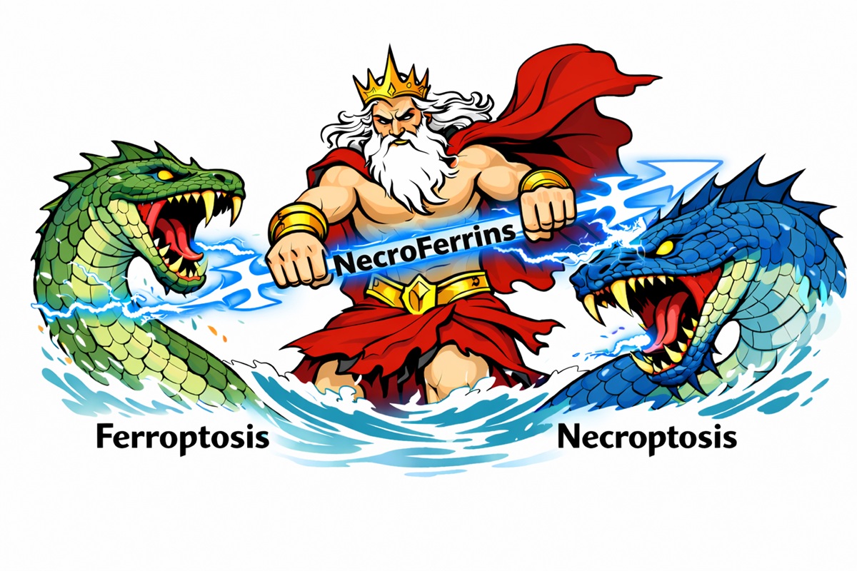
NecroFerrins as dual therapeutic inhibitors targeting necroptosis and ferroptosis
Over the past two decades, increasing attention has been devoted to regulated forms of cell death that occur independently of apoptosis. Among these, necroptosis and ferroptosis exhibit a necrotic-like morphology, yet are defined by distinct molecular and ...
More.Over the past two decades, increasing attention has been devoted to regulated forms of cell death that occur independently of apoptosis. Among these, necroptosis and ferroptosis exhibit a necrotic-like morphology, yet are defined by distinct molecular and biochemical signatures. Both pathways have attracted considerable interest due to growing evidence implicating them in the pathogenesis of a wide range of acute and chronic disorders. Notably, the simultaneous engagement of multiple regulated necrosis pathways has been reported in many disease contexts, highlighting the limitations of single-target therapeutic approaches. In this light, the design of multi-target-directed ligands, that is, embracing a polypharmacological strategy, has emerged as a promising direction for the development of future therapies. In this review, we propose the concept of NecroFerrins, a class of small molecules that simultaneously inhibit necroptosis and ferroptosis. Within this class, we identify RIPROStatins as a distinct subclass of RIPK1 inhibitors that additionally possess radical-trapping antioxidant activity. The objective of this review is also to stimulate translational research on complex diseases using polypharmacological drugs acting as necrosis inhibitors.
Less.Claire Delehouzé, Stéphane Bach
DOI:https://doi.org/10.70401/fos.2026.0041 - August 05, 2026
Oxidative stress and inflammation: A panoply of pathways but a paucity of perspective
Reactive oxygen species (ROS) are typically elevated at inflammatory sites due to the influx of large numbers of neutrophils and other myeloid cells that produce high levels of oxygen free radicals as microbicidal agents. Reactive oxygen intermediates are ...
More.Reactive oxygen species (ROS) are typically elevated at inflammatory sites due to the influx of large numbers of neutrophils and other myeloid cells that produce high levels of oxygen free radicals as microbicidal agents. Reactive oxygen intermediates are also generated as a byproduct of cellular metabolism, due to mitochondrial damage, exposure to toxins or radiation, and are also frequently increased during inflammatory reactions. However, although oxidative stress is often implicated as an initiator or amplifier of inflammation, particularly in the context of cancer, how reactive oxygen modulates inflammatory signalling remains poorly understood. Here we discuss the diversity of pathways and molecules implicated in promoting or suppressing inflammation in response to ROS. As we shall see, reactive oxygen has been reported to influence inflammatory outputs through direct effects on phosphatases that regulate nuclear factor kappa B (NF-κB) activation, through activation of the nuclear factor erythroid 2-related factor 2 (Nrf2) or hypoxia-inducible factor (HIF)-1 transcription factors that can directly or indirectly promote inflammatory gene expression, by provoking DNA damage leading to activation of the cyclic guanosine monophosphate (GMP)-adenosine monophosphate (AMP) synthase (cGAS)/stimulator of interferon genes (STING) pathway, or through promoting assembly of inflammasomes. Thus, there are a multitude of routes by which ROS can influence inflammatory responses. How reactive oxygen promotes inflammation in specific contexts is likely to be influenced by several factors, including the source of ROS, whether intracellular or extracellular, the responding cell type, as well as the extent of deviation from normal homeostatic setpoints. Understanding how reactive oxygen shapes inflammation has important implications for therapeutic intervention in multiple disease states.
Less.Aoife Costigan, Seamus J. Martin
DOI:https://doi.org/10.70401/fos.2026.0040 - August 03, 2026
Hub of life and death: Global regulation of ferroptosis by inter-organelle crosstalk
Ferroptosis is an iron-dependent form of programmed cell death defined by the lethal accumulation of lipid peroxides. Accumulating evidence indicates that ferroptosis plays a critical role in a wide range of pathological processes, including tumor progression, ...
More.Ferroptosis is an iron-dependent form of programmed cell death defined by the lethal accumulation of lipid peroxides. Accumulating evidence indicates that ferroptosis plays a critical role in a wide range of pathological processes, including tumor progression, ischemia-reperfusion injury, and neurodegenerative diseases. The key determinants of cellular susceptibility to ferroptosis are the homeostasis of iron and lipid metabolism, whose maintenance relies heavily on the precise regulation of inter-organelle communication networks. In this review, we focus on the spatial biology of ferroptosis regulation. From three core dimensions, iron homeostasis modulation, lipid metabolism remodeling, and redox balance maintenance, we systematically dissect the mechanisms by which signal crosstalk and functional coordination among multiple organelles (lysosomes, endoplasmic reticulum, mitochondria, plasma membrane, nucleus, lipid droplets, Golgi apparatus, and peroxisome) govern ferroptosis initiation, signal amplification, and cellular defense responses. Comprehensive elucidation of this multi-organellar coordinated regulatory network will not only advance our fundamental understanding of ferroptosis execution but also provide a holistic perspective for the development of organelle-targeted therapeutic strategies against human diseases.
Less.Yunjie Cai, ... Yongyou Zhang
DOI:https://doi.org/10.70401/fos.2026.0039 - July 30, 2026