Current evidence for a role of clonally expanded somatic mutations in atherosclerosis and chronic kidney disease-associated vascular aging

Lara G. Merino
#

,
Fabiana Stefani
#
,
Quentin Giraud
#
,
Maria Eriksson
*
*Correspondence to:
Maria Eriksson, Department of Medicine Huddinge, Karolinska Institute, Huddinge, Sweden.
E-mail: maria.eriksson.2@ki.se
Geromedicine. 2026;2:202606. 10.70401/Geromedicine.2026.0029
Received: February 11, 2026Accepted: July 07, 2026Published: July 07, 2026
Abstract
Somatic mutations accumulate in tissues throughout life and are increasingly being recognized as contributors to aging. While their role in cancer has been extensively characterized, their occurrence and functional impact in the cardiovascular system remain poorly understood, partly due to technical limitations in detecting low-frequency and cell-specific variants. Recent advances in single-cell sequencing have begun to unveil how somatic mutations accumulate both in vascular resident cells, including vascular smooth muscle cells, and in circulating hematopoietic cells, such as those associated with clonal hematopoiesis of indeterminate potential. Both resident vascular cells and infiltrating immune cells harboring clonally expanded somatic mutations may influence vascular pathology. This mini-review summarizes the current knowledge on somatic mutation accumulation in the vasculature, focusing on recent studies and the techniques applied for their detection. In addition, we discuss emerging evidence for the functional relevance of somatic mutations in aging and age-related vascular disease.
Keywords
Somatic mutations, atherosclerosis, chronic kidney disease, vascular smooth muscle cells, clonal hematopoiesis of indeterminate potential, sequencing
1. Introduction
Aging is a complex, multifactorial biological process marked by a progressive, time-dependent decline in tissue function and increased risk of age-associated diseases. The aging process is thought to be driven by a series of highly interconnected cellular and molecular processes, collectively referred to as the ‘hallmarks of aging’[1,2]. Genomic instability is one of these hallmarks and encompasses a broad spectrum of alterations in DNA structure and sequence arising from both endogenous and exogenous sources of damage. This instability can lead to permanent changes in the DNA sequence, known as somatic mutations.
Somatic mutations have been shown to occur and accumulate progressively throughout life in a plethora of tissues[3-7]. While somatic mutations have been extensively described and characterized in neoplastic diseases, increasing evidence indicates that they may drive or contribute to diseases other than cancer.
One mechanism by which somatic mutations can contribute to disease development is the clonal expansion of mutated cells. Clonal expansion can be driven by the mutations themselves, leading to those mutations being defined as drivers. In contrast, passenger mutations are those that expand either due to co-occurrence with a driver mutation in the same cell or due to extrinsic mechanisms that promote cell expansion independent of the presence of the mutation. The latter would be referred to as bystander clonality[8]. In this context, common mutations can be detected using bulk sequencing approaches, due to the existence of cellular clones. However, the detection of somatic mutations present in only one or a few cells remains a technical and bioinformatic challenge. To address this limitation, several next-generation sequencing strategies have been developed to identify rare and ultra-rare genetic variants both in single cells and bulk tissues. While each technique has specific advantages and limitations, their development has substantially advanced our ability to detect and characterize somatic mutations both in healthy and diseased tissues[9-11].
Clonal propagation has been shown to be relevant in cardiovascular disease, providing a context in which somatic mutations could arise, propagate, and contribute to disease. For example, age-associated somatic mutations in hematopoietic stem cells lead to a non-neoplastic expansion of mutant leukocytes, a process named clonal hematopoiesis of indeterminate potential (CHIP). Importantly, leukocytes carrying CHIP mutations can promote atherosclerosis development[12].
Focusing on the vascular wall, pioneering studies performed as early as 1973 already suggested that atherosclerotic lesions could be of a clonal nature[13]. Since then, multiple studies have confirmed the clonal dynamics of the vascular wall in injury and disease, due to the development of refined lineage tracing approaches and single-cell genomics[14,15]. Such evidence identifies the vasculature as a suitable environment for mutation development and propagation.
Furthermore, circulating and resident cells are highly interconnected. During atherosclerosis development, circulating monocytes adhere to the endothelium and infiltrate the intimal layer of the arteries, where they differentiate into macrophages and foam cells that accumulate oxidized low-density lipoprotein (LDL). In parallel, vascular smooth muscle cells (VSMCs) undergo clonal expansion, contributing to fibrous cap formation and to the foam cell pool. Importantly, recruited macrophages can modulate the clonality of proliferating VSMCs[16,17]. Lastly, recent single-cell studies suggest that modulated VSMCs could contribute to the inflammatory milieu of the plaque through altered secretory programs, as well as promote extracellular matrix remodeling[18], highlighting the extensive crosstalk between circulating and resident cells in atherosclerosis.
This mini-review examines current knowledge on the occurrence of somatic mutations in atherosclerosis and chronic kidney disease (CKD). In addition, it highlights emerging evidence supporting a functional role for somatic mutations in the pathogenesis of atherosclerosis and in CKD–associated early vascular aging.
2. Somatic Mutations Etiology and Role in Aging
Somatic mutations arise post-zygotically and accumulate throughout life[19]. While their role in cancer is well established, their contribution to non-cancer pathologies has only begun to be appreciated, largely due to the technical challenges that, until recently, limited the detection of somatic mutations in non-cancerous tissues. However, with the development of novel next-generation sequencing technologies, it has been possible to characterize the somatic mutation burden across multiple tissues[3] and determine its correlation with age[9-11].
Ultradeep targeted sequencing of cancer-associated genes in sun-exposed skin and esophagus revealed an age-associated accumulation of mutation-carrying non-cancerous clones[20,21]. Further studies applied whole-genome sequencing (WGS) to cell clones derived from single progenitor cells isolated from the small intestine, colon, and liver[7]; fat, kidney, and epidermis[5], and skeletal muscle progenitors[4], confirming an increase in somatic mutation numbers with age in these tissues. In addition, bulk and single-cell WGS approaches applied to neurons[22-24], oligodendrocytes[25], and cardiomyocytes[26], among other cell types, demonstrated that post-mitotic cells can also accumulate mutations in an age-dependent manner.
Overall, somatic mutations have been estimated to accumulate at a rate of 10-50 single nucleotide variants (SNVs) per cell per year[19]. Somatic mutations were traditionally attributed to DNA replication errors and their imperfect repair. However, the assessment of somatic mutagenesis in neurons revealed that these largely post-mitotic cells display mutation rates and spectra that overlap with those of mitotically active tissues, and that mutation burden does not scale directly with cell division rates[23,27]. These observations indicate that somatic mutations can arise independently of cell division, for example, through DNA damage associated with transcription in neurons[27] or through endogenous oxidative stress, as reflected by characteristic SNV patterns detected in cardiomyocytes and neurons[24,26].
In addition to advances in somatic mutation detection, evidence for a functional role of somatic mutations in aging and age-associated disease, outside of cancer, is beginning to emerge. The rate of somatic mutation accumulation has been shown to inversely correlate with lifespan across several species[28], and enhanced DNA repair mechanisms that preserve genomic integrity have been observed in the exceptionally long-lived bowhead whale[29].
Somatic mutation accumulation has also been suggested to contribute to the development of Alzheimer’s disease[24] and osteoarthritis[30]. Further supporting a functional role for somatic mutations in aging, a recent study from our group demonstrated that increased somatic mutation burden in muscle progenitors leads to impaired muscle repair and function following injury[31]. In a second study, we also identified the Hutchinson–Gilford progeria syndrome (HGPS) causing mutation and its associated protein, progerin, as a potential contributor to premature vascular aging in patients with CKD[32].
In line with the findings of the studies mentioned above, clonal expansion of mutated cells is therefore considered to be important for the mutations to exert detrimental effects. In the field of vascular biology, several cell types, including VSMCs, endothelial cells (ECs), and macrophages, have been shown to present different degrees of clonal expansion during injury and disease[33]. Therefore, the vascular system comprises an interesting context for the study of somatic mutation development and propagation.
3. Clonal Expansion in the Arterial and Hematopoietic Systems
3.1 Clonal expansion within the arterial wall
Under physiological conditions, VSMCs in the arterial media are largely quiescent. Their primary functions include regulating blood pressure, maintaining vascular tone, and producing extracellular matrix components that provide structural integrity to the vessel wall. In response to injury, however, VSMCs exhibit local heterogeneity, with regional variation in gene expression profiles[34], consistent with their diverse embryonic origins[35,36]. The aortic vasculature is of polyclonal origin[15,37], and under normal conditions, limited VSMC renewal at low rates compensates for minor cell loss. However, in pathological contexts (i.e. injury or vascular disease), a small subset of VSMCs can rapidly proliferate in an oligoclonal manner and thereby form clones (Figure 1). In the same settings, VSMCs can further dedifferentiate and acquire alternative phenotypes, a process referred to as VSMC phenotypic switching, which is extensively reviewed elsewhere[38]. Clonal expansion of VSMCs has been reported in numerous cardiovascular pathologies such as atherosclerosis, blood vessel injury, aneurysm and CKD-associated vascular disease[13-15,17,32,37,39].
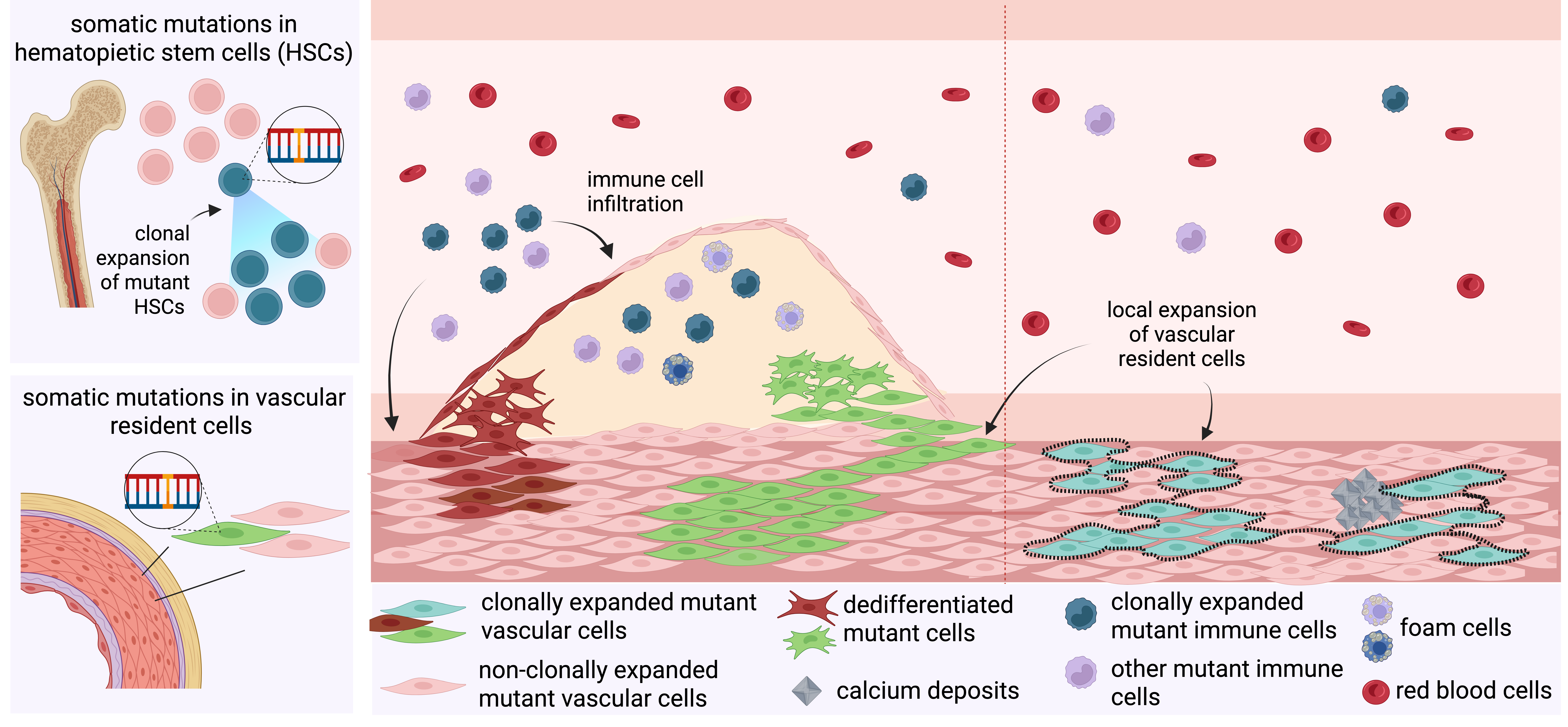
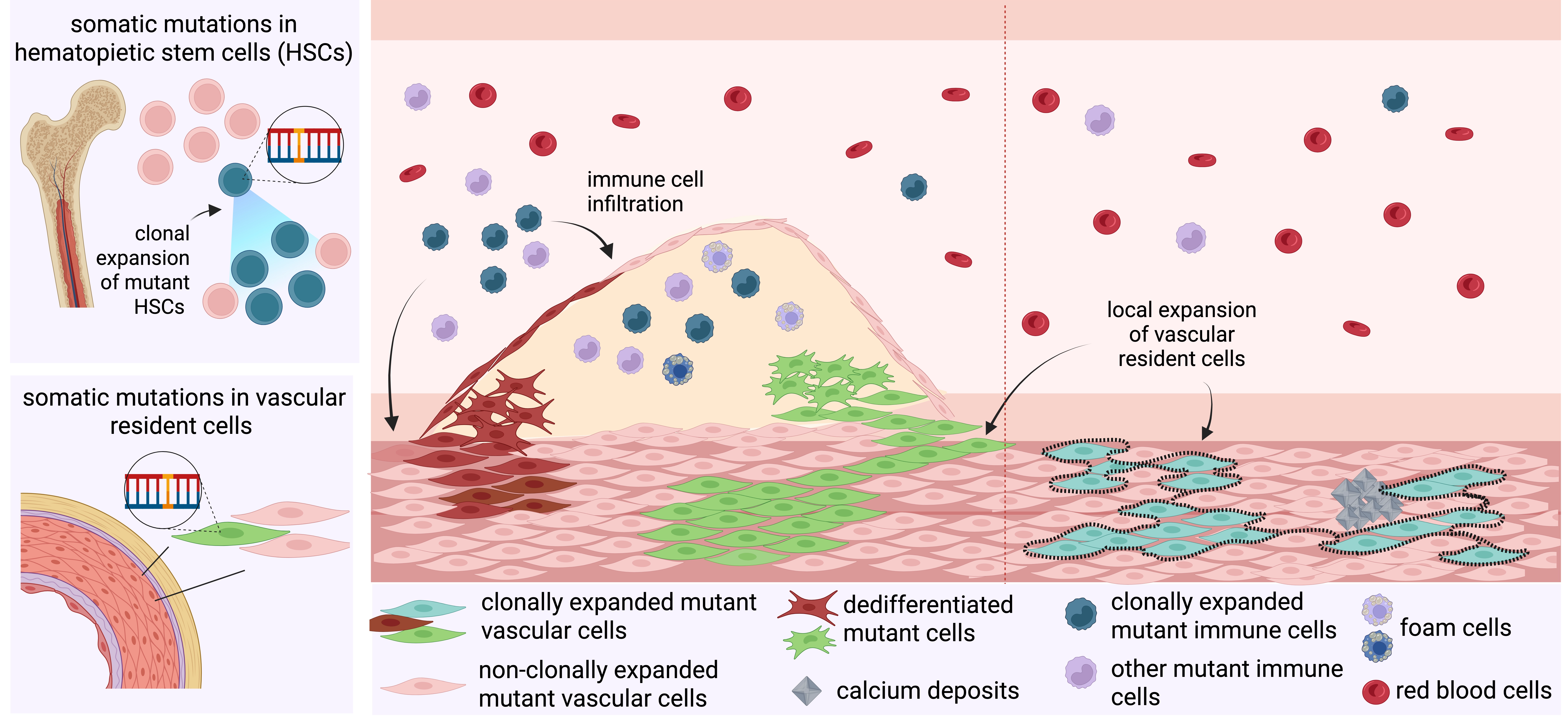
Atherosclerosis is among the conditions in which the dynamics of VSMC proliferation have been more extensively explored. Monoclonality of the plaque was first proposed by Benditt and Benditt by studying X chromosome inactivation in plaques from women[13]. Modern lineage-tracing studies have confirmed that plaques commonly arise through oligoclonal expansion of a small number of medial smooth muscle cells (SMCs) whose progeny diversify into multiple plaque phenotypes[14,15,17,37]. Mechanistic work in mice shows that SMC fate and clonal architecture can be shaped by local cues such as macrophage-SMC crosstalk and integrin-β3 signaling, and that these processes are influenced by age[15,17].
Overall, the literature supports a model in which medial VSMC clonality occurs in several vascular diseases, impacting their development and progression. Endothelial repair is likewise mediated mainly by local EC proliferation rather than by substantial input from migrating cells[40]. Furthermore, it must be noted that, upon severe or extensive damage, non-VSMC cells, such as adventitial progenitors, can also be recruited to diseased sites[41]. Altogether, these observations suggest that the vascular wall represents a particularly relevant tissue for studying the dynamics and consequences of somatic mutation occurrence and propagation.
3.2 Clonal expansion of circulating cells
Somatic mutations and clonal expansions of mutant cells have been described to occur in circulating hematopoietic cells in a process named CHIP. CHIP defines the presence of clonally expanded hematopoietic cells carrying a specific somatic driver mutation in an individual with no evidence of a hematologic neoplasm[42-44]. By convention, CHIP is called when an advantageous somatic variant is present at a variant-allele fraction (VAF) of ≥ 2% in peripheral blood or bone-marrow DNA (corresponding to 4% of cells assuming a heterozygous mutation). CHIP prevalence rises steeply with age. Estimates typically report ~10-20% prevalence or higher in individuals ≥ 70 years[42-44]. The most common CHIP driver mutations occur in genes encoding epigenetic regulators and spliceosome or signaling factors, such as DNMT3A, TET2, ASXL1, JAK2, TP53, PPM1D, and SF3B1[42-44]. Importantly, many expanded clones identified by whole-genome approaches lack a recognized candidate driver mutation[45]. CHIP has an associated cardiovascular risk that is comparable to, and in some studies exceeds, that conferred by traditional cardiovascular disease risk factors[42,46]. CHIP has also emerged as an independent mortality predictor in patients with coronary artery disease[42,46,47] and CKD[48]. Mechanistic and multi-omics studies have provided plausible biological mechanisms for these epidemiologic observations, which have been mostly focused on qualitative changes in myeloid function and heightened systemic inflammation. For instance, TET2 mutations have been identified as drivers of LDL receptor up-regulation, increased macrophage lipid accumulation, and inflammasome activation[12,47]. Similarly, Dnmt3a- and Tet2-driven CHIP worsened cardiac dysfunction in mice challenged with angiotensin II infusion by upregulation of inflammatory pathways[49]. In line with this, reduced activation of IL-6 signaling reduces the risk of CHIP-associated cardiovascular disease in CHIP carriers[50]. scRNA-seq of patient monocytes revealed upregulation of osteogenic genes, and Tet2-/- bone-marrow transplant experiments in mice showed that mutant myeloid cells drive mesenchymal calcification via secreted mediators[51].
Recently, some studies have indicated that CHIP may also directly promote clonal infiltration and expansion of blood monocyte-derived macrophages within plaques[47,52-55]. Compared to their wild-type counterparts, JAK2-mutant immune cells were overrepresented in atherosclerotic lesions. Mutant cells were also more abundant within lesions than in the bloodstream, a shift attributed to increased endothelial adhesion and local proliferation[56]. TP53-mutant CHIP is also known to promote greater aortic plaque size and macrophage accumulation via enhanced proliferation of p53-deficient plaque macrophages[57]. These findings suggest that somatic mutations can also propagate in the vascular wall through infiltration of CHIP-mutant cells, a feature that will be further discussed later.
4. Somatic Mutations in Atherosclerosis and Early Vascular Aging in Chronic Kidney Disease
4.1 Recent findings on somatic mutation accumulation in the vascular wall
One of the earliest studies of somatic mutations in the vascular wall identified a mutation in TGFBR2 in cellular patches from carotid atherosclerotic plaques[58,59]. Over the past decade, advances in next-generation sequencing technologies have accelerated research into somatic mutations, also enabling a more comprehensive characterization of the somatic mutational landscape in vascular tissues. These developments have, in turn, begun to advance our understanding of cardiovascular disease[60]. Recent studies investigating somatic mutations have employed diverse experimental designs and sequencing approaches, providing important new evidence for the accumulation of somatic mutations in the vascular wall and their potential contribution to disease development. Here, we discuss these findings, which are summarized in Table 1.
Table 1. Summary of recent studies investigating somatic mutations in vascular resident cells
| Study | Tissue | Method | Key Findings | Possible limitations |
| Abascal et al.; Nature; 2021[23] | Human bladder and intestinal SMCs | Single molecule duplex-sequencing (Nanoseq) | • High sensitivity • Low error rate • Single molecule resolution | • Bulk DNA input • No per-cell information • No direct assessment in the vasculature |
| Wang et al.; Cardiovascular Research; 2022[61] | Mouse VSMCs from atherosclerotic lesions | Bulk WGS | • Assessment for somatic mutagenesis in atherosclerosis • Whole genome coverage | • Amplification-induced artifacts • Bulk input from multiple lesions (dilution of rare or lesion specific variants) • No per-cell information • Limited time for mutation accumulation |
| Dederichs et al.; ATVB; 2024[63] | Human myeloid and lymphoid cells from blood and atherosclerotic lesions | Targeted sequencing and ddPCR | • CHIP mutations in lesions • Similar variant allele frequency in blood monocytes and lesion macrophages | • Bulk DNA input • No per-cell information • Restricted to specific mutations |
| Steffensen et al.; JCI; 2025[62] | Human atherosclerotic lesions and buffy coat | WES | • Detection of clonal mutations across different plaque regions • CHIP mutations in the lesion | • Bulk DNA input • No per-cell information • Amplificatoon-induced artifcats • Only exome analysed |
| Von Scheidt et al.; EHJ; 2025[47] | Human blood and atherosclerotic lesions + controls | Targeted DNA sequencing | • Identification of TET2 mutation in atherosclerotic lesions • TET2 mutation is associated with plaque severity and mortality | • Bulk DNA input • No per-cell information • No information on mutations other than CHIP |
| Revêchon et al.; Nature aging; 2025[32] | Human CKD blood and epigastric arteries + controls | Targeted screening with ddPCR | • High resolution (< 0,4% allele frequency) • Disease-related clonal expansion of a specific somatic mutation | • Restricted to one specific mutation (LMNA1824T) • No per-cell information |
SMCs: smooth muscle cells; VSMCs: vascular smooth muscle cells; WGS: whole genome sequencing; CHIP: clonal hematopoiesis of indeterminate potential; WES: whole exome sequencing; CKD: chronic kidney disease; ddPCR: digital droplet polymerase chain reaction; TET2: tet methylcytosine dioxygenase 2.
The first of these recent studies suggesting that VSMCs could accumulate somatic mutations employed a highly accurate duplex sequencing–based method, which reduces error rates and enables detection of DNA variants in single molecules[23]. Here, visceral SMCs isolated from bladder and colon microdissections were shown to accumulate mutations at a rate of 20.7 SNVs per cell per year. While the study did not directly investigate vascular SMCs, it showed that SMCs can accumulate somatic mutations over time, and application of the same low error rate sequencing method to vascular tissues could provide high-resolution information on the vascular wall mutational landscape.
Wang et al.[61] directly assessed whether somatic mutations in VSMCs could contribute to atherogenesis in mice. By applying WGS to purified VSMCs from atherosclerotic plaques located in the aortic arch and its associated branches, the authors showed that plaque VSMCs harbored a modestly increased number of somatic mutations, compared with VSMCs from lesion-adjacent non-diseased aorta, where few somatic mutations were detected. The prior purification of VSMCs before sequencing provides evidence that this cell type can accumulate somatic mutations in a disease context. However, most of the identified mutations occurred in genes with low expression in plaque VSMCs and were therefore considered unlikely to exert functional effects at the protein level. Accordingly, the authors concluded that somatic mutations did accumulate in murine VSMCs during atherosclerosis but were unlikely to be key drivers of the disease in their mouse model.
In a recent study by Steffensen et al.[62], large-scale whole-exome sequencing (WES) of human carotid plaques, non-atherosclerotic arteries, and buffy coat identified an average of 37.8 mutations per plaque, with clonal frequencies ranging from 1% to 30.6%. Interestingly, plaque microdissection prior to sequencing indicated that some mutations were detected across different plaque regions in the same plaque, suggesting clonal expansion across different arterial layers (including the media and subcore intima). While no mutational hotspots were identified, over-representation analysis on the mutated genes identified an enrichment in terms related to the contractile apparatus, and the genes were shown to be highly expressed in both contractile and modulated VSMCs in available atherosclerosis scRNA-seq datasets. Out of approximately 492 mutated genes, 21 were found to be cancer genes. This suggests that some mutations could be drivers that provide a selective advantage, while others are passengers or bystander that accompany cells expanding due to the clonal nature of atherosclerotic development.
This work also showed infiltration of atherosclerotic lesions by hematopoietic cell clones, which comprised substantial fractions of the plaque cell population alongside locally expanded arterial clones. NOTCH2 and TET2 genes are notable examples of mutated genes detected both in buffy coat and atherosclerotic plaques. These findings build on the study from Dederichs et al.[63], which previously reported the presence of CHIP mutated clones in atherosclerotic plaques and heart tissue using targeted sequencing and digital droplet PCR (ddPCR).
The infiltration of CHIP-mutated cells was further observed in the study from von Scheidt et al.[47], with TET2 mutations being detected in coronary and carotid atherosclerotic plaques and in the myocardium. The authors described CHIP as a predictor of mortality in coronary artery disease and showed that patients with CHIP TET2 mutations have a more severe plaque phenotype. However, no correlation between the degree of mutation infiltration in the tissue and plaque severity was established.
Altogether, these 3 studies[47,62,63] point towards a new role of CHIP mutations in atherosclerosis development.
Further supporting the occurrence of clonally expanded somatic mutations in VSMCs, our recent work identified the LMNA c.1824C>T somatic mutation in the epigastric arteries of CKD patients (mean variant allele frequency 11%, maximum 50%)[32]. Interestingly, this mutation is the cause of HGPS, a premature aging disorder in which patients die from severe cardiovascular disease[64,65]. The vascular phenotype of HGPS closely resembles that of physiological vascular aging with VSMC loss, calcification, and fibrosis[66], a phenotype that is also observed in CKD arteries. In HGPS, the LMNA c.1824C>T mutation leads to the expression of an aberrant form of Lamin A, called progerin. Progerin-positive cells were identified as VSMCs due to their medial location and positivity for smooth muscle actin. However, the mutation was also detected in the blood from CKD patients, although at a lower allele frequency than in the artery (from 0.012% to 0.047%). This opens the possibility of a potential interplay between circulating and resident cells, where circulating cells may contribute to the detected mutant VSMC population, although this would require transdifferentiation. The overall number of progerin-expressing cells correlated with cellular and vascular pathology. Notably, these cells formed clusters, displaying signs of clonality. In addition, mice carrying the mouse equivalent of the LMNA c.1824C>T mutation in only a fraction of VSMCs displayed clonal propagation of the mutated cells as well as signs of vascular pathology. These observations support a contribution of progerin to CKD-associated vascular aging.
4.2 Current challenges and limitations in the study of somatic mutations in the vascular wall
The studies presented above have provided valuable evidence that somatic mutations can accumulate within the vascular wall in atherosclerosis and CKD. However, given the emerging nature of this field, we discuss some of the advantages and potential technical limitations of each experimental approach, and how they may influence study interpretation (summarized in Table 1).
Abascal et al.[23] established that somatic mutation accumulation occurred in tissue biopsies composed primarily of SMCs using a highly accurate duplex sequencing strategy. Nevertheless, this approach does not allow mapping mutations to individual cells, limiting the ability to assess the mutational burden of each cell and the co-occurrence of mutations within the same cell. This also precludes linking somatic mutations to a cell-specific transcriptional profile.
The study from Wang et al.[61] concluded that despite showing an increased number of somatic mutations in murine VSMCs during atherosclerosis, their findings did not support a functional role for somatic mutations in disease development. Nevertheless, some nuances may influence the interpretation of these results. First, WGS has limited resolution to detect low frequency variants, as distinguishing them from sequencing errors remains challenging. This could underestimate the number of mutations in both the plaque and the non-lesional aorta, hindering the detection of rare but functionally relevant variants. Second, the joint collection of the aortic arch and the associated branches may further dilute rare or plaque-specific clonal mutations if multiple plaques are present in the tissue. Lastly, the ApoE-/- mouse is characterized by a relatively rapid expansion of VSMCs, in contrast to the slower plaque development observed in humans, which limits sensitivity for studying somatic mutations acquired during aging or long-term vascular disease. Altogether, while this study showcased that the atherosclerosis process could lead to somatic mutation accumulation specifically in VSMCs, further research is needed to understand the functional consequences.
The work from Steffensen et al.[62] provided a description of somatic mutations in human carotid atherosclerotic plaques using modern sequencing techniques, but similarly to the Wang study, further work is needed to fully ascertain the extent of somatic mutations in the plaque. While WES has allowed significant advances in the understanding of CHIP due to its higher sequencing depth, it still poses some limitations. By performing WES, only the exome is characterized, leaving most of the genome not analyzed. Since exons have been shown to be more protected from mutagenesis, potentially through transcription-coupled repair[4], this could lead to underestimating the real mutation number. In addition, as with WGS, the error rate from WES could further hinder the detection of low frequency clones. This reduced sensitivity could explain the observation that control arteries in this study have no detectable mutations.
Overall, these three studies underscore the relevance of using single-cell methods to be able to distinguish whether plaque mutations are derived from infiltrating blood cells or resident cells from the vascular wall, and, in the latter case, which specific vascular cell type. If combined with single-cell transcriptome information for the mutated cells, this will also shed light on how mutations affect cell functionality and therefore disease development.
Lastly, our recent work[32] described the occurrence of the HGPS disease-causing mutation (LMNA c.1824C>T) in epigastric arteries from CKD patients and its correlation with vascular pathology. It should be noted that in our study we used a targeted approach to specifically screen for the progeria mutation. However, our study is limited to one mutation, and additional mutations arising as a consequence of the uremic environment need to be assessed to better characterize the mutational landscape of CKD arteries.
Altogether, these studies provide compelling evidence that somatic mutations accumulate within the vascular wall and underscore the emergence of a rapidly evolving field of research. Further development in sequencing technologies, particularly highly accurate and uniform WGS at single-cell resolution and the integration of single-cell DNA and RNA sequencing data from the same cell, will enable a more comprehensive characterization of the vascular mutational landscape. Such approaches have the potential to identify mutations at single-cell resolution, determine their cellular origin, and directly link genetic alterations to transcriptional changes and cellular phenotypes, thereby advancing our understanding of vascular aging and disease development.
5. Conclusions and Future Perspectives
While somatic mutagenesis is emerging as a field of increasing interest, the occurrence and contribution of somatic mutations to cardiovascular disease are only starting to be explored. Contrasting results across studies are likely due to differences in experimental design and sequencing approaches. Multiple CHIP-associated mutations have been shown to accumulate with age and correlate with cardiovascular risk, atherosclerosis, and mortality. However, while it is now established that the TET2 and DNMT3A mutations promote the upregulation of inflammatory pathways in atherosclerosis, only a limited number of studies have provided evidence for functional implications of these mutations in cardiovascular disease by modulating cellular infiltration. Moreover, the spectrum of genes implicated in CHIP may extend beyond canonical drivers, underscoring the need for higher-resolution sequencing technologies combined with more robust functional studies.
CHIP-mutant cells have now been detected within atherosclerotic lesions, where they influence lesion progression and stability. Nevertheless, novel functions of these infiltrating cells beyond their pro-inflammatory and paracrine effects are starting to emerge and merit further study. Early studies inferring transdifferentiation of cells based on lineage marker expression have been difficult to interpret, highlighting the need for high-resolution lineage-tracing approaches to determine whether, and under what conditions, infiltrating hematopoietic clones adopt alternative vascular cell fates.
In addition to circulating cells, somatic mutations may also arise within resident vascular cells and propagate either as an effect of the mutation itself or through the intrinsic clonal nature of the vascular wall, particularly in disease conditions. In this context, single-cell resolution approaches are essential to link specific mutations to individual cellular phenotypes, identify co-occurring mutations, and place them within gene regulatory networks known to affect and influence cardiovascular pathology[67]. In the future, multi-omic strategies combining single-cell sequencing with spatial information could provide insights into the localization and distribution of mutant cells within the tissue and how they affect tissue architecture.
Overall, a more comprehensive understanding of tissue and cell-specific mutational landscapes, as well as the biological mechanisms driving somatic mutation acquisition and clonal expansion in cardiovascular disease, will be important to further advance an area that now represents one of the frontiers in cardiovascular research.
Acknowledgements
We used AI-assisted technologies, including ChatGPT (GPT-5.5, OpenAI) and Mistral Large 3, for the purpose of language editing. The authors have reviewed, edited, and approved the final content and take full responsibility for the content of this manuscript.
Authors contribution
Merino LG, Giraud Q: Writing-original draft, writing-review & editing.
Stefani F: Visualization, writing-original draft, writing-review & editing.
Eriksson M: Conceptualization, writing-review and editing.
Conflicts of interest
Not applicable.
Ethical approval
Not applicable.
Consent to participate
Not applicable.
Consent for publication
Not applicable.
Availability of data and materials
Not applicable.
Funding
The study is supported by grants from the Swedish Research Council (2023-02847), the European Research Council (ERC Advanced grant 2022-101097871) and the Swedish Cancer Foundation (254500Pj).
Copyright
© The Author(s) 2026.
References
-
1. López-Otín C, Blasco MA, Partridge L, Serrano M, Kroemer G. The hallmarks of aging. Cell. 2013;153(6):1194-1217.[DOI]
-
2. López-Otín C, Blasco MA, Partridge L, Serrano M, Kroemer G. Hallmarks of aging: An expanding universe. Cell. 2023;186(2):243-278.[DOI]
-
6. Manders F, van Boxtel R, Middelkamp S. The dynamics of somatic mutagenesis during life in humans. Front Aging. 2021;2:802407.[DOI]
-
7. Blokzijl F, de Ligt J, Jager M, Sasselli V, Roerink S, Sasaki N, et al. Tissue-specific mutation accumulation in human adult stem cells during life. Nature. 2016;538(7624):260-264.[DOI]
-
8. Stratton MR, Campbell PJ, Futreal PA. The cancer genome. Nature. 2009;458(7239):719-724.[DOI]
-
10. Yu Z, Coorens THH, Uddin MM, Ardlie KG, Lennon N, Natarajan P, et al. Genetic variation across and within individuals. Nat Rev Genet. 2024;25(8):548-562.[DOI]
-
11. Coorens TH, Oh JW, Choi YA, Lim NS, Zhao B, Voshall A, et al. The somatic mosaicism across human tissues network. Nature. 2025;643(8070):47-59.[DOI]
-
13. Benditt EP, Benditt JM. Evidence for a monoclonal origin of human atherosclerotic plaques. Proc Natl Acad Sci U S A. 1973;70(6):1753-1756.[DOI]
-
14. Chappell J, Harman JL, Narasimhan VM, Yu H, Foote K, Simons BD, et al. Extensive proliferation of a subset of differentiated, yet plastic, medial vascular smooth muscle cells contributes to neointimal formation in mouse injury and atherosclerosis models. Circ Res. 2016;119(12):1313-1323.
-
16. Misra A, Rehan R, Lin A, Patel S, Fisher EA. Emerging concepts of vascular cell clonal expansion in atherosclerosis. Arterioscler Thromb Vasc Biol. 2022;42(3):e74-e84.[DOI]
-
18. Yurdagul A Jr. Crosstalk between macrophages and vascular smooth muscle cells in atherosclerotic plaque stability. Arterioscler Thromb Vasc Biol. 2022;42(4):372-380.[DOI]
-
19. Ren P, Zhang J, Vijg J. Somatic mutations in aging and disease. GeroScience. 2024;46(5):5171-5189.[DOI]
-
23. Abascal F, Harvey LMR, Mitchell E, Lawson ARJ, Lensing SV, Ellis P, et al. Somatic mutation landscapes at single-molecule resolution. Nature. 2021;593(7859):405-410.[DOI]
-
24. Miller MB, Huang AY, Kim J, Zhou Z, Kirkham SL, Maury EA, et al. Somatic genomic changes in single Alzheimer’s disease neurons. Nature. 2022;604(7907):714-722.[DOI]
-
28. Cagan A, Baez-Ortega A, Brzozowska N, Abascal F, Coorens THH, Sanders MA, et al. Somatic mutation rates scale with lifespan across mammals. Nature. 2022;604(7906):517-524.[DOI]
-
30. Ren P, Zheng C, Pang Y, Qiang Y, Sun S, Wang Q, et al. Single-cell analysis of the somatic mutational landscape in human chondrocytes during aging and in osteoarthritis. Nat Aging. 2025;5(12):2417-2431.[DOI]
-
33. Lin A, Brittan M, Baker AH, Dimmeler S, Fisher EA, Sluimer JC, et al. Clonal expansion in cardiovascular pathology. JACC Basic Transl Sci. 2024;9(1):120-144.[DOI]
-
38. Chen R, McVey DG, Shen D, Huang X, Ye S. Phenotypic switching of vascular smooth muscle cells in atherosclerosis. J Am Heart Assoc. 2023;12(20):e031121.[DOI]
-
42. Jaiswal S, Fontanillas P, Flannick J, Manning A, Grauman PV, Mar BG, et al. Age-related clonal hematopoiesis associated with adverse outcomes. N Engl J Med. 2014;371(26):2488-2498.[DOI]
-
44. Xie M, Lu C, Wang J, McLellan MD, Johnson KJ, Wendl MC, et al. Age-related mutations associated with clonal hematopoietic expansion and malignancies. Nat Med. 2014;20(12):1472-1478.[DOI]
-
46. Jaiswal S, Libby P. Clonal haematopoiesis: Connecting ageing and inflammation in cardiovascular disease. Nat Rev Cardiol. 2020;17(3):137-144.[DOI]
-
47. von Scheidt M, Adkar SS, Krefting J, Hoermann G, et al. Clonal haematopoiesis of indeterminate potential and mortality in coronary artery disease. Eur Heart J. 2026;47(4):453-469.[DOI]
-
55. Tall AR, Fuster JJ. Clonal hematopoiesis in cardiovascular disease and therapeutic implications. Nat Cardiovasc Res. 2022;1(2):116-124.[DOI]
-
65. De Sandre-Giovannoli A, Bernard R, Cau P, Navarro C, Amiel J, Boccaccio I, et al. Lamin a truncation in Hutchinson-Gilford progeria. Science. 2003;300(5628):2055.[DOI]
-
66. Herzog MJ, Müller P, Lechner K, Stiebler M, Arndt P, Kunz M, et al. Arterial stiffness and vascular aging: Mechanisms, prevention, and therapy. Sig Transduct Target Ther. 2025;10:282.[DOI]
Copyright
© The Author(s) 2026. This is an Open Access article licensed under a Creative Commons Attribution 4.0 International License (https://creativecommons.org/licenses/by/4.0/), which permits unrestricted use, sharing, adaptation, distribution and reproduction in any medium or format, for any purpose, even commercially, as long as you give appropriate credit to the original author(s) and the source, provide a link to the Creative Commons license, and indicate if changes were made.
Publisher’s Note
Science Exploration remains a neutral stance on jurisdictional claims in published
maps
and institutional affiliations. The views expressed in this article are solely those
of
the author(s) and do not reflect the opinions of the Editors or the publisher.
Share And Cite


Science Exploration Style
Merino LG, Stefani F, Giraud Q, Eriksson M. Current evidence for a role of clonally expanded somatic mutations in atherosclerosis and chronic kidney disease-associated vascular aging. Geromedicine. 2026;2:202606. https://doi.org/10.70401/Geromedicine.2026.0029
Tips
Copy completed.
Submit a Manuscript
Author Instructions
Cite this Article
Article Metrics
0
View
0
Download
Cited
Article Updates

- Abstract
- Keywords
- 1. Introduction
- 2. Somatic Mutations Etiology and Role in Aging
- 3. Clonal Expansion in the Arterial and Hematopoietic Systems
- 4. Somatic Mutations in Atherosclerosis and Early Vascular Aging in Chronic Kidney Disease
- 5. Conclusions and Future Perspectives
- Acknowledgements
- Authors contribution
- Conflicts of interest
- Ethical approval
- Consent to participate
- Consent for publication
- Availability of data and materials
- Funding
- References
- Copyright
 Get citation
Science Exploration Style

Merino LG, Stefani F, Giraud Q, Eriksson M. Current evidence for a role of clonally expanded somatic mutations in atherosclerosis and chronic kidney disease-associated vascular aging. Geromedicine. 2026;2:202606. https://doi.org/10.70401/Geromedicine.2026.0029
copy
Share Link
copy
Subscribe