Why the term ‘erythrocyte aging’ is preferable to ‘erythrocyte senescence’

Anton Tkachenko
1,*

,
Ondrej Havranek
1,2
*Correspondence to:
Anton Tkachenko, BIOCEV, First Faculty of Medicine, Charles University, Vestec 25250, Czech Republic.
E-mail: anton.tkachenko@lf1.cuni.cz
Geromedicine. 2026;2:202620. 10.70401/Geromedicine.2026.0030
Received: March 31, 2026Accepted: July 08, 2026Published: July 09, 2026
Abstract
Over decades, nomenclature in erythrocyte cell death research has been unrefined. Recently, several attempts have been made to adapt the nomenclature suggested by the Nomenclature Committee on Cell Death (NCCD) guidelines to erythrocytes. Within the framework of the standardization of terms used in erythrocyte cell death studies, erythrocyte senescence and eryptosis, a regulated cell death of erythrocytes, have been defined and the formal distinction between both events has been recently established. Differences in their physiological role have been underscored, and markers that can be used to distinguish between erythrocyte senescence and eryptosis have been identified. As a further step, we aim to dissect cellular senescence in nucleated cells and erythrocytes, identifying their hallmarks, key triggers, molecular mechanisms, immunity-related effects, as well as physiological aspects. Available evidence suggests that cellular senescence in nucleated cells can be considered an active biological program, while erythrocyte senescence rather reflects a more passive, gradual accumulation of cellular damage. Fundamental mechanism-based and physiological differences between senescence in nucleated cells and erythrocytes highlighted in this mini-review indicate that the use of the term ‘erythrocyte senescence’ should be discouraged as misleading. Instead, the term ‘erythrocyte aging’ might be more appropriate.
Keywords
Aging, CD47, cell death, reactive oxygen species, senescence
1. Introduction
Five iterations of the Nomenclature Committee on Cell Death (NCCD) guidelines have consolidated the field of cell death by providing and constantly updating the unified criteria for defining and identifying distinct cell death modalities, setting up the nomenclature standards[1-5]. The resulting unification of the scientific language has significantly contributed to accelerated drug discovery by defining specific molecular markers (instead of vaguely distinguishable morphological signs) of each distinct regulated cell death (RCD) mode and by standardizing recommended assays for their evaluation[5]. Although each particular RCD is characterized by a unique set of regulatory and executive molecules involved, cell death-regulating signaling cascades frequently overlap and are tightly interconnected, creating a signaling network capable of launching a program of cellular suicide as an adaptive cellular response to moderate stresses[6] (in contrast to uncontrollable cellular demise, i.e. accidental cell death (ACD)[2], triggered by extreme, unendurable stresses). At the same time, cellular senescence is considered an alternative-to-cell-death mechanism used by eukaryotic nucleated cells to respond to stresses that exceed their capacity for compensation[7]. Cellular senescence is linked primarily to an irreversible cell cycle arrest and the ability to secrete pro-inflammatory signaling molecules, referred to as the senescence-associated secretory phenotype (SASP). The NCCD defines cellular senescence as a non-RCD-related event, which is specifically characterized by a permanent loss of the ability to proliferate and by the acquisition of SASP[3]. Interestingly, this term is frequently used for erythrocytes[8-10], circulating enucleated cells which are terminally differentiated and lack the capability to divide and express proteins (i.e. they cannot acquire SASP)[11]. However, in the field of erythrocyte research, the concept of erythrocyte senescence is mostly limited to age-related changes in erythrocytes that facilitate their gradual clearance by macrophages[9,11], which is in contradiction with the definition provided by the NCCD. In general, the NCCD remains skeptical towards the application of the term ‘cell death’ to erythrocytes and to their ability to elicit cell death-related effects (e.g., immunogenic), debating and questioning their status as viable cells[1,3]. However, accumulating evidence indicates that erythrocytes don’t undergo just a mechanical disintegration, but that they are capable of dying in several programmed ways. In our recent reviews, we have tried to adapt the principles of the NCCD nomenclature for erythrocyte cell death research (e.g., eryptosis[12] and necroptosis of erythrocytes, i.e. erythronecroptosis[13]), underscoring that the RCD-related general organization of an adaptive cellular response to stress might be similar between erythrocytes and nucleated cells (though definitely less diverse and abundant), making them commit suicide if adaptation fails. Thus, RCD-promoting signals in erythrocytes might lead to different lethal outcomes[13-15]. Interestingly, a growing body of evidence suggests that these morphologically distinct outcomes (non-lytic eryptosis and lytic erythronecroptosis) are mutually exclusive[12-14,16]. Moreover, the discovery of spectosis, another lytic RCD of red blood cells (RBCs), which occurs in response to the complement membrane attack complex (MAC) and is mediated by miniNLRP3 (a truncated form of NLRP3) recruitment and consequent caspase-8-dependent β-spectrin degradation, is another breakthrough in erythrocyte cell death research[17]. MiniNLRP3 recruits apoptosis-associated speck-like protein containing a CARD (ASC) and caspase-8 to form a protein complex resembling one of the PANoptosomes, suggesting that spectosis shares features of PANoptosis of nucleated cells[15,18]. Most importantly, spectosis elicits properties of innate immune cell death, since it culminates in the release of heme, an erythrocyte-associated damage-associated molecular pattern (DAMP) molecule[17,18]. Pro-inflammatory effects of heme and other erythrocyte-derived DAMPs are well-documented[19,20]. The discovery of spectosis clearly demonstrates the RCD-associated release of immunogenic heme from erythrocytes. Therefore, these recent advances in the field of erythrocyte cell death support the idea of reconsidering the status of RBC death by the NCCD.
In agreement with the issues outlined above, we believe that it is also required to adjust the terminology for erythrocyte senescence studies based on the NCCD recommendations. In particular, in our recently published review, we addressed a well-known confusion in the literature regarding the almost interchangeable use of the terms ‘erythrocyte senescence’ and ‘eryptosis’, clearly demonstrating the distinctness of these events[11]. Erythrocyte senescence was defined as a scheduled, gradual, and physiological acquisition of structural changes that promote macrophage-mediated RBC clearance. At the same time, eryptosis was suggested to represent a rapid, emergency response to potentially lethal stresses to prevent lytic destruction of erythrocytes. It has been hypothesized that eryptosis is caused by similar stressors as senescence. However, to trigger eryptosis, these stressors should be strong enough to promote significant phosphatidylserine externalization and consequent fast phagocytosis of erythrocytes by macrophages within minutes. Therefore, detectable phosphatidylserine externalization was discouraged from being interpreted as a marker of erythrocyte senescence[11]. Furthermore, we interpreted erythrocyte senescence as a passive process associated with life-long gradual damage. However, it does not correspond to cellular senescence as it is defined by the NCCD[3], but rather should be interpreted as aging.
Therefore, to ensure terminology accuracy in erythrocyte cell death research, we aim to dissect and compare the processes of cellular senescence in nucleated and enucleated cells.
2. Hallmarks of Cellular Senescence in Nucleated Cells: Cell Cycle Arrest and Senescence-Associated Secretory Phenotype
As stated above, cellular senescence is defined as an irreversible cell cycle arrest with the preservation of cellular viability and metabolic activity[3,21,22]. Canonically, cellular senescence is triggered by DNA damage and telomere shortening (genotoxic stress and proliferative exhaustion, respectively), oxidative and mitochondrial stresses linked to excessive generation of reactive oxygen species (ROS), epigenetic reprogramming, or oncogene activation[22,23]. For instance, DNA damage and telomere shortening with further telomere uncapping promote activation of DNA damage response (DDR) proteins, which trigger p53-dependent activation of p21, an inhibitor of cyclin-dependent kinases (CDKs). p21-mediated CDK inhibition results in the inability to form CDK–cyclin complexes, culminating in cellular senescence[24,25]. ROS promote DNA damage causing the DDR to induce cellular senescence. Additionally, ROS trigger mitochondrial dysfunction, which results in mitochondrial reactive oxygen species (mitROS) generation for further exacerbation of oxidative stress and inefficient adenosine triphosphate (ATP) synthesis, eventually leading to AMP-activated protein kinase (AMPK) recruitment. In its turn, AMPK activates the p53/p21 pathway to trigger cellular senescence[26,27]. In addition to this pathway, cellular senescence is induced by the p16/Rb pathway, which is activated by a variety of stimuli, including oncogene activation (e.g., Ras)[28,29]. As a result, cellular proliferation is blocked (Figure 1). At the same time, senescent cells remain metabolically active and capable of secreting a wide array of regulatory molecules (cytokines, chemokines, enzymes, or growth factors), affecting adjacent cells. Collectively, these molecules are referred to as SASP[30-32]. DNA damage is considered to be an important (but not always essential) condition for inducing SASP, which is primarily regulated by CCAAT/enhancer-binding protein β (C/EBPβ) and nuclear factor kappa B (NF-κB) transcription factors[33]. The most potent pro-inflammatory SASP molecules are tumor necrosis factor-α (TNF-α), interleukin-6 (IL-6), IL-8, IL-1, IL-18, etc.[34]. Cellular senescence has been also linked to metabolic reprogramming. Energy metabolism is affected primarily due to mitochondrial dysfunction, which leads to a higher AMP/ATP ratio, and glycolysis activation[35]. Additionally, cellular senescence is associated with disrupted lipid and amino acid metabolism[36,37], as well as lysosomal dysfunction, which is reflected in the use of senescence-associated beta-galactosidase (SA-β-gal) as a common biomarker of cellular senescence[38,39].
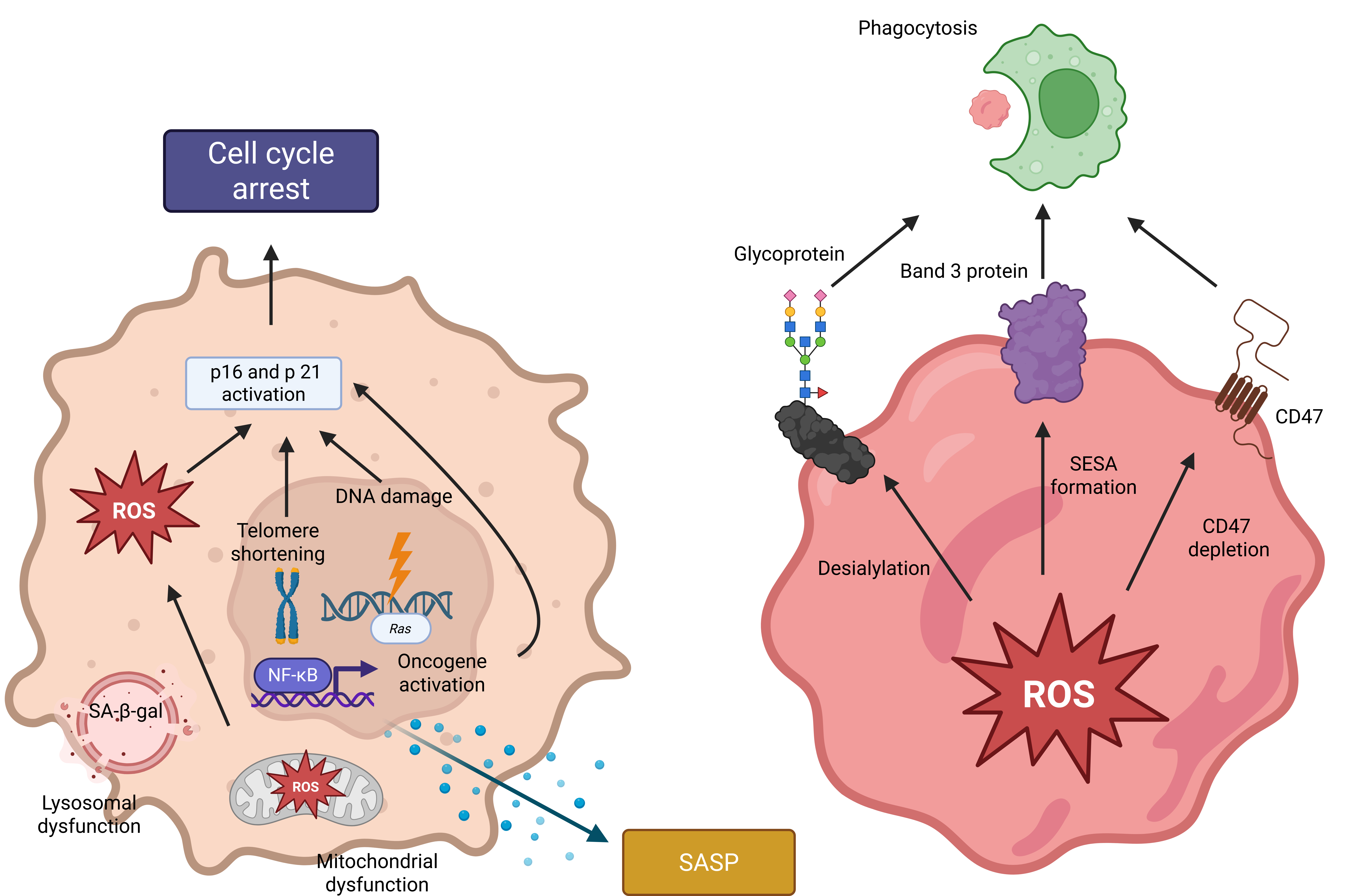
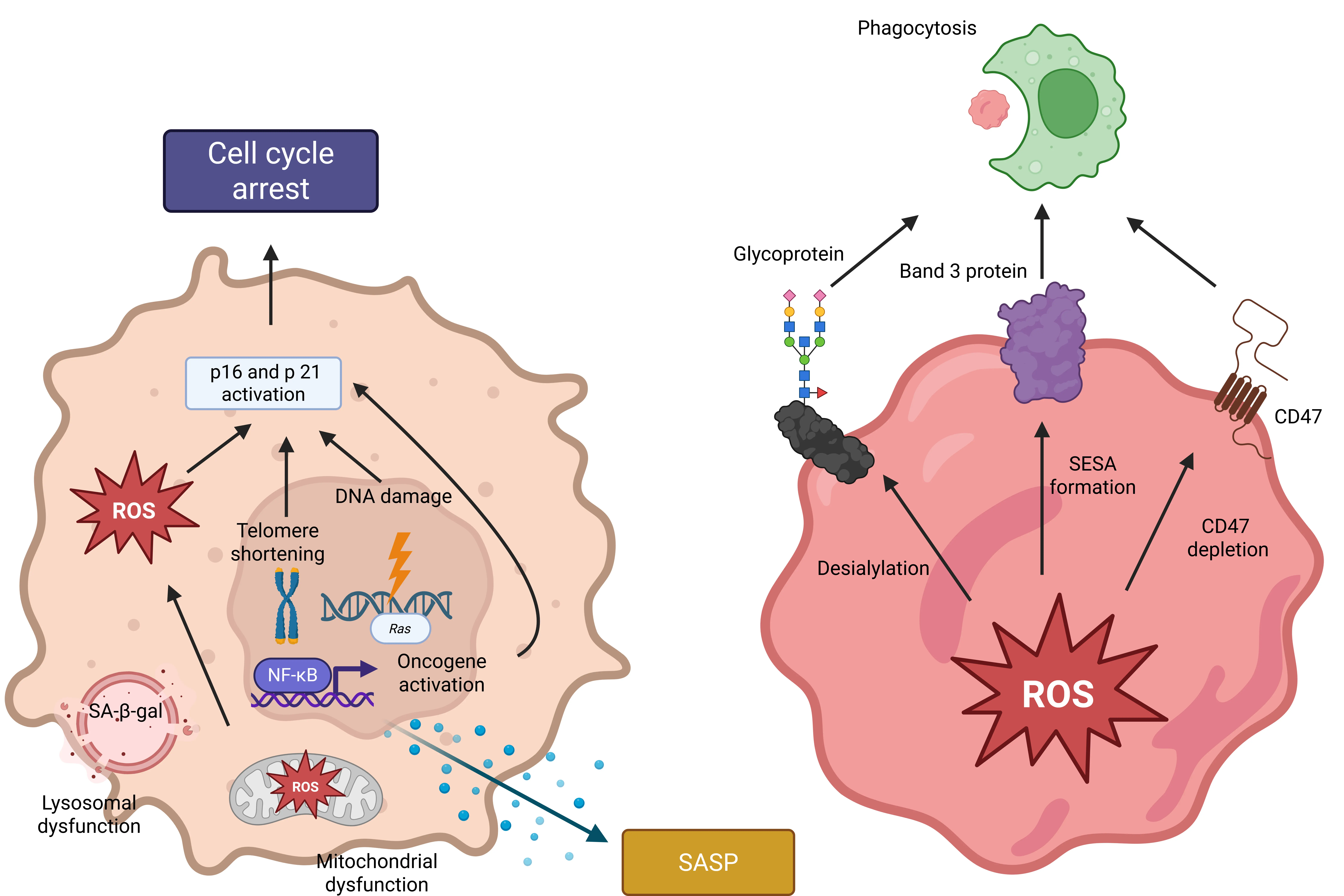
Figure 1. Senescence in nucleated cells and erythrocytes. Cellular senescence in nucleated cells (left image) is triggered by DNA damage, telomere shortening, oncogene activation, and oxidative stress to ensure a permanent cell cycle arrest (via activation of p16 and p21 - inhibitors of cyclin-dependent kinases), preserving cellular viability. Cellular senescence is linked to the secretion of pro-inflammatory factors (SASP). On the contrary, aging-dependent oxidative stress in erythrocytes triggers modifications of band 3 protein to form SESA, CD47 depletion, and surface desialylation, which facilitate phagocytosis-mediated clearance of senescent erythrocytes (right image). Created in BioRender. Havranek, O. (2026) https://BioRender.com/b39wb5n. ROS: reactive oxygen species; SA-β-gal: senescence-associated beta-galactosidase; SASP: senescence-associated secretory phenotype; SESA: senescent erythrocyte-specific antigen; ROS: reactive oxygen species.
Physiologically, cellular senescence aims at restricting the proliferation of damaged cells, but preserving their functionality[40]. Moreover, it contributes to maintaining homeostasis by its anti-tumor function, e.g., ceasing proliferation of damaged cells which could otherwise potentially undergo malignant transformation[41,42]. Cellular senescence is implicated in the regulation of embryogenesis, tissue remodeling, fibrosis, etc.[43]. Cytokines and chemokines associated with cellular senescence regulate inflammation[43]. At the same time, pro-inflammatory cellular senescence-associated SASP acts as a driving force in a broad range of aging-related diseases[44]. Although cellular senescence prevents malignant transformation, it acts as a double-edged sword in already developed cancer. On one hand, it halts the growth of tumors by inhibiting proliferation. On the other hand, SASP can fuel inflammation, promoting tumor progression[45].
To conclude, cellular senescence in nucleated cells is associated with cell cycle arrest, SASP, metabolic reprogramming, mitochondrial and lysosomal dysfunction, as well as resistance to apoptosis.
3. Hallmarks of Cellular Senescence in Enucleated Erythrocytes: ROS-Mediated Damage to RBC Macromolecules to Trigger Phagocytosis
The lifespan of erythrocytes is approximately 120 days. During this period, erythrocytes accumulate damage, which eventually triggers gradual expression of “eat-me” signals sensed by macrophages to ensure physiological clearance of these aged RBCs by phagocytosis[9,11]. Morphologically and rheologically, erythrocyte aging is characterized by the transition of RBCs from biconcave-shaped entities to the spherical ones, which results in reduced deformability[46,47]. In addition, aged RBCs have a smaller volume and surface area[48]. Decreased deformability can be also linked to a higher membrane stiffness associated with an increase in the cholesterol/phospholipid ratio[47]. Additionally, old erythrocytes tend to aggregate, forming rouleaux, which might be attributed to their thinner glycocalyx layer[49]. Aged erythrocytes contain higher levels of oxidatively modified or glycated hemoglobin[11,50]. All RBC changes outlined above affect the functionality of erythrocytes, compromising the effectiveness of oxygen delivery.
To ensure turnover of erythrocytes, such dysfunctional cells should be eliminated from circulation. Indeed, accumulation of senescent signals promotes phagocytosis of old erythrocytes. Expression of the senescent erythrocyte-specific antigen (SESA), CD47 depletion, and surface desialylation can be mentioned among the most common ‘death labels’ of erythrocytes[11,51-53] (Figure 1). SESA represents oxidatively modified or clustered band 3 protein (i.e. anion exchanger 1, AE1), which acts as a ligand for autologous immunoglobulin G (IgG) proteins[54]. When these antibodies bind to SESA exposed on the cellular surface, selective phagocytosis of aged erythrocytes is promoted[55]. In addition to AE1, CD47 is another protein crucially involved in determining the lifespan and survival of erythrocytes. Physiologically, CD47 prevents phagocytosis of erythrocytes, serving as a “don’t eat me” signal for macrophages through the stimulation of signal regulatory protein alpha (SIRPα), an inhibitory transmembrane glycoprotein expressed on the surface of immune cells capable of phagocytosis[56]. The CD47/SIRPα binding acts as a negative signal for macrophages, ensuring survival of CD47-expressing cells, but this decreases in older erythrocytes due to gradual, age-dependent CD47 depletion[57]. At the same time, there is some evidence that age-dependent conformational changes of CD47 facilitate its interaction with thrombospondin-1 (TSP-1), recognized as a pro-phagocytic signal, suggesting that not only CD47 depletion but also its structural alterations associated with RBC aging are involved in the clearance of old erythrocytes[58]. Interestingly, erythrocyte stiffness developed in response to senescence-associated aldehyde-mediated cross-linking of erythrocyte membrane proteins can override pro-survival CD47/SIRPα signaling through myosin-II hyperactivation in phagocytes. In particular, CD47 was found to protect rigid but more spherical stomatocytes from being engulfed by macrophages to a higher degree than discocytes[59]. Moreover, survival of erythrocytes is regulated by the content of sialic acids. Sialic acids represent negatively charged carbohydrate components of the exterior glycoproteins, which determine the overall negative charge of the erythrocyte’s surface and ensure electrostatic repulsion from other cells to prevent aggregation[60]. Aged erythrocytes might lose up to 30 % of their sialic acids, and this surface desialylation promotes phagocytosis[61,62]. Importantly, changes in redox homeostasis associated with overproduction of ROS and insufficiency of the antioxidant system[63] have been shown to contribute to the generation of all of the above-mentioned RBC senescence-associated signals[11]. Additionally, oxidative damage to AE1, which is the major factor in SESA formation, is also implicated in microvesicle generation, another hallmark of erythrocyte aging[64]. This process has been hypothesized to remove damaged molecules (that act as senescent signals) from aged erythrocytes to increase their lifespan and to prevent their preliminary clearance[65]. As erythrocytes senesce, band 4.1 protein, a cytoskeletal protein composed of 4.1a and 4.1b polypeptides, undergoes modifications. As a result of non-enzymatic and oxidative stress-mediated deamidation, the 4.1b protein is gradually converted to the 4.1a form. Therefore, the 4.1a/4.1b ratio increases as erythrocytes age[47,66]. Interestingly, D’Alessandro et al. demonstrated that erythrocyte aging and oxidative stress-mediated RBC storage lesions might be attributable to a ferroptosis-like process critically dependent on six-transmembrane epithelial antigen of prostate 3 (STEAP3), a gene encoding a metalloreductase catalyzing the conversion of Fe3+ to pro-ferroptotic Fe2+, and modulated by ferroptosis-related genes (LPCAT3, EPXH2, FADS1, and FADS2)[67]. Although the occurrence of fully-fledged ferroptosis (a distinct Fe2+-driven RCD) in enucleated cells is highly debatable, this study suggests that Fenton reaction-driven iron-mediated ferroptosis-like processes might be involved in oxidative stress that triggers RBC senescence. Furthermore, Ningtyas et al. showed that senescent erythrocytes (judged by phosphatidylserine externalization and FasR upregulation, which might also be indicative of eryptotic RBCs) were more prone than the younger ones to form complexes with platelets readily cleared by splenic phagocytic cells[68]. As recently shown, aged erythrocytes exhibit certain metabolic signatures of senescence. In particular, Jamshidi et al. identified ergothioneine depletion, as well as accumulation of ophthalmate and glycerophosphocholine, as metabolic hallmarks of RBC aging[69]. Reisz et al. reported that stored RBCs were characterized by lower citrulline and spermine levels and higher levels of arginine, ornithine, and spermidine[70].
It should be mentioned that although erythrophagocytosis of aged erythrocytes is considered a physiological and relatively “immunologically silent” event, massive erythrophagocytosis of storage-damaged or prematurely aged RBCs can be pro-inflammatory due to heme-mediated induction of ferroptosis in macrophages[71]. Moreover, aged CD47-depleted RBCs promoted NLRP3 inflammasome activation and secretion of proinflammatory cytokines (e.g., IL-1β, IL-6, IL-12p40, and interferon gamma (IFN-γ)) in macrophages following erythrophagocytosis[72]. These findings demonstrate that the clearance of aged erythrocytes might be involved in immune regulation.
Additionally, RBC aging occurring in vivo should be distinguished from RBC storage-related aging ex vivo (alterations commonly known as RBC storage lesions). Packed RBCs are stored for 42 days, while the lifespan of circulating erythrocytes is 120 days[73]. In contrast to physiologically aged erythrocytes, stored RBCs lose the ability to generate sufficient amounts of ATP and 2,3-DPG and experience K+ deficiency due to its leakage[74]. Moreover, stored erythrocytes are more prone to microvesiculation compared to RBCs that age in circulation[64]. At the same time, oxidative stress is shared as a pivotal driver of aging-associated damage both in vivo and ex vivo.
Thus, aging-associated changes in erythrocytes in vivo, primarily mediated by oxidative stress (SESA generation, CD47 depletion, and loss of sialic acids), play a major role in labeling them for phagocytosis. In addition, aged erythrocytes might be cleared through forming platelet-erythrocyte complexes and as a result of ferroptosis-like destruction. At the same time, it should be noted that elucidation of the mechanisms and markers of RBC senescence is associated with technical challenges linked to isolating and studying truly old circulating RBCs, which jeopardizes the progress in the field.
4. Erythrocytes Become Aged Not Senescent
The major differences between cellular senescence in nucleated and enucleated cells are summarized in Table 1. Aging of erythrocytes does not meet both major NCCD criteria for cellular senescence: permanent cell cycle arrest and SASP[3]. Nucleus-free erythrocytes cannot divide and lack the cell-cycle arrest machinery, including key cell cycle regulators like p21 and p16 (recommended as first-line markers for verifying cellular senescence by the guidelines for experiments on cellular senescence)[75]. At the same time, while cellular senescence is classically defined by cell cycle arrest, its obligatory character remains a subject of active debate in the current literature. In particular, there is accumulating evidence that a wide array of post-mitotic cells with no proliferative potential (e.g., neurons, adipocytes, cardiomyocytes, or osteocytes) exhibit multiple senescence pathways (DDR, SASP, senescence-associated mitochondrial dysfunction, epigenetic reprogramming, etc.)[76]. Thus, the lack of cell cycle arrest alone is not sufficient to exclude the occurrence of cellular senescence in erythrocytes. However, other typical senescence signaling pathways are likewise absent or non-functional in erythrocytes. The absence of the protein-synthesizing apparatus does not allow production and secretion of molecules associated with SASP. Although there is some evidence that erythrocytes can scavenge and accumulate cytokines and chemokines[77], erythrocytes express neither SASP molecules, nor SASP-regulating transcription factors (C/EBPβ and NF-κB). From a physiological and/or evolutionary standpoint, the loss of the ability to generate and secrete SASP molecules by mature erythrocytes might be explained by the loss of organelles (enucleation, the expulsion of mitochondria, etc.) when RBCs mature to be able to stock more hemoglobin molecules to boost oxygen-carrying efficiency. This is supported by the fact that nucleated erythroid precursors in the bone marrow are capable of undergoing cellular senescence with p16/p21-mediated proliferative arrest and acquisition of true SASP[78]. Moreover, p21 was reported to be required for terminal erythroid differentiation beyond its cell cycle arrest-regulating role[79]. At the same time, reticulocytes (enucleated circulating direct RBC precursors) are devoid of p16 and p21[80], suggesting that the presence of the nucleus is a key factor that determines the capacity of undergoing true cellular senescence. However, erythrocytes are known to generate extracellular vesicles (EVs) as a mechanism of getting rid of damaged molecules (e.g., oxidatively damaged or glycated hemoglobin, oxidatively modified AE1, etc.) to prolong their lifespan[65,81]. Interestingly, it is generally accepted that secretion of EVs by senescent nucleated cells is considered to be one of the essential elements of SASP[82,83]. Notably, both in aged RBCs and senescent nucleated cells, EV generation aims at extending the lifespan of cells. A growing body of evidence suggests that senescent nucleated cell-derived EVs promote secondary senescence and amplify inflammation[84]. The effects of aged RBC-derived EVs are underexplored. However, the cargo of these EVs is mostly represented by hemoglobin molecules, which are well-known erythrocyte-derived DAMPs[20]. Therefore, this mechanism might mediate immune regulation by aged erythrocytes. Does EV secretion by aged erythrocytes serve as a functional analogue to EV-mediated SASP? Currently, this question remains to be answered, and further research in this direction should be encouraged.
Table 1. Hallmarks of cellular senescence of nucleated cells and erythrocyte senescence.
| Features | Cellular senescence in nucleated cells | Erythrocyte senescence |
| Cell cycle arrest | Present[3,21,22] | Absent (erythrocytes are terminally differentiated and cannot divide a priori)[11] |
| SASP | Present[3,21,22] | Absent[11] |
| Pivotal triggers | DNA damage[22,23,33], telomere shortening[22-25], oncogene activation[22,23], ROS[23] | Oxidative stress-induced damage[11] |
| Major markers | p21[22,23], p16[22,23], SASP factors[3,22,23,33,34], SA-β-gal[3,38,76], HMGB1[76], and SADS[76] | SESA formation[11,54], CD47 depletion[11,57,58], and loss of sialic acids[11,52,62] |
| Clearance | Cells remain vital and metabolically active[40] | Cells are phagocytized[55,59] |
| Mitochondrial dysfunction | Present[35,76] | Absent[12] |
| Lysosomal dysfunction | Present[38,39] | Absent[12] |
| Effects on inflammation | Predominantly pro-inflammatory[33,34] | Immunologically silent[12] |
| Oxidative stress | Involved[23] | Crucial[11] |
| CD47 expression | Upregulated[92,93] | Downregulated[11,57,58] |
CD: cluster of differentiation; HMGB1: high-mobility group box 1; ROS: reactive oxygen species; SA-β-gal: senescence-associated beta-galactosidase; SADS: senescence-associated decondensation of satellites; SASP: senescence-associated secretory phenotype; SESA: senescent erythrocyte-specific antigen.
Importantly, senescent nucleated cells generate a wide array of pro-inflammatory cytokines (as SASP), indicating that cellular senescence elicits pro-inflammatory effects and contributes to chronic inflammation[85]. At the same time, RBCs are largely immunologically silent under physiological conditions[12]. Moreover, aging-associated modifications of erythrocytes tag them for phagocytosis, which ensures their elimination. However, recent evidence provided by Klei et al. suggests that aged erythrocytes may also undergo physiological hemolysis in the spleen, which might be associated with immunity-regulating effects (e.g., through RBC-derived DAMPs)[86]. On the contrary, senescent cells remain viable and are resistant to apoptosis[87]. This difference in the cell fate of senescent nucleated and enucleated cells underscores a key discrepancy in the physiological functions of both events. Furthermore, triggers and markers of senescence in nucleated cells and erythrocytes don’t coincide. Enucleated erythrocytes cannot experience DDR, telomere shortening, and oncogene activation due to the lack of a DNA-containing nucleus. Although DNA damage is a pivotal driver of cellular senescence, mitochondrial dysfunction-associated senescence (MiDAS) triggered by ATP and NAD+ depletion[88], ROS-induced senescence activated by the p16/Rb pathway without causing DNA damage[89], and paracrine senescence driven by IL-1α and IL-8 in an NF-κB-dependent fashion[90] or by HMGB1 through JAK/STAT and NF-κB signaling[91] can be DDR-independent. Nevertheless, the pathways mentioned above are non-functional in mature erythrocytes. Therefore, only oxidative stress is shared as a driving force of senescence in both types of cells. At the same time, oxidative stress is the major trigger of erythrocyte senescent signals (SESA formation, CD47 depletion, and loss of sialic acids), while in nucleated cells, oxidative stress acts as an auxiliary factor. For instance, ROS can oxidatively damage CD47 molecules and reduce their number on the surface of erythrocytes (which cannot be counteracted by overexpression in erythrocytes). At the same time, nucleated senescent cells are known to overexpress CD47 to evade ROS-mediated loss of CD47 and subsequent phagocytosis[92,93], which is one of the mechanisms to maintain their viability. Furthermore, in contrast to nucleated cells, mitochondrial and lysosomal dysfunctions, as well as epigenetic reprogramming, don’t contribute to senescence of organelle-free erythrocytes.
Taken together, it seems that senescence of nucleated cells and RBCs are largely non-related cell states.
5. Conclusions
As highlighted through our summary, cellular senescence in nucleated cells is physiologically fundamentally different from erythrocyte senescence. Although this process in erythrocytes cannot be considered totally passive and is regulated (including the mechanisms that aim to reduce senescence-associated alterations like EV secretion), to avoid confusion, it could be suggested that the term ‘erythrocyte senescence’ should not be used, as it might be misleading. Instead, the term ‘erythrocyte aging’ might be more appropriate as it better reflects the physiological processes occurring in erythrocytes.
Authors contribution
Tkachenko A, Havranek O: Conceptualization, formal analysis, project administration, resources, supervision, visualization, writing-original draft, writing-review & editing.
Conflicts of interest
The author declares no conflicts of interest.
Ethical approval
Not applicable.
Consent to participate
Not applicable.
Consent for publication
Not applicable.
Availability of data and materials
Not applicable.
Funding
This study was supported by the Ministry of Health, Czech Republic (DRO-VFN00064165), the National Institute for Cancer Research (EXCELES-LX22NPO5102), and the Ministry of Education, Youth and Sports, Czech Republic via Charles University (the Cooperation program).
Copyright
© The Author(s) 2026.
References
-
1. Kroemer G, El-Deiry WS, Golstein P, Peter ME, Vaux D, Vandenabeele P, et al. Classification of cell death: Recommendations of the nomenclature committee on cell death. Cell Death Differ. 2005;12(Suppl 2):1463-1467.[DOI]
-
6. Galluzzi L, Bravo-San Pedro JM, Kepp O, Kroemer G. Regulated cell death and adaptive stress responses. Cell Mol Life Sci. 2016;73(11):2405-2410.[DOI]
-
7. Galluzzi L, Myint M. Cell death and senescence. J Transl Med. 2023;21(1):425.[DOI]
-
12. Tkachenko A, Alfhili MA, Alsughayyir J, Attanzio A, Al Mamun Bhuyan A, Bukowska B, et al. Current understanding of eryptosis: Mechanisms, physiological functions, role in disease, pharmacological applications, and nomenclature recommendations. Cell Death Dis. 2025;16(1):467.
-
13. Tkachenko A, Havranek O. Erythronecroptosis: An overview of necroptosis or programmed necrosis in red blood cells. Mol Cell Biochem. 2024;479(12):3273-3291.[DOI]
-
14. Tkachenko A, Havranek O. Cell death signaling in human erythron: Erythrocytes lose the complexity of cell death machinery upon maturation. Apoptosis. 2025;30(3):652-673.[DOI]
-
21. Hernandez-Segura A, Nehme J, Demaria M. Hallmarks of cellular senescence. Trends Cell Biol. 2018;28(6):436-453.[DOI]
-
22. Ajoolabady A, Pratico D, Bahijri S, Tuomilehto J, Uversky VN, Ren J, et al. Hallmarks of cellular senescence: Biology, mechanisms, regulations. Exp Mol Med. 2025;57(7):1482-1491.[DOI]
-
23. Kumari R, Jat P. Mechanisms of cellular senescence: Cell cycle arrest and senescence associated secretory phenotype. Front Cell Dev Biol. 2021;9:645593.[DOI]
-
24. Victorelli S, Passos JF. Telomeres and cell senescence-size matters not. EBioMedicine. 2017;21:14-20.[DOI]
-
25. Eppard M, Passos JF, Victorelli S. Telomeres, cellular senescence, and aging: Past and future. Biogerontology. 2024;25(2):329-339.[DOI]
-
27. Jin P, Feng XD, Huang CS, Li J, Wang H, Wang XM, et al. Oxidative stress and cellular senescence: Roles in tumor progression and therapeutic opportunities. MedComm. 2024;3(4):e70007.[DOI]
-
29. Villot R, Poirier A, Bakan I, Boulay K, Fernández E, Devillers R, et al. ZNF768 links oncogenic RAS to cellular senescence. Nat Commun. 2021;12:4841.[DOI]
-
31. Karras A, Lioulios G, Kantartzi K, Fylaktou A, Panagoutsos S, Stangou M, et al. Measuring the senescence-associated secretory phenotype. Biomedicines. 2025;13(9):2062.[DOI]
-
32. Dong Z, Luo Y, Yuan Z, Tian Y, Jin T, Xu F. Cellular senescence and SASP in tumor progression and therapeutic opportunities. Mol Cancer. 2024;23(1):181.[DOI]
-
34. Han Z, Wang K, Ding S, Zhang M. Cross-talk of inflammation and cellular senescence: A new insight into the occurrence and progression of osteoarthritis. Bone Res. 2024;12(1):69.[DOI]
-
36. Garza-Martínez L, Fitzgerald-Bocarsly P. The cellular senescence–metabolism axis: Emerging insights into T cell dynamics in the context of biological aging. Cell Commun Signal. 2025;24(1):24.[DOI]
-
38. Tan JX, Finkel T. Lysosomes in senescence and aging. EMBO Rep. 2023;24(11):EMBR202357265.[DOI]
-
40. Herranz N, Gil J. Mechanisms and functions of cellular senescence. J Clin Invest. 2018;128(4):1238-1246.[DOI]
-
41. Campisi J. Cellular senescence as a tumor-suppressor mechanism. Trends Cell Biol. 2001;11:S27-S31.[DOI]
-
42. Hinds P, Pietruska J. Senescence and tumor suppression. F1000Research. 2017;6:2121.[DOI]
-
43. Saito Y, Yamamoto S, Chikenji TS. Role of cellular senescence in inflammation and regeneration. Inflamm Regen. 2024;44(1):28.[DOI]
-
45. Colucci M, Sarill M, Maddalena M, Valdata A, Troiani M, Massarotti M, et al. Senescence in cancer. Cancer Cell. 2025;43(7):1204-1226.[DOI]
-
46. Hoehn RS, Jernigan PL, Chang AL, Edwards MJ, Pritts TA. Molecular mechanisms of erythrocyte aging. Biol Chem. 2015;396(6-7):621-631.[DOI]
-
48. Waugh RE, Narla M, Jackson CW, Mueller TJ, Suzuki T, Dale GL, et al. Rheologic properties of senescent erythrocytes: Loss of surface area and volume with red blood cell age. Blood. 1992;79(5):1351-1358.[PubMed]
-
49. Neu B, Sowemimo-Coker SO, Meiselman HJ. Cell-cell affinity of senescent human erythrocytes. Biophys J. 2003;85(1):75-84.[DOI]
-
50. Gwozdzinski K, Pieniazek A, Gwozdzinski L. Red blood cells in normal and pathological states: Redox reactions of hemoglobin. Molecules. 2026;31(3):444.[DOI]
-
54. Lutz HU. Naturally occurring anti-band 3 antibodies in clearance of senescent and oxidatively stressed human red blood cells. Transfus Med Hemother. 2012;39(5):321-327.[DOI]
-
57. Van Bruggen R. CD47 functions as a removal marker on aged erythrocytes. ISBT Sci Ser. 2013;8(1):153-156.[DOI]
-
60. Zhu W, Zhou Y, Guo L, Feng S. Biological function of sialic acid and sialylation in human health and disease. Cell Death Discov. 2024;10:415.[DOI]
-
63. Tkachenko A, Havránek O. Redox status of erythrocytes as an important factor in eryptosis and erythronecroptosis. Folia Biol. 2023;69(4):116-126.[DOI]
-
70. Reisz JA, Earley EJ, Nemkov T, Key A, Stephenson D, Keele GR, et al. Arginine metabolism is a biomarker of red blood cell and human aging. Aging Cell. 2025;24(2):e14388.[DOI]
-
71. Youssef LA, Rebbaa A, Pampou S, Weisberg SP, Stockwell BR, Hod EA, et al. Increased erythrophagocytosis induces ferroptosis in red pulp macrophages in a mouse model of transfusion. Blood. 2018;131(23):2581-2593.[DOI]
-
72. Li Q, Zhao F, Zhang J, Yang Q, Lu G, Xia R, et al. NLRP3 inflammasome activation regulates aged RBC clearance. Inflammation. 2018;41(4):1361-1371.[DOI]
-
75. Ogrodnik M, Carlos Acosta J, Adams PD, d’Adda di Fagagna F, Baker DJ, Bishop CL, et al. Guidelines for minimal information on cellular senescence experimentation in vivo. Cell. 2024;187(16):4150-4175.[DOI]
-
76. von Zglinicki T, Wan T, Miwa S. Senescence in post-mitotic cells: A driver of aging? Antioxid Redox Signal. 2021;34(4):308-323.[DOI]
-
79. Hsieh FF, Barnett LA, Green WF, Freedman K, Matushansky I, Skoultchi AI, et al. Cell cycle exit during terminal erythroid differentiation is associated with accumulation of p27Kip1 and inactivation of cdk2 kinase. Blood. 2000;96(8):2746-2754.[DOI]
-
82. Wallis R, Mizen H, Bishop CL. The bright and dark side of extracellular vesicles in the senescence-associated secretory phenotype. Mech Ageing Dev. 2020;189:111263.[DOI]
-
83. Oh C, Koh D, Bin Jeon HB, Kim KM. The role of extracellular vesicles in senescence. Mol Cells. 2022;45(9):603-609.[DOI]
-
85. Davalos AR, Coppe JP, Campisi J, Desprez PY. Senescent cells as a source of inflammatory factors for tumor progression. Cancer Metastasis Rev. 2010;29(2):273-283.[DOI]
-
89. Davalli P, Mitic T, Caporali A, Lauriola A, D’Arca D. ROS, cell senescence, and novel molecular mechanisms in aging and age-related diseases. Oxid Med Cell Longev. 2016;2016:3565127.[DOI]
-
90. Chou LY, Ho CT, Hung SC. Paracrine senescence of mesenchymal stromal cells involves inflammatory cytokines and the NF-κB pathway. Cells. 2022;11(20):3324.[DOI]
Copyright
© The Author(s) 2026. This is an Open Access article licensed under a Creative Commons Attribution 4.0 International License (https://creativecommons.org/licenses/by/4.0/), which permits unrestricted use, sharing, adaptation, distribution and reproduction in any medium or format, for any purpose, even commercially, as long as you give appropriate credit to the original author(s) and the source, provide a link to the Creative Commons license, and indicate if changes were made.
Publisher’s Note
Science Exploration remains a neutral stance on jurisdictional claims in published
maps
and institutional affiliations. The views expressed in this article are solely those
of
the author(s) and do not reflect the opinions of the Editors or the publisher.
Share And Cite


Science Exploration Style
Tkachenko A, Havranek O. Why the term ‘erythrocyte aging’ is preferable to ‘erythrocyte senescence’. Geromedicine. 2026;2:202620. https://doi.org/10.70401/Geromedicine.2026.0030
Tips
Copy completed.
Submit a Manuscript
Author Instructions
Cite this Article
Article Metrics
0
View
0
Download
Cited
Article Updates

- Abstract
- Keywords
- 1. Introduction
- 2. Hallmarks of Cellular Senescence in Nucleated Cells: Cell Cycle Arrest and Senescence-Associated Secretory Phenotype
- 3. Hallmarks of Cellular Senescence in Enucleated Erythrocytes: ROS-Mediated Damage to RBC Macromolecules to Trigger Phagocytosis
- 4. Erythrocytes Become Aged Not Senescent
- 5. Conclusions
- Authors contribution
- Conflicts of interest
- Ethical approval
- Consent to participate
- Consent for publication
- Availability of data and materials
- Funding
- References
- Copyright
 Get citation
Science Exploration Style

Tkachenko A, Havranek O. Why the term ‘erythrocyte aging’ is preferable to ‘erythrocyte senescence’. Geromedicine. 2026;2:202620. https://doi.org/10.70401/Geromedicine.2026.0030
copy
Share Link
copy
Subscribe