Beyond chronological age: Sex and IL-6 are associated with natural killer cell and monocyte counts in a Sicilian cohort spanning adulthood to extreme longevity

Giorgio Bertolazzi
1,#

,
Anna Calabrò
2,#
,
Giulia Accardi
2
,
Anna Aiello
2
,
Calogero Caruso
2,*
,
Anna Maria Corsale
3,4
,
Marta Di Simone
3,5
,
Rosalia Caldarella
6
,
Serena Meraviglia
3,4,†
,
Giuseppina Candore
2,†
*Correspondence to:
Calogero Caruso, Laboratory of Immunopathology and Immunosenescence, Department of Biomedicine, Neuroscience and Advanced Diagnostics, University of Palermo, Palermo 90134, Italy.
E-mail: calogero.caruso@unipa.it
Geromedicine. 2026;2:202637. 10.70401/Geromedicine.2026.0031
Received: May 24, 2026Accepted: July 09, 2026Published: July 10, 2026
Abstract
Aims: This study aimed to characterise the natural killer (NK; CD3-CD16+CD56+) and monocyte compartments in 54 participants by analysing absolute cell counts in relation to age, sex, cytomegalovirus (CMV) serostatus, an established marker of immune ageing, and interleukin-6 (IL-6) values, as an indicator of inflamm-ageing.
Methods: The analysis was conducted in a cohort of Sicilians (28 females and 26 males) aged 19-110 years, including 20 adults (19-64 years), 15 older adults (68-88 years), 11 long-living individuals (93-104 years), and 8 semi- and supercentenarians (105-110 years; hereafter referred to as the oldest centenarians). Multiple negative binomial regression models were used to assess the independent effects of age, sex, CMV serostatus, and IL-6 concentrations on NK and monocyte counts.
Results: Although NK cells and monocytes are generally reported to increase with age, adjustment for covariates revealed that NK cell counts showed only a non-significant age-related trend. In contrast, no statistically significant independent association with age was detected for monocytes. A statistically significant sex effect emerged, with men exhibiting higher NK cell counts than women. Monocyte counts were significantly and positively associated with IL-6 levels.
Conclusion: These findings suggest that previously reported associations of advanced age, particularly extreme longevity, with higher NK-cell and monocyte counts may not be independent of sex, in the case of NK cells, or of chronic low-grade inflammation, as reflected by IL-6 levels, in the case of monocytes. Given the observational design, these results should be interpreted as adjusted associations rather than evidence of mediation or causality.
Keywords
Age, cytomegalovirus, sex, interleukin-6, extreme longevity, monocytes, natural killer
1. Introduction
The immune system of semi- and supercentenarians (hereafter collectively referred to as the oldest centenarians) shows distinctive characteristics that may contribute to their ability to reach exceptional ages, while maintaining relatively preserved health status[1-6]. Investigating the immunophenotypic profile of these individuals provides a valuable framework for understanding adaptive responses to age-associated immune remodelling, including the long-term impact of chronic antigenic stimulation by persistent infections such as cytomegalovirus (CMV), a well-known modulator of immune ageing, largely due to its lifelong persistence and its profound ability to remodel immune architecture[4,5,7-9].
In previous investigations performed in the same Sicilian cohort of 28 females and 26 males[4,10,11], we examined both the relative frequencies and absolute numbers of multiple immune cell populations, including Tαβ and Tγδ lymphocytes, CD3-CD56+CD16+ natural killer (NK) cells, as well as leukocytes and pro-inflammatory markers, including interleukin (IL)-6. Collectively, our observations across Tαβ, Tγδ, and NK cells support the concept that immune ageing is not a uniform process of decline but rather reflects a dynamic pattern of differential adaptation. The age-related increase of CD8+ TEMRA cells (CD3+CD45RA+CCR7-), Vδ1 γδ T cells, and CD3-CD16+CD56+ NK cells may represent adaptive immune strategies that allow centenarians, particularly those reaching extreme ages, to cope with lifelong antigenic pressure while preserving immune competence[4,6,10,11]. It is noteworthy that these findings revealed pronounced inter-individual heterogeneity in markers of immunosenescence, which appeared to be shaped by both chronological age and CMV serostatus.
To further refine the characterisation of lymphocyte remodelling in extreme old age, a more recent analysis evaluated absolute CD3-CD19+ B-cell counts in the same cohort of controls and centenarians, examining their relationships with age, sex, CMV serostatus, and IL-6 blood levels by multiple negative binomial regression models. B cells showed a non-significant trend towards age-related decline and no sex differences, whereas high anti-CMV antibody titers were significantly associated with lower B-cell numbers, and IL-6 levels showed a borderline inverse association[12]. It is noteworthy that, in older individuals, CMV seropositivity has been associated not only with a higher burden of age-related diseases but also with reduced vaccine responsiveness and increased susceptibility to infections[13-17].
Since in the aforementioned studies, the oldest centenarians exhibited higher circulating levels of NK cells and monocytes, and given that multiple negative binomial regression models applied to B-lymphocyte counts showed no significant effect of age on B-cell values, we applied the same modelling approach in the present study to assess the independent effects of sex, age, CMV serostatus, and IL-6 levels on circulating NK-cell and monocyte counts. The present analysis examined whether previously reported age-related differences in NK cell and monocyte counts remained detectable after adjustment for sex, CMV serostatus, and IL-6 levels.
2. Methods
2.1 Participants and age validation
The study cohort comprised 20 adults (10 men and 10 women; age range 19.5-63.6 years), 15 older adults (8 males and 7 females; age range 68.5-87.3 years), 11 long-living individuals (LLIs) (7 males and 4 females; age range 93.3-104.7 years), and 8 oldest centenarians (1 male and 7 females; age range 105.7-110.3 years). All LLIs and the oldest centenarians were seropositive for CMV, whereas CMV seropositivity was observed in 78% of older adults and 63% of younger adults[4]. All study procedures had been conducted in accordance with the ethical principles set out in the Declaration of Helsinki as revised in subsequent amendments, as well as with national and institutional research ethics standards. The study protocol was approved by the Ethics Committee of the University Hospital of Palermo (Nutrition and Longevity study, approval No. 032017). For further details on the recruitment procedures, see the work[18]. Age validation for all enrolled centenarians was performed through a multi-step verification process. Identity cards and tax codes of participants were cross-checked for internal consistency and further verified against those of their offspring caregivers, as well as against reported marriage dates and additional family records. For semi-supercentenarians and supercentenarians, age validation was further supported by the Italian National Institute of Statistics semi-supercentenarian survey[19]. In addition, individuals aged 108 years or older were identified and recruited through the “Supercentenari d’Italia” database[20]. A detailed description is provided in the work[21].
2.2 Blood collection and sample handling; CMV serology; haematological and biochemical analyses; flow cytometric analysis
Blood samples were obtained by venipuncture in the morning following an overnight fasting period of 12 hours. Blood collection and sampling were conducted as previously described in the studies[4,18]. All participants, particularly the older individuals, were explicitly informed that the fasting requirement did not include restriction of fluid intake. Differential leukocyte counts, comprising monocytes, were obtained from whole-blood samples using an automated XN-2000 haematology analyser (Sysmex) and reported as absolute cell numbers. Serum concentrations of IL-6 were determined by an electrochemiluminescence-based immunoassay (Roche Diagnostics)[4]. Serum concentrations of anti-CMV IgG antibodies were quantified using a chemiluminescent immunoassay (DiaSorin) as previously described[4]. Participants were classified into two categories: CMV IgG levels ≥ 180 U/mL and < 180 U/mL, with the latter category including CMV-seronegative individuals. For details, see the work[12]. Flow cytometric assessments had been performed on freshly collected whole-blood samples following red blood cell lysis. Cells had been stained with fluorochrome-conjugated monoclonal antibodies obtained from BD Biosciences, including CD3–FITC, CD16/56–PE, and CD45–PerCP/Cy5.5. Data acquisition was carried out using a FACSCantoTM flow cytometer (BD Biosciences) and the gating strategy is illustrated in Figure S1. A detailed description is provided in the study[11].
2.3 Statistical analysis
To examine the relationships between circulating NK cell and monocyte counts and selected covariates (age, sex, CMV serostatus, and IL-6 concentrations), multiple negative binomial regression models were fitted using the glm.nb function from the MASS package (R version 4.5.0). Models employed a log link function, and the dispersion parameter was estimated by maximum likelihood for each fitted model (θ1 = 4.3, θ2 = 14.3). This modelling strategy was selected to appropriately account for the distributional characteristics of count data and to address potential overdispersion. Model adequacy was formally assessed to ensure the reliability of the estimates. The negative binomial framework extends conventional regression methods by allowing the simultaneous evaluation of multiple independent predictors on a count-based outcome variable, thereby enabling the independent contribution of each covariate to be estimated while adjusting for the effects of the others. Model goodness of fit was evaluated using the poisgof function from the epiDisplay R package. Model fit was assessed using a goodness-of-fit test, with P > 0.1 indicating no evidence of lack of fit. For regression coefficients, statistical significance was defined as a two-sided P-value < 0.05. Within this analytical framework, regression coefficients estimate the change in the logarithm of the expected cell counts associated with a one-unit increase in each continuous explanatory variable. To further explore relationships among covariates, non-parametric analyses, including bootstrap-based t-tests and Spearman’s rank correlation, were applied. Data visualization was performed using the ggplot2 package (version 4.0.3), and all statistical analyses were conducted with R software (version 4.5.0).
3. Results
We used two separate multivariable regression models to evaluate whether sex, age, CMV serostatus, and IL-6 levels were associated with absolute blood counts of NK cells (defined by flow cytometry as CD3-CD16+CD56+ cells) and monocytes (Table 1). For NK cells, adjustment for covariates identified a marked and statistically significant effect of sex, with men displaying higher NK cell counts than women (Figure 1). Although NK cell values tended to increase with age, this trend did not reach statistical significance. No significant associations were observed with CMV antibody titres or IL-6 levels (Figure S2a,b,c). Regarding blood monocyte count values, the multivariable model identified a strong and statistically significant association with IL-6, with higher IL-6 concentrations corresponding to increased monocyte counts (Figure 2). No significant relationships were detected with sex, age, or CMV antibody titres (Figure S3a,b,c).
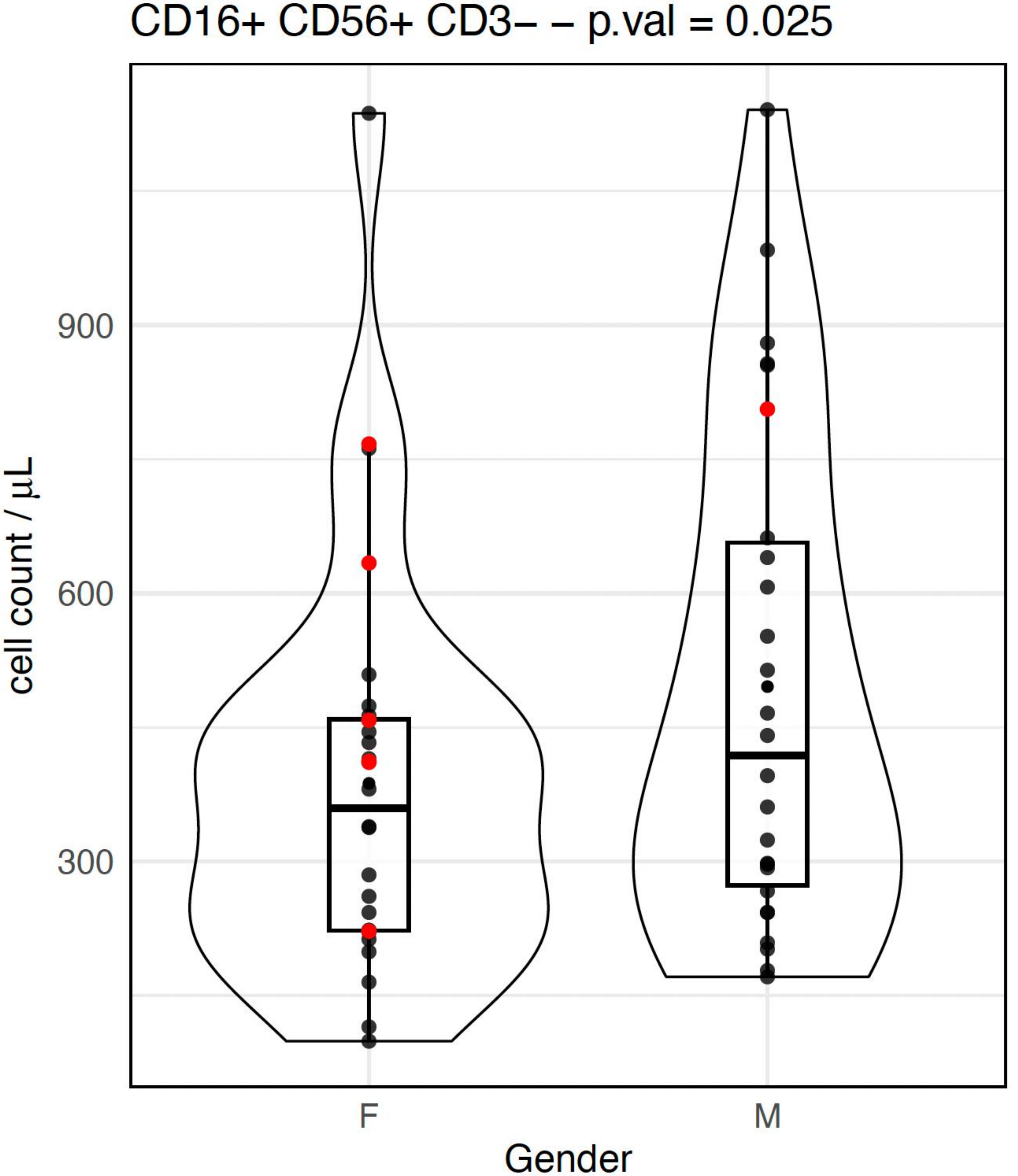
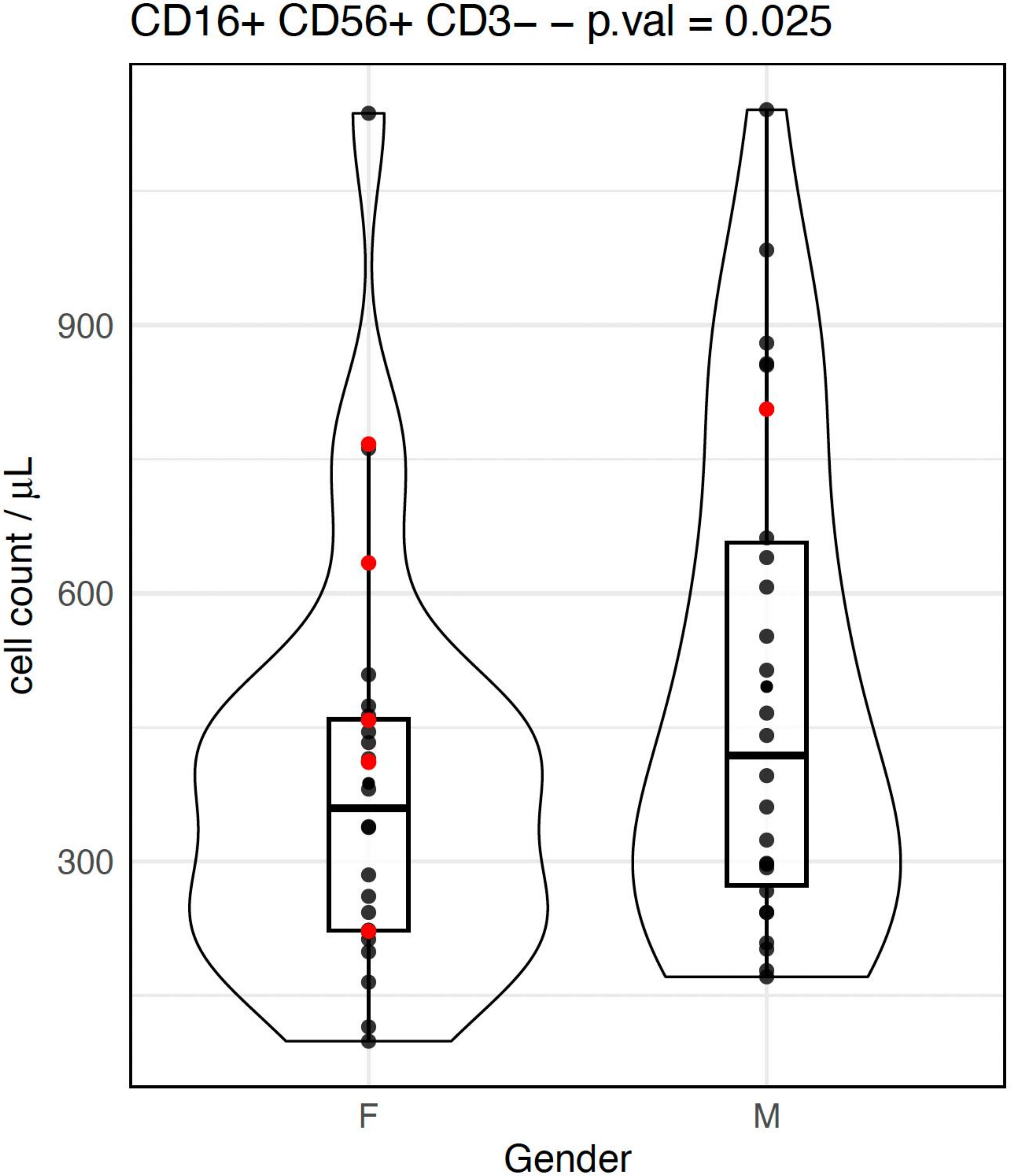
Figure 1. Blood NK cell count (CD3-CD16+CD56+) values stratified by sex. Violin plots illustrate the distribution of NK cell counts in women and men. Each dot represents an individual healthy donor; red dots indicate the oldest centenarians (≥ 105 years), and black dots indicate younger participants. Multivariable regression analysis revealed a statistically significant effect of sex (P = 0.025), with higher NK cell counts in men than in women. NK: natural killer.
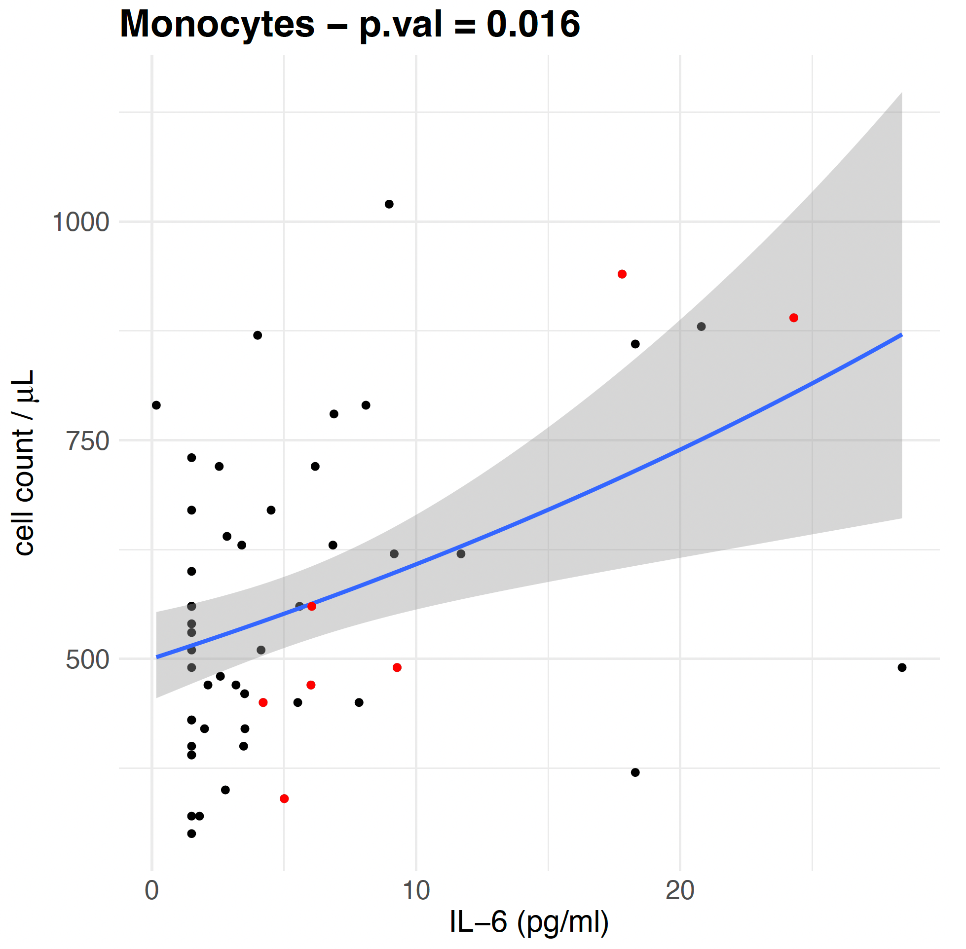
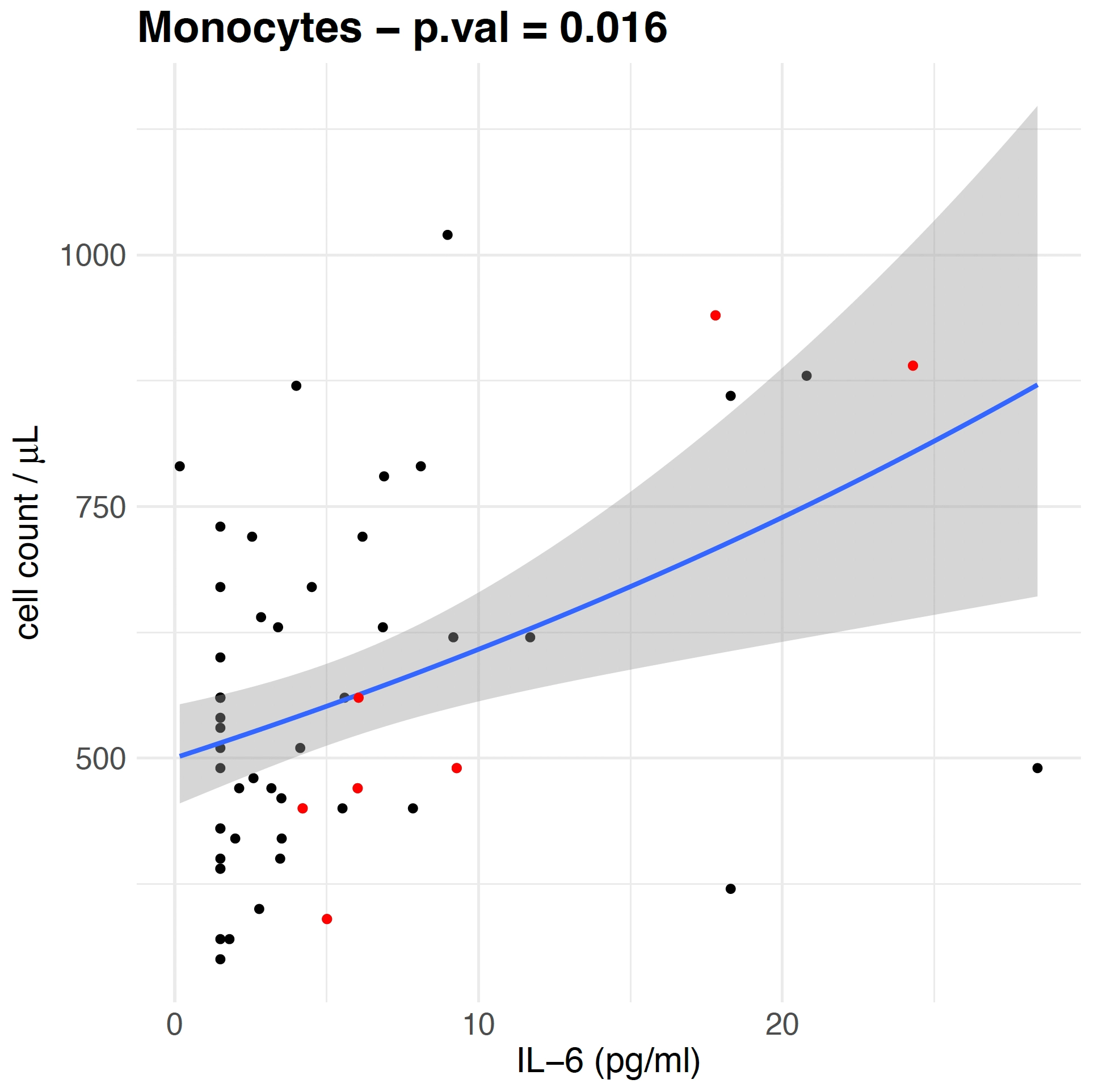
Figure 2. Association between circulating IL-6 levels and blood monocyte count values. Scatter plot depicting the relationship between monocyte counts and IL-6 concentrations in all participants (n = 54). Each dot represents an individual healthy donor; red dots indicate the oldest centenarians (≥ 105 years), and black dots indicate younger participants. The solid blue line represents the fitted trend from the negative binomial regression model, and the shaded area denotes the 95% model-based confidence interval. Multivariable regression analysis showed a statistically significant association (P-value = 0.016), with higher IL-6 levels corresponding to increased monocyte counts. IL-6: interleukin 6.
Table 1. Results of the multiple negative binomial regression models examining the associations of sex, age, CMV serostatus, and IL-6 concentration with circulating NK cell and monocyte counts.
| Coefficient | IRR | IRR CI | Standard Error | P-Value | |
| NK CELLS | |||||
| (intercept) | 295.74 | 197.3-449.8 | 0.201 | ||
| Female | 0.74 | 0.57-0.96 | 0.134 | 0.025 | * |
| Age | 1.00 | 0.99-1.01 | 0.003 | 0.089 | N.S. |
| Anti-CMV ≥ 180 | 1.21 | 0.85-1.71 | 0.176 | 0.290 | N.S. |
| IL-6 | 1.00 | 0.98-1.03 | 0.013 | 0.650 | N.S. |
| MONOCYTES | |||||
| (intercept) | 468.34 | 374.0-589.1 | 0.112 | ||
| Female | 0.94 | 0.81-1.1 | 0.075 | 0.460 | N.S. |
| Age | 1.00 | 0.99-1.0 | 0.002 | 0.270 | N.S. |
| Anti-CMV ≥ 180 | 0.88 | 0.73-1.07 | 0.098 | 0.210 | N.S. |
| IL-6 | 1.02 | 1.00-1.03 | 0.007 | 0.016 | * |
Results are presented as incidence rate ratios (IRRs = exp(β)), with 95% confidence intervals (95% CI). Standard errors correspond to the estimated standard errors of the regression coefficients (β) on the log scale, as obtained from the fitted negative binomial models. P values refer to Wald tests for the regression coefficients. *: Significant associations (P < 0.05); N.S.: not significant. In bold type: data statistically significant; IRR: incidence rate ratios; CI: confidence intervals; NK: natural killer; CMV: cytomegalovirus; IL-6: interleukin 6.
4. Discussion
In the present study, we re-analysed previously published data on circulating NK cells[11] and monocytes[4] in a Sicilian cohort spanning a wide age range, with particular attention to LLIs and the oldest centenarians. Building on our previous observations showing increased circulating levels of NK cells and monocytes in individuals reaching extreme ages, we applied multiple negative binomial regression models to assess the independent contribution of age, sex, CMV serostatus, and IL-6 concentrations to NK and monocyte counts. This modelling strategy was selected because it is particularly appropriate for count-based immunological outcomes and allows the simultaneous evaluation of several biologically relevant predictors[12]. To the best of our knowledge, no previous study, whether or not including the oldest centenarians, has analysed circulating NK and monocyte counts by simultaneously considering these four variables.
In previous investigations, chronological age was positively associated with peripheral NK cell percentages, with the highest values observed in the most advanced-age centenarians. Sex-stratified analyses indicated that this age-related increase persisted in both sexs, with a stronger statistical association in men[10]. However, in the present study, this relationship was no longer statistically significant after adjustment for key confounders, including sex, CMV serostatus, and circulating IL-6 levels, in a multivariable model. This attenuation likely reflects collinearity among these variables, mirroring the biological interdependence of ageing-related processes rather than a mere statistical artefact. Indeed, in our cohort, age had been closely associated with both CMV seropositivity and elevated IL-6 levels, supporting their contribution to immune ageing[12]. Notably, despite the predominance of women among the oldest centenarians, NK cell counts were significantly lower in women (Table 1). This finding is not unexpected and may be explained by a combination of genetic and hormonal factors, as well as a functional trade-off whereby NK cells in women tend to be more active or functionally mature despite lower numbers. A key mechanism underlying this difference involves the X-linked gene KDM6A (also known as UTX), an epigenetic regulator[22]. Despite X-chromosome inactivation, immune-related genes may escape complete silencing, resulting in a functional double gene dosage in women[23,24]. This leads to higher UTX expression in female NK cells, which enhances antiviral activity but is associated with reduced cell numbers[22]. Conversely, lower UTX expression in men is linked to higher NK cell counts but reduced production of interferon-γ, a critical antiviral cytokine[22]. Regarding the contribution of CMV infection, which is not found in our study, Solana et al.[25] distinguished between the impact of ageing and that of persistent CMV infection on the NK-cell compartment. According to their interpretation, ageing appears to be mainly responsible for the redistribution of NK-cell subsets defined by CD56 and CD16 expression, whereas CMV contributes primarily to the expansion of long-lived or “memory-like” NK cells characterised by the expression of NKG2C and CD57. Thus, NK-cell remodelling in older individuals reflects the interplay between biological ageing, reduced production of newly generated NK cells, and chronic antigenic exposure, particularly to CMV. The reduced output of new NK cells may explain the decline in CD3-CD56+CD16- cells, since these represent a more immature population more directly dependent on continuous bone-marrow output. Conversely, CD16+ NK cells are more mature peripheral cells that may be maintained for longer through prolonged survival and homeostatic proliferation, processes now known to be linked, at least in part, to age-related epigenetic changes in haematopoietic stem cells, as well as to chronic CMV exposure. Although biologically important, this aspect cannot be verified because of the absence of the determination, in our study, of CD57+ or NKG2C+ NK cells. Consequently, a specific effect of CMV on these subsets cannot be detected in our dataset.
In our previous analysis, an age-related increase in circulating monocyte counts was observed only in men[4]. However, after adjustment for key confounders in a multivariable model, monocyte counts were no longer associated with age but remained significantly and positively correlated with serum IL-6 concentrations.This finding suggests that the association between age and monocyte expansion is attenuated after adjustment for IL-6, indicating that IL-6 may account for part of the observed association. Indeed, chronic low-grade inflammation is sustained by a self-reinforcing loop in which circulating monocytes produce IL-6, which in turn promotes the survival, recruitment, and activation of additional monocytes and other immune cells. In physiological conditions, monocyte numbers and IL-6 levels are tightly regulated within a homeostatic range. Monocytes continuously traffic to tissues, where they contribute to immune surveillance, repair, and maintenance, while low basal levels of IL-6 support these functions[26-30]. With ageing, however, this balance may shift toward a state of chronic activation, in which elevated IL-6 levels drive monocyte expansion and functional reprogramming. Therefore, our findings support the view that inflamm-ageing, rather than age itself, is a primary determinant of increased monocyte counts, with potential sex-specific modulation.
Metcalf et al.[31] analysed classical, intermediate, and non-classical monocyte subsets in healthy young adults and older individuals (≥ 65 years), demonstrating that under unstimulated conditions, monocyte subsets did not exhibit significant age-related alterations. In contrast, following stimulation with toll-like receptor 4 (TLR4), TLR7/8, and retinoic acid-inducible gene I agonists, marked transcriptional and functional differences emerged between younger and older subjects. These findings support the concept that monocyte ageing may be manifested primarily at the functional and transcriptional levels rather than as a simple quantitative alteration in basal cell counts. However, this study included neither LLIs nor the oldest centenarians and did not specifically address circulating monocyte counts or their independent association with age, sex, CMV serostatus, and IL-6 concentrations, therefore it is not comparable with the results of the present study.
Several limitations of this study should be considered. First, its cross-sectional design inherently restricts the ability to infer causality or to capture temporal changes and trajectories associated with ageing, although it remains appropriate for identifying associations and describing prevalence patterns. Another limitation is the lack of detailed characterization of NK cell subsets. This was a deliberate methodological decision, as the primary aim of the study was to assess total CD3-CD16+CD56+ NK cell counts, known to increase in centenarians[10] across a broad age spectrum. Consequently, more refined immunophenotyping was beyond the scope of the present work. Future studies will be needed to elucidate age-related changes within specific NK cell subsets. Furthermore, the study cohort also exhibited a marked sex imbalance, consistent with the demographic profile of extreme longevity in Italy, where women substantially outnumber men. Data from national registries indicate that among individuals aged ≥ 107 years, men account for only a small minority, resulting in a women-to-men ratio exceeding 10:1[20]. This imbalance inevitably constrains the robustness of sex-stratified analyses and calls for caution when interpreting sex-specific findings. Finally, the number of very old participants, particularly the oldest-old individuals, was limited. However, this reflects the rarity of such populations since supercentenarians represent only a small fraction of centenarians[20].
Future research should address these limitations through carefully designed collaborative studies, ideally involving groups with specific expertise in semi- and supercentenarian research. However, given the profoundly ecological and context-dependent nature of both genetic and immunological determinants of extreme longevity, recruitment should not simply be expanded across heterogeneous geographical and socio-environmental settings. Pooling biologically and epidemiologically distinct populations would risk obscuring meaningful differences and generating misleading averages rather than clarifying the mechanisms underlying immune ageing within specific ecological contexts. Rather, larger studies should preserve the territorial, demographic, and exposomic coherence of the populations under investigation, to avoid diluting or confounding context-specific biological signals. Within this framework, increased sample size, improved male representation, and more detailed analyses of immune cell subsets remain important objectives. Ultimately, larger, preferably longitudinal studies, based on well-characterised and ecologically comparable populations, will be essential to validate and refine the biological significance of the associations observed in this study[32-36].
5. Conclusion
Our findings indicate that no independent association between age and innate immune cell populations was detected after adjustment for chronic inflammation and sex, suggesting that the observed age-related differences are associated with these interconnected biological factors rather than chronological age per se. These results underscore the importance of adopting an integrated framework that considers the interplay between sex, inflamm-ageing, and chronic antigenic stimulation in shaping immune remodelling during extreme longevity. These observations may help refine future immunogerontological research frameworks and support the development of more individualized approaches to the study of immune ageing.
Supplementary materials
The supplementary material for this article is available at: Supplementary materials.
Acknowledgements
We thank Alessandro Delucchi, a representative of the European Supercentenarian Organisation and of Longevity Quest, data provider to the Buck Institute for Research on Aging, for the identification of the semi- and supercentenarians by http://www.supercentenariditalia.it. The authors declare that ChatGPT Plus was used solely for language polishing during the manuscript preparation process. The authors take full responsibility for the integrity, originality, and accuracy of the work.
Authors contribution
Bertolazzi G: Conceptualization, methodology, formal analysis, data curation.
Candore G: Conceptualization, supervision, funding acquisition.
Caruso C: Conceptualization, methodology, investigation, writing-original draft, funding acquisition, supervision.
Calabrò A: Conceptualization, formal analysis, data curation, writing-original draft, writing-review & editing.
Corsale AM, Di Simone M: Methodology, investigation.
Meraviglia S: Methodology, formal analysis, supervision.
Accardi G: Investigation, data curation, writing-original draft, writing-review & editing.
Caldarella R: Investigation.
Aiello A: Investigation, writing-original draft, writing-review & editing.
Conflicts of interest
The authors declare no conflicts of interest.
Ethical approval
The study was conducted in accordance with the Declaration of Helsinki and approved by the Ethics Committee of “Paolo Giaccone”, University Hospital, which approved the study protocol (Nutrition and Longevity, Approval No. 032017, March 01, 2017).
Consent to participate
Informed consent was obtained from all subjects involved in the study.
Consent for publication
Not applicable.
Availability of data and materials
Data supporting the findings of this study are available from supplementary materials and the corresponding author upon reasonable request.
Funding
None.
Copyright
© The Author(s) 2026.
References
-
5. Anaya JM, Lozada-Martinez ID, Acosta-Ampudia Y, Tobón G. Immunosenescence and human healthspan. Lessons from centenarians. Curr Opin Immunol. 2026;100:102777.[DOI]
-
6. Plaza-Florido A, Carrera-Bastos P, Pérez-Prieto I, Fiuza-Luces C, Radom-Aizik S, del Pozo Cruz B, et al. The long-lived immune system of centenarians. Nat Rev Immunol. 2026.[DOI]
-
7. Looney RJ, Falsey A, Campbell D, Torres A, Kolassa J, Brower C, et al. Role of cytomegalovirus in the T cell changes seen in elderly individuals. Clin Immunol. 1999;90(2):213-219.[DOI]
-
8. Vescovini R, Telera AR, Pedrazzoni M, Abbate B, Rossetti P, Verzicco I, et al. Impact of persistent cytomegalovirus infection on dynamic changes in human immune system profile. PLoS One. 2016;11(3):e0151965.[DOI]
-
9. Hassouneh F, Goldeck D, Pera A, van Heemst D, Slagboom PE, Pawelec G, et al. Functional changes of T-cell subsets with age and CMV infection. Int J Mol Sci. 2021;22(18):9973.[DOI]
-
12. Bertolazzi G, Calabrò A, Accardi G, Aiello A, Caruso C, Corsale AM, et al. B cell levels in centenarians, semi-supercentenarians, and supercentenarians: Descriptive analysis by age, sex, cytomegalovirus status, and interleukin-6. J Ageing Longev. 2026;6(1):9.[DOI]
-
13. Tu W, Rao S. Mechanisms underlying T cell immunosenescence: Aging and cytomegalovirus infection. Front Microbiol. 2016;7:2111.[DOI]
-
16. Klenerman P, Oxenius A. T cell responses to cytomegalovirus. Nat Rev Immunol. 2016;16(6):367-377.[DOI]
-
18. Aiello A, Accardi G, Aprile S, Caldarella R, Carru C, Ciaccio M, et al. Age and sex-related variations of molecular and phenotypic parameters in a cohort of Sicilian population: From young to centenarians. Aging Dis. 2021;12(7):1773.[DOI]
-
19. Centenari in Italia, una popolazione in aumento [Internet]. Rome: Istituto Nazionale di Statistica; 2022 [cited 2024 Nov 12]. Available from: https://www.istat.it/wp-content/uploads/2022/06/STAT-TODAY_CENTENARI-2021.pdf
-
20. Persone viventi più longeve in Italia [Internet]. Italy: Supercentenari d’Italia; 2014 [cited 2026 Apr 14]. Available from: https://www.supercentenariditalia.it/persone-viventi-piu-longeve-in-italia
-
23. Consortium G, Tukiainen T, Villani AC, Yen A, Rivas MA, Marshall JL, et al. Landscape of X chromosome inactivation across human tissues. Nature. 2017;550(7675):244-248.[DOI]
-
24. Souyris M, Cenac C, Azar P, Daviaud D, Canivet A, Grunenwald S, et al. TLR7 escapes X chromosome inactivation in immune cells. Sci Immunol. 2018;3(19):eaap8855.[DOI]
-
25. Solana R, Campos C, Pera A, Tarazona R. Shaping of NK cell subsets by aging. Curr Opin Immunol. 2014;29:56-61.[DOI]
-
28. Abraham S, Parekh J, Lee S, Afrin H, Rozenblit M, Blenman KRM, et al. Accelerated aging in cancer and cancer treatment: Current status of biomarkers. Cancer Med. 2025;14(9):e70929.[DOI]
-
29. Gabay C. Interleukin-6 and chronic inflammation. Arthritis Res Ther. 2006;8:S3.[DOI]
-
33. Argentieri MA, Amin N, Nevado-Holgado AJ, Sproviero W, Collister JA, Keestra SM, et al. Integrating the environmental and genetic architectures of aging and mortality. Nat Med. 2025;31(3):1016-1025.[DOI]
Copyright
© The Author(s) 2026. This is an Open Access article licensed under a Creative Commons Attribution 4.0 International License (https://creativecommons.org/licenses/by/4.0/), which permits unrestricted use, sharing, adaptation, distribution and reproduction in any medium or format, for any purpose, even commercially, as long as you give appropriate credit to the original author(s) and the source, provide a link to the Creative Commons license, and indicate if changes were made.
Publisher’s Note
Science Exploration remains a neutral stance on jurisdictional claims in published
maps
and institutional affiliations. The views expressed in this article are solely those
of
the author(s) and do not reflect the opinions of the Editors or the publisher.
Share And Cite


Science Exploration Style
Bertolazzi G, Calabrò A, Accardi G, Aiello A, Caruso C, Corsale AM, et al. Beyond chronological age: Sex and IL-6 are associated with natural killer cell and monocyte counts in a Sicilian cohort spanning adulthood to extreme longevity. Geromedicine. 2026;2:202637. https://doi.org/10.70401/Geromedicine.2026.0031
Tips
Copy completed.
Submit a Manuscript
Author Instructions
Cite this Article
Article Metrics
0
View
0
Download
Cited
Article Updates

 Get citation
Science Exploration Style

Bertolazzi G, Calabrò A, Accardi G, Aiello A, Caruso C, Corsale AM, et al. Beyond chronological age: Sex and IL-6 are associated with natural killer cell and monocyte counts in a Sicilian cohort spanning adulthood to extreme longevity. Geromedicine. 2026;2:202637. https://doi.org/10.70401/Geromedicine.2026.0031
copy
Share Link
copy
Subscribe