Regulation of miR-29 family by resistance training: Implications for muscular and cognitive aging

Paola Pinto-Hernandez
1,2,3,*

,
David Fernandez-Vivero
1,2
,
Manuel Fernandez-Sanjurjo
1,2
,
Loretta Francesca Cosco
4
,
Pasqualina Buono
4,5
,
Benjamín Fernandez-Garcia
1,2
,
Cristina Tomas-Zapico
1,2
,
Eduardo Iglesias-Gutierrez
1,2,*
*Correspondence to:
Paola Pinto-Hernandez. Department of Functional Biology, Area of Physiology, University of Oviedo, Oviedo 33006, Asturias, Spain.
E-mail: paola.pinto.hernandez@ki.se
Eduardo Iglesias-Gutierrez. Department of Functional Biology, Area of Physiology, University of Oviedo, Oviedo 33006, Asturias, Spain. E-mail: iglesiaseduardo@uniovi.es
Eduardo Iglesias-Gutierrez. Department of Functional Biology, Area of Physiology, University of Oviedo, Oviedo 33006, Asturias, Spain. E-mail: iglesiaseduardo@uniovi.es
Geromedicine. 2027;3:202628. 10.70401/Geromedicine.2026.0043
Received: April 19, 2026Accepted: October 13, 2026Published: October 13, 2026
Abstract
During aging, modifiable lifestyle factors, such as physical activity, play an increasingly important role. Specifically, it is known that physical fitness declines relatively predictably with age and that resistance training can be a highly effective strategy for preventing this decline, both at muscular and cognitive levels. Several studies have described that one of the mechanisms mediating some of the beneficial effects of physical exercise during aging is the regulation of miRNAs, and in particular, the miR-29 family appears to play a relevant role. In this review, we analyse, firstly, the relevance of the miR-29 family in aging and, secondly, the protective effects of resistance exercise in delaying age-related dysfunction through the regulation of miR-29.
Keywords
Aging, sarcopenia, resistance exercise, skeletal muscle, miRNAs, miR-29 family
1. Introduction
Population aging represents a major global challenge, characterized by the progressive accumulation of cellular and tissue alterations that reduce the organism’s ability to maintain physiological resilience[1]. Consequently, individuals experience increased susceptibility to metabolic, cardiovascular, neurodegenerative, and musculoskeletal disorders, as well as cancer, which together constitute the leading causes of morbidity and mortality worldwide[2]. According to the United Nations estimates, more than 1.5 billion people will be over 65 years old by 2050[3], underscoring the urgent need to understand the molecular mechanisms that drive functional decline during aging.
Although non-modifiable risk factors, such as age-related hormonal changes and genetic determinants, are linked to the development of these conditions, the impact of modifiable lifestyle-associated factors, such as nutrient availability and physical activity, has been recognized during aging[4]. In fact, fitness levels decline in a relatively predictable manner with advancing age, mainly due to the loss of muscle mass and strength after 50 years. This negatively impacts quality of life, and it is associated with reduced functional capacity and independence, as well as an increased risk of diseases, such as neurodegenerative diseases and dementia[5-7], highlighting the importance of interventions that mitigate losses in both physical and cognitive function. Although low-intensity aerobic exercise has traditionally been recommended for older adults, a recent meta-analysis has shown that the most effective interventions included resistance exercise (RE)[8], which helps preserve musculoskeletal integrity[9] and mitigate cognitive decline in aging[10].
The beneficial effects of exercise are increasingly linked to epigenetic mechanisms, which become progressively dysregulated with age. Among the epigenetic regulators, microRNAs (miRNAs) have attracted particular attention. These small non-coding RNAs (ncRNAs) modulate gene expression through sequence-dependent interactions with target mRNAs[11,12] and their expression patterns are strongly influenced by both aging and lifestyle factors, such as diet and physical activity[2]. Within the miRNA landscape, the miR-29 family occupies a central role in regulating key biological processes associated with aging[13]. The miR-29 family is widely evolutionarily conserved, and the seed sequence, which plays an essential role in mRNA target recognition, is identical in all mammals, including primates and rodents[14]. This family is composed of three members: miR-29a, miR-29b, and miR-29c, that are expressed from two genomic loci: miR-29a/b-1 (located on chromosome 7 for H. sapiens and chromosome 6 for M. musculus) and miR-29b-2/c (located on chromosome 1 for H. sapiens and M. musculus). MiR-29 family members are prominently expressed across multiple tissues and developmental stages, where they regulate metabolic and structural homeostasis. Thus, elevated levels have been linked to impaired insulin secretion in pancreatic β cells, dysregulated gluconeogenesis in the liver, and insulin resistance in skeletal muscle and adipose tissue[14]. Structurally, miR-29 members function as fibromiRs; specifically their lower levels correlate with the upregulation of multiple extracellular matrix (ECM) genes[15]. Given its dual involvement in metabolic regulation and tissue structural integrity, the miR-29 family represents a promising regulatory axis through which RE could potentially exert its protective effects against age-related functional decline. This study investigates, first, the relevance of the miR-29 family in the aging process and, second, the protective effects of RE in delaying age-related dysfunction through miR-29 regulation.
2. Role of miR-29 Family During Aging
Two well-cited review articles have identified several hallmarks of aging[16,17], including epigenetic alterations as a central feature. Within these epigenetic mechanisms, ncRNAs, and particularly miRNAs, have emerged as key regulators of aging processes. Indeed, one of the first miRNAs to be characterized, lin-4, regulates lifespan in C. elegans[18]. Since then, their contribution to lifespan modulation has been deeply explored, not only through the study of the post-transcriptional repression of genes involved in longevity pathways, cellular stress responses, and metabolic regulation[19], but also because the components of the miRNA biogenesis machinery itself become dysregulated with aging[1]. MiRNAs also regulate tissue- and cell-type-specific aging phenotypes in mammals, particularly in tissues relevant for exercise performance, such as the liver, heart, and the main tissue involved: skeletal muscle.
2.1 Impact of miR-29 on muscle regeneration and aging-driven sarcopenia
During aging, skeletal muscle homeostasis is disrupted, shifting the balance between growth and atrophy, ultimately resulting in a loss of muscle mass and function, characteristic of sarcopenia. The expression of numerous miRNAs is dysregulated during this process, and the miR-29 family appears to play a relevant role[20]. During postnatal skeletal muscle growth and muscle differentiation, the expression of this family is upregulated in mice[21]. Members of the miR-29 family target Eln, Col1a1 and Col3a1, which encode ECM proteins, thereby potentially reducing the accumulation of fibrotic tissue in the muscle[22-24]. The involvement of miR-29 is further demonstrated in muscle fibrosis associated with muscular dystrophy[25,26], where miR-29 levels are often downregulated, suggesting that restoration of its expression could improve this condition. This miRNA family additionally promotes muscle regeneration by activating satellite cells and contributing to myogenesis. It does this, in part, by targeting negative regulators like YY1, HDAC4, and RYBP[27]. However, during aging, muscle cells become senescent, unable to contribute to muscle growth, and elevated and sustained levels of miR-29c can inhibit the proliferation of muscle progenitor cells (Figure 1)[21,25,26,28]. Thus, the miR-29 family appears to exert a context-dependent role: beneficial during regeneration, yet potentially detrimental to muscle mass preservation when chronically elevated during aging.
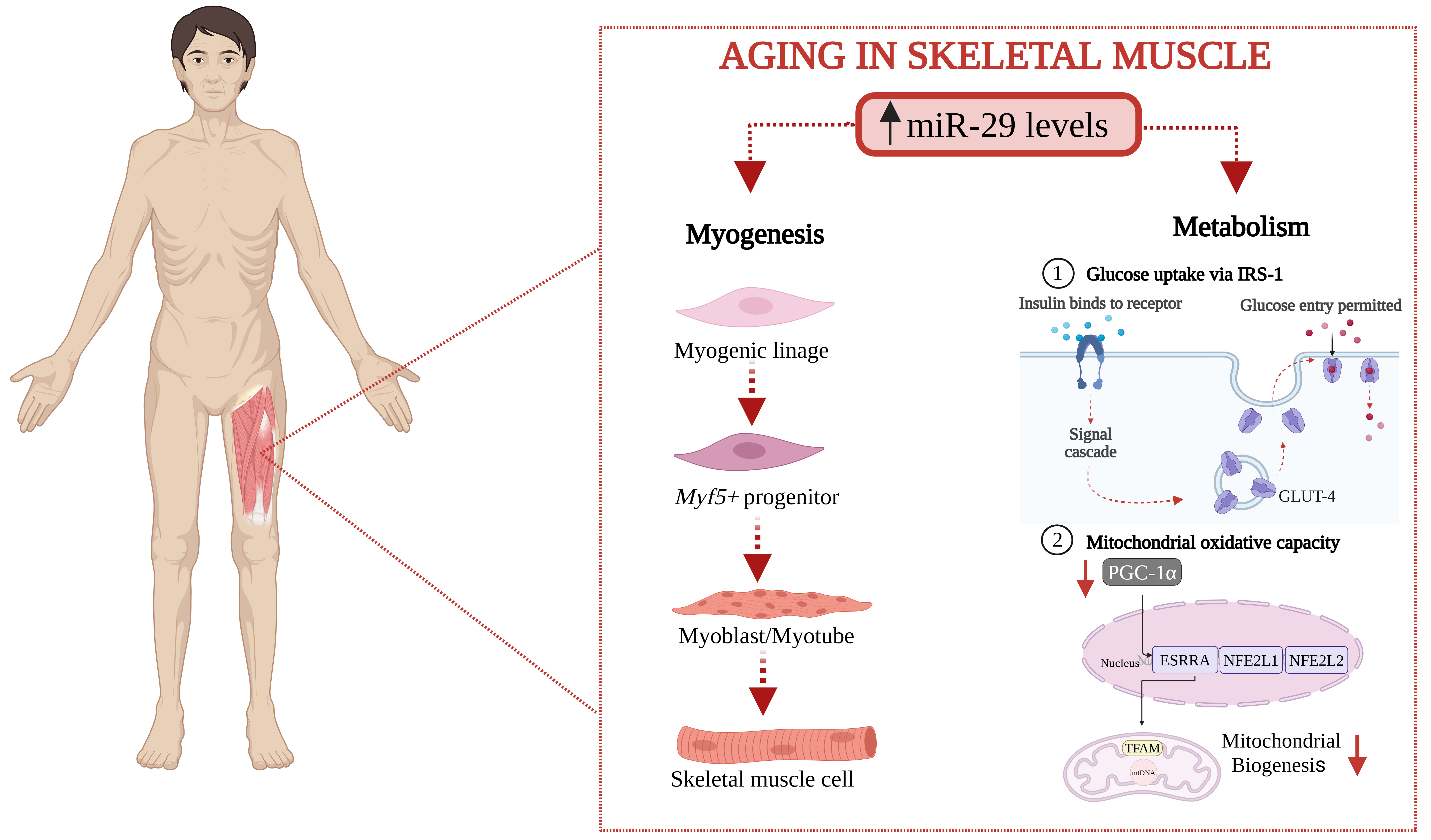
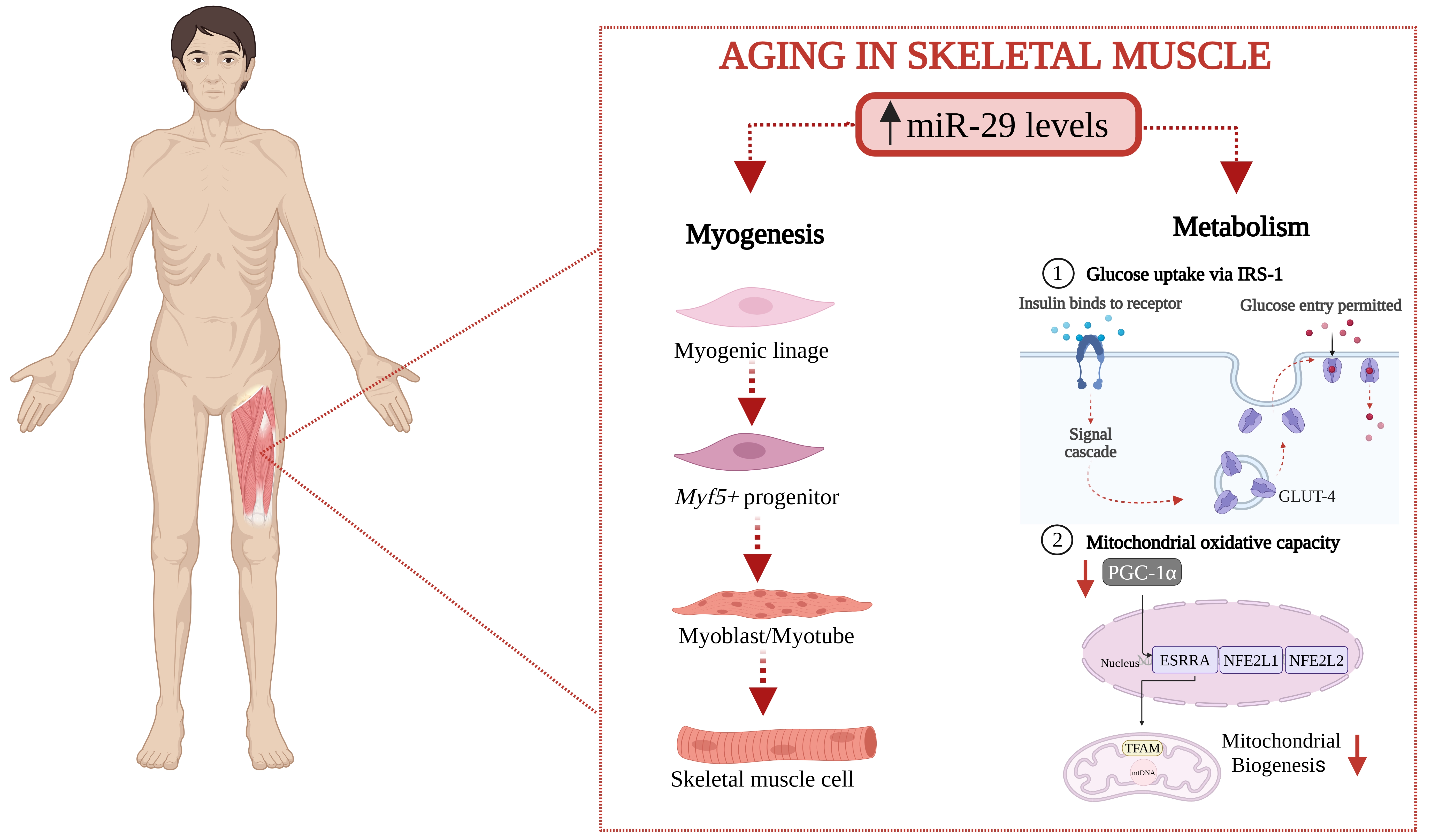
Figure 1. Consequences of elevated miR-29 family expression levels in skeletal muscle during aging. Sustained miR-29 levels inhibit myogenesis and myoblast differentiation. Furthermore, regarding glucose metabolism, the miR-29 family negatively affects glucose uptake via IRS-1 and decreases the transcriptional coactivator PGC1α, thereby reducing mitochondrial oxidative capacity. Created in BioRender. Pinto, P. (2026) https://BioRender.com/zg9nu60. IRS-1: insulin receptor substrate 1; PGC1α: peroxisome proliferator-activated receptor gamma coactivator 1-alpha; GLUT-4: glucose transporter 4; ESRRA: estrogen-related receptor alpha; NFE2L1: nuclear factor, erythroid 2 like 1; NFE2L2: nuclear factor, erythroid 2 like 2.
2.2 Metabolic functions of miR-29 in skeletal muscle
Beyond these structural and regenerative impairments, aging skeletal muscle also undergoes profound metabolic alterations that further contribute to functional decline. Aged muscle exhibits persistent inflammation, reduced mitochondrial oxidative capacity, impaired lipid utilization, a shift towards a more glycolytic metabolism, and a progressive decline in insulin sensitivity[29]. Importantly, many of these metabolic disturbances are also linked to miR-29 family dysregulation (Figure 1). Indeed, in the skeletal muscle of type 2 diabetic patients and murine models of this condition, the expression of miR-29 family members is upregulated. This overexpression impairs insulin signalling via IRS-1, reduced glucose uptake, and diminished palmitate oxidation[14]. Regarding mitochondrial oxidative capacity, miR-29a and miR-29c overexpression led to a repression of the transcriptional coactivator PGC1α (PPARGC1a), decreasing mitochondrial biogenesis, and, consequently, the oxidative capacity of skeletal muscle (Figure 1)[30]. Similarly, in terms of inflammation, the absence of miR-29a-3p, miR-29b-3p, and miR29c-3p alleviates skeletal muscle senescence and systemic inflammation[31].
In addition to the information found in the literature, we have performed an in-silico analysis to explore the functional context of the miR-29 family members most highly expressed in human skeletal muscle, i.e., miR-29a-3p, miR-29b-3p, and miR-29c-3p. Thus, miRNA target interactions were obtained from miRWalk v2.0[32] using a stringent prediction score threshold (≥ 0.95), resulting in 3,078 predicted miRNA–gene interactions, of which 168 were experimentally validated (miRTarBase v9.0)[33]. These interactions corresponded to 1,215 unique target genes, including 49 genes with at least one experimentally validated miRNA interaction (miRTarBase v9.0)[33]. To specifically explore muscle-related functions, a high-confidence protein-protein interaction (PPI) network was constructed by integrating tissue enrichment information (TISSUES 2.0 database score ≥ 3 for muscle)[34] using STRING v.12 and Cytoscape v.3.10.4[35]. As shown in Figure 2a, this approach identified a network comprising 97 closely interconnected muscle-associated proteins. Functional annotation revealed that these proteins are predominantly involved in muscle cell structure, differentiation, and maintenance, as well as in metabolic pathways related to glucose and polysaccharide metabolism and insulin signalling (Figure 2b). The biological relevance of this regulatory network was further supported by human phenotype enrichment analysis using the Monarch database[36], which linked these proteins primarily to muscle structure and impaired muscle function (Figure 2b).
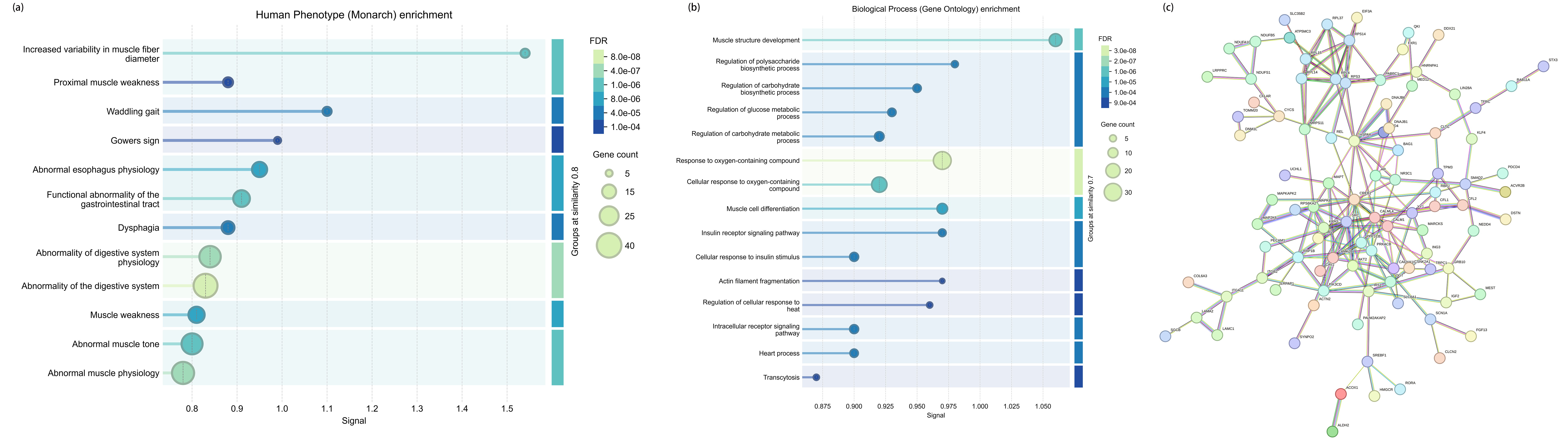
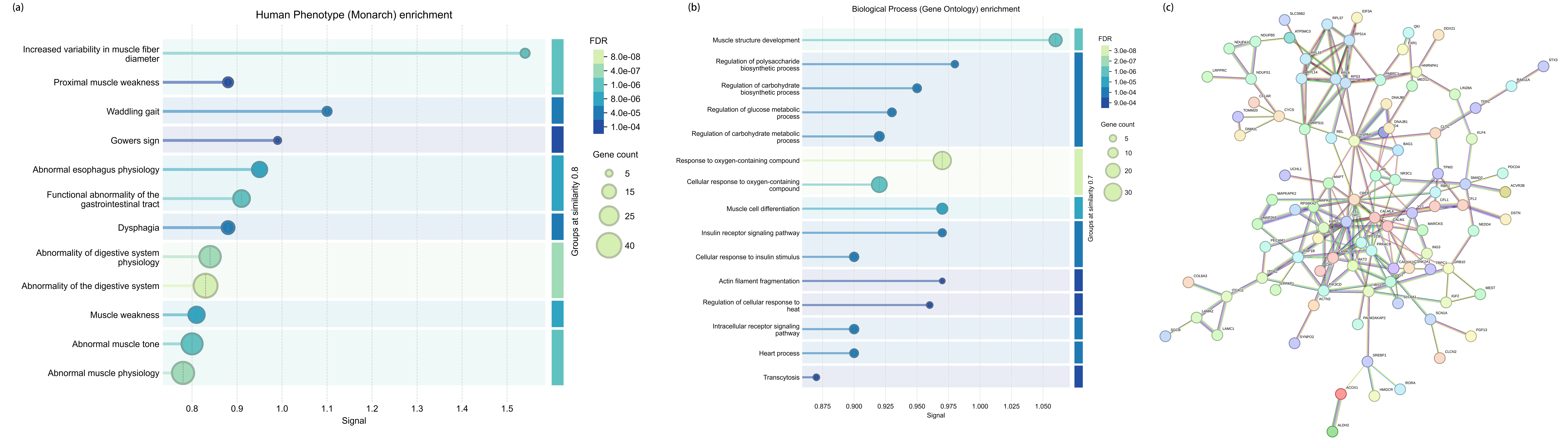
Figure 2. Functional analysis of the miR-29 family in human muscle tissue. (a) Validated and predicted target genes of miR-29a-3p, miR-29b-3p, and miR-29c-3p obtained from miRWalk v.2.0 using a stringent threshold (score ≥ 0.95). Only proteins with high confidence (score ≥ 0.7) PPI network that are also enriched in muscle tissue (TISSUES database score ≥ 3) are represented in the network using STRING v.12 and Cytoscape v.3.10.4. Nodes represent proteins, and edges denote protein-protein associations; (b) Representative GO Biological Process enrichment analysis of the proteins comprising the PPI network available in STRING v.12; (c) Representative human phenotype enrichment analysis for the same protein set, performed using the Monarch database available in STRING v.12. Enriched terms are displayed according to their signal, calculated as weighted mean of the observed/expected ratio and -log(FDR) with related terms clustered together. PPI: protein-protein interaction; GO: gene ontology; FDR: false discovery rate.
Overall, experimental findings and in-silico analyses consistently highlight that the miR-29 family not only influences the structural remodelling of muscle architecture that characterizes muscle aging but also intersects with the metabolic pathways that determine fuel selection, insulin responsiveness, and overall muscle energetic efficiency.
2.3 Role of miR-29 family in cognitive aging and neurodegeneration
During aging, some members of the miR-29 family are progressively upregulated in the brain[37] However, its downregulation has been implicated in neurodegenerative processes, particularly Alzheimer’s disease (AD). Thus, in postmortem brain tissue from patients with late-onset AD, a significant reduction in the miR-29a/b1 cluster correlates with increased expression of β-secretase 1 (BACE1), leading to enhanced amyloid precursor protein (APP) cleavage and subsequent amyloid-beta (Aβ) peptide accumulation[38]. Beyond the amyloidogenic pathway, the miR-29 family has also been reported to be an important survival factor for neuronal cells, where its negative regulation upregulates the expression of proapoptotic genes such as VDAC1[39,40] and dysregulates Neuronal Navigator 3 (NAV3), impairing cortical axon guidance[41].
Additionally, miR-29 members modulate neuroinflammation by regulating glial cell activation. In microglia, an age-associated increase in miR-29a and miR-29b reduces the expression of IGF-1 and CX3CL1, contributing to a pro-inflammatory profile in these cells[37,42], while miR-29c-3p inhibition triggers proinflammatory cytokine release[43]. In astrocytes, miR-29a-3p preserves mitochondrial function and limits cellular damage by targeting the pro-apoptotic gene Puma[44]. Furthermore, miR-29 is highly expressed in cerebellar Purkinje cells, where its deletion increases Hip1 expression, leading to phenotypic features of spinocerebellar ataxia[45].
Taken together, reduced miR-29 levels appear to accelerate the progression of several neurodegenerative processes, including AD and spinocerebellar ataxia. Therefore, finding preventive strategies that can raise the levels of this family during the disease could be of therapeutic value.
2.4 Role of miR-29 in skeletal muscle-brain axis
One aspect of particular interest is interorgan communication within the muscle-brain axis in response to exercise. Accumulating evidence suggests that exercise stimuli modulate the expression of specific miRNAs abundant in muscle, subsequently altering their profiles in plasma and serum[46]. These circulating alterations have several implications for the integrity of the central nervous system (CNS), highlighting a close reciprocal relationship between neuromuscular health and cognitive function. Regarding the miR-29 family, murine models of muscle atrophy have demonstrated elevated levels of plasma extracellular vesicle (EV) miR-29b-3p, which has been proposed to be internalized by neural cells. Specifically, EV-miR-29b-3p derived from C2C12 myotubes is actively taken up by SH-SY5Y neurons, subsequently reducing the expression of key genes essential for neuronal differentiation and survival, such as BCL-2 and LAMC1[47].
Consequently, modulating miR-29 expression in skeletal muscle through non-pharmacological interventions, such as resistance training, could fundamentally regulate its secretion in EVs, affecting the CNS in a beneficial way during aging.
3. Resistance Exercise as Beneficial Modulator of Aging Physiology: Role of miR-29 Family
Regular physical exercise is one of the most effective interventions for promoting healthy aging[48]. The principal hallmarks of aging encompass genomic instability, telomere attrition, epigenetic dysregulation, loss of proteostasis, impaired macroautophagy, dysregulated nutrient-sensing signalling, mitochondrial dysfunction, increased cellular senescence, ECM remodelling, stem cell degeneration, altered intercellular communication, chronic inflammation, dysbiosis, and psychosocial isolation[17]. Within this landscape, the miR-29 family seems to play an important role in many of these processes. Specifically, chronic DNA damage during aging upregulates miR-29 via a p53-dependent pathway to accelerate senescence[49], whereas its downregulation is required to maintain fibroblast quiescence and prevent aberrant ECM remodelling[50]. Furthermore, miR-29 directly targets peroxisome proliferator-activated receptor gamma coactivator 1-alpha (PGC-1α) in cardiac tissue, making its precise regulation essential to prevent mitochondrial dysfunction and metabolic dysregulation[51]. Accumulating evidence indicates that exercise also modulates many of these processes, thereby mitigating the development of age-related diseases[52]. Consistent with these mechanistic insights, current guidelines for adults aged ≥ 65 years advocate multicomponent exercise programmes, in which resistance training is a key element with increasing relevance[53].
3.1 Resistance exercise counteracts hallmarks of aging
In older adults, RE induces an extensive molecular reprogramming, that underlies its well-documented benefits for physical and psychological health[54,55].
In skeletal muscle, RE enhances muscle size and strength via activation of key anabolic pathways, including phosphoinositide 3-kinase (PI3K)/protein kinase B (Akt)/mechanistic target of rapamycin (mTOR), while promoting ECM remodelling that improves collagen organization and tissue quality[56]. In this sense, lifelong football training, a sport with a relevant strength component[57] increases the expression of key markers involved in oxidative metabolism and in the DNA repair and senescence suppression pathways in skeletal muscle[58]. Furthermore, it increases the expression of muscular biomarkers involved in the auto-lysosomal and proteasome-mediated protein degradation machinery, cellular growth and differentiation processes, and in proteasome promotion and autophagy processes in skeletal muscle from lifelong trained elderly subjects compared to untrained controls, promoting healthy aging[59]. Interestingly, although current evidence suggests that regular exercise may help attenuate age-related telomere attrition, its effects on telomere length and telomerase activity appear to be tissue-specific and vary according to exercise type[60-62]. Thus, RE tends to increase both parameters in skeletal muscle and blood cells compared with inactive individuals; however, other exercise modalities, such as aerobic exercise or high-intensity interval training (HIIT), elicit greater effects[63,64].
In addition, RE contributes to a more favourable metabolic profile by improving glucose and lipid metabolism, enhancing whole-body insulin sensitivity and glycemic control in older adults, and promoting a healthier body composition[65]. Similarly, football training improves lipid and glucose metabolism in young, healthy individuals and in elderly subjects, promoting the increase in the expression of key biomarkers involved in cellular energy metabolism, i.e., PPARγ, adiponectin, AMPKα1/α2, TFAM, NAMPT, PGC1α, SIRT1, and in glucose and lipid oxidative metabolism[58,66].
These adaptations help to restore tissue homeostasis and mitigate functional decline during aging, partly mediated by the release of some exerkines[67], including regulatory miRNAs, like the miR-29 family[68].
3.2 Resistance exercise affects miR-29 family expression in skeletal muscle
MiRNAs have been identified as key mediators of several processes involved in skeletal muscle adaptations to exercise. Although their roles in both acute and chronic endurance exercise have been studied[69], the temporal dynamics of miRNA expression in skeletal muscle in response to RE remain insufficiently understood. In addition, most of the studies have focused on miRNAs highly expressed in skeletal muscle, so called myomiRs, such as miR-1, miR-133a, miR-133b, miR-206, miR-208, miR-208b, miR-486, and miR-499. These myomiRs regulate myoblast proliferation and myogenic regeneration and differentiation following a single bout of RE. Specifically, McCarthy et al. reported a decreased expression of miR-1 and miR-133a by ~50% in the mouse plantaris muscle of an animal model of muscle hypertrophy following 7 days of functional overload[70]. In humans[71], resistance exercise has also been shown to modulate the expression of various myomiRs, since in young men, an acute bout of resistance exercise coupled with ingestion of amino acids resulted in a significant decrease in miR-1 expression in skeletal muscle, in agreement with the loss of miR-1 expression in response to a hypertrophic stimulus observed in the mouse. Furthermore, Small et al. demonstrated that the muscle-enriched miR-486 modulated PI3K/Akt signaling by targeting phosphatase and tensin homolog (PTEN) and forkhead box O1a (Foxo1a)[72]. However, several studies have also identified non-myomiRs enriched in skeletal muscle that are dynamically regulated by RE, including the miR-29 family[68,73].
Regarding human studies, there is very limited evidence on the relationship between RE and the expression of miR-29 family members at muscle level, particularly in the elderly. Indeed, there is only one study, carried out by Rivas and colleagues[74] who explored the expression of the miR-29 family in the vastus lateralis of healthy young and old men after an acute RE protocol consisting of 3 sets of bilateral knee extension exercise (10 repetitions at 80% of 1 RM) and 3 sets of bilateral leg press exercise (10 repetitions at 80% of 1 RM). A downregulation of miR-29a-3p and miR-29c-3p was detected 6 h after the session, but only in young men, suggesting a blunted miRNA adaptive response to RE in older individuals. Interestingly, Davidsen and colleagues analysed the expression of the miR-29 family in the vastus lateralis of healthy young men (18-30 years old), stratified as low versus high responders, to 12 weeks of RE training. Their findings showed that low responders had reduced muscular miR-29a expression, which may reflect a compensatory response to insufficient activation of growth- and remodelling-related genes[73].
Taken together, it seems that the miR-29 family might act as a molecular switch for muscle adaptation with a different kinetics depending on the RE stimulus. The acute decrease after RE in young people could be related to an increase in anabolic pathways (mTOR), while its elevated levels in high responders to RE training could be essential for structural remodelling. In older adults, the absence of this acute dynamic could suggest that elevated miR-29 levels could be a hallmark of an attenuated anabolic response and the development of sarcopenia. However, a major limitation is that the reported findings are based solely on acute resistance exercise protocols and primarily on young, exclusively male cohorts. Consequently, extrapolating these results to older individuals or female populations remains highly challenging. This gap is particularly critical given that sex hormones directly influence epigenetic adaptations and circulating EV-miRNA profiles following exercise. Furthermore, cyclical fluctuations in ovarian hormones during the menstrual cycle have been shown to significantly alter baseline plasma miRNA levels[75,76]. Therefore, further research integrating age- and sex-diverse cohorts is strictly necessary to properly evaluate the definitive role of the miR-29 family in long-term training conditions. To bridge these clinical gaps, animal models have been utilized, offering insights to further explore how the miR-29 family responds to RE. Previous data from our research group showed that miR-29a-3p was downregulated in the soleus muscle of young male mice after 4-weeks of RE training. Furthermore, the expression levels of this miRNA in the quadriceps, metabolically more glycolytic, were negatively correlated with resistance performance gain in response to RE training, meaning that the greater the resistance improvement with training, the lower the miR-29a-3p expression in this muscle. Moreover, we have described that systemic miR-29a/b1 knock-out mice showed an impaired resistance capacity, which may be related to the altered expression of key regulatory genes related to substrate availability, transport, and metabolic use[68].
In general, while the available human literature on miR-29 in response to resistance exercise remains critically limited, particularly regarding older populations and women, the data suggest that the miR-29 family may play an important role in skeletal muscle adaptive capacity. Although exercise modulates its expression, an attenuated response is observed in older individuals, and the kinetics appear to depend on whether the stimulus is acute or chronic. Mechanistically, these adaptations are hypothesized to be driven by the activation of key anabolic pathways, such as PI3K/Akt/mTOR and IGF-1, which promote muscle growth, remodelling, and performance enhancement. However, given the limited robustness of current translational data, future research must move beyond descriptive association studies and critically analyse the precise, causal mechanisms underlying miR-29 regulation during chronic resistance training in both older men and women.
3.3 Resistance exercise crosstalk mediated by extracellular miR-29 family: an emerging exerkine
The regulatory relevance of miRNAs in skeletal muscle tissue in response to RE is not limited to the intracellular compartment. Skeletal muscle can also release and take up miRNAs, largely packaged within EVs, which are increasingly recognized as mediators of intercellular communication[77,78]. EV-carried miRNAs can mirror, complement, or diverge from intramuscular miRNA changes, providing an additional layer of regulation[77]. It has been shown that both acute and chronic RE can modulate the EV-miRNA profile[79]. However, the information is more limited regarding the effect of RE training on the modulation of miR-29 circulating levels. Ghodrat and colleagues[80] explored whole-blood miR-29b expression in response to a combination of RE and HIIT exercise for 8-weeks, recruiting overweight to moderately obese females with type 2 diabetes (T2D), aged 45-65 years. Regarding the training protocol, it included upper and lower body RE and it was applied at a progressive intensity, from 40 to 70% 1RM: first and second weeks, 20 reps with 40% of 1RM; third and fourth weeks, 15 reps with 50% of 1RM; fifth and sixth weeks, 12 reps with 60% of 1RM; and seventh and eighth weeks, 10 reps with 70% of 1RM. The HIIT protocol was a low-volume HIIT (7-10 intervals) involving 60 s running bouts on the treadmill at 80-90% VO2max, interspersed with 60 s of active recovery at 20-30% VO2max. The training group was separated in two different categories: same day (SD) group, who trained RE followed by HIIT exercise 3 days/week, and different day (DD) group, who trained RE and HIIT exercise on different days (6 days/week). A sedentary group was also included. They observed that circulating miR-29b levels were significantly lower in the DD group compared to the sedentary group. Furthermore, using an in vitro C2C12 model validation, they reported that transfection of miR-29b mimic resulted in decreased protein levels of IGF-1, a factor involved in the IGF1-PI3K-AKT signalling pathway[81], the dysregulation of which is one of the reasons for the muscle atrophy observed during T2D[82]. Indeed, Li et al.[81] demonstrated that miR-29b, but not miR-29a or miR-29c, controls muscle atrophy in vitro, showing that miR-29b inhibition resulted in increased expression levels of IGF1 and PI3K, suggesting a role in skeletal muscle cells in rats.
While identifying the precise tissue origin remains a major challenge in circulating EV-miRNA profiling, our previous investigations alongside independent studies have demonstrated that the miR-29 family can be actively secreted in EVs by cultured skeletal muscle cells in response to electrostimulation[68]. Lautaoja-Kivipelto et al.[84] also analysed this response after a 24-hour low-frequency electrical pulse stimulation (EPS) protocol (1 Hz, 2 ms, 12 V) using short RNA sequencing in EVs obtained from C2C12 cells; the baseline presence of miR-29a-3p was robustly verified in the secreted EVs post-stimulation. Furthermore, in a more complex three-dimensional “myobundle” model consisting of multinucleated, striated myofibers and a pool of Pax7(+) myogenic satellite cell precursors, Vann et al.[85] examined the secreted miRNA EV profile under chronic electrical stimulation at both low and high frequencies. MiR-29a-3p expression was significantly elevated in EVs following both stimulation regimens, reinforcing our previous data that skeletal muscle tissue can actively export miR-29a-3p via EVs during contractile activity. These findings highlight a relevant role of miR-29a-3p in the systemic response to exercise, acting as a myokine.
Considering all these data together, miRNAs, and particularly the miR-29 family, could have a role in skeletal muscle adaptation to RE, although it is complex, context-dependent, and not yet fully understood. The limited available evidence, both in humans and animal models, shows that RE decreases the expression of members of the miR-29 family in muscle tissue, especially miR-29a and miR-29c, although a blunted response is observed in older individuals, and the kinetics appear dependent on the acute or chronic stimulus. This modulation could be associated with improved muscle growth, remodelling, and performance, likely through the enhancement of key anabolic pathways such as IGF-1/mTOR/PI3K-AKT. However, this hypothesis requires further causal validation in diverse human populations. In this sense, the reduction of members of the miR-29 family could be considered a favourable adaptive mechanism, while attenuated responses could contribute to a reduced capacity for RE adaptation.
On the other hand, at the circulating level, whole-blood miR-29b kinetics do not reflect the intramuscular response but rather adds a new systemic layer of regulation. This compartmental divergence aligns with findings by D’Souza et al.[86], who reported a different pattern of expression among plasma, EV, and vastus lateralis miRNAs of young men following acute exercise. This discrepancy could be dually driven: first, by the selective exercise-induced packaging of specific miRNAs into EVs independently of cellular concentrations; and second, by methodological differences. Specifically, whole-blood assays, such as that by Ghodrat et al.[80], capture both protein-bound and EV-encapsulated miRNAs, thereby reflecting a significant contribution from non-skeletal muscle tissues (e.g., endothelial or immune cells) and establishing a more complex physiological crosstalk during exercise. Therefore, the contrast between decreased muscular miR-29 after acute RE and increased circulating whole-blood levels following chronic HIIT and RE combined protocols highlights the multi-factorial nature of miR-29 regulation. To resolve these discrepancies, future research must move beyond descriptive associations and focus on elucidating the precise causal and translational mechanisms of this exercise-induced crosstalk, particularly regarding EV-mediated transport.
Nevertheless, moving beyond descriptive associations to establish the precise causal and translational mechanisms of this EV-mediated axis remains a critical priority for future research.
4. Conclusions
Resistance exercise has emerged as a powerful non-pharmacological intervention to counteract the progressive loss of structural integrity, metabolic flexibility, and regenerative capacity that occurs in every tissue during aging, with skeletal muscle being particularly vulnerable to these changes. Accumulating evidence points to the miR-29 family as a key epigenetic regulator at the intersection of these processes. Furthermore, findings from human and animal studies suggest that RE can influence miR-29 expression in skeletal muscle, as well as its circulating levels.
Future research should aim to clarify the temporal and tissue-specific regulation of the miR-29 family in response to RE, including its role in EVs mediated inter-organ communication. Longitudinal studies combining muscle biopsies, circulating miRNA profiling, and functional outcomes will be essential to determine whether the miR-29 family can serve as a biomarker of training adaptation or a therapeutic target to enhance healthy aging. Ultimately, elucidating how RE reshapes miR-29-dependent regulatory networks may open new avenues to mitigate sarcopenia and preserve metabolic health in the aging population.
Authors contribution
Pinto-Hernandez P: Writing-original draft, visualization
Fernandez-Vivero D: Writing-original draft, methodology.
Fernandez-Sanjurjo M, Fernandez-Garcia B, Tomas-Zapico C: Visualization.
Cosco LF, Buono P: Writing-original draft.
Iglesias-Gutierrez E: Writing-original draft, supervision, writing-review & editing.
All authors have read and agreed to the published version of the manuscript.
Conflicts of interest
The authors declare no conflicts of interest.
Ethical approval
Not applicable.
Consent to participate
Not applicable.
Consent for publication
Not applicable.
Availability of data and materials
Not applicable.
Funding
This work was supported by the Fundación Tatiana Pérez de Guzmán el Bueno (Convocatoria de ayudas a proyectos de investigación in neuroscience, 2020; Grant No. P-121-FTPGB 2020) to C.T.-Z. and E.I.-G. P.P.H. received support from the Alfonso Martín Escudero Foundation (Spain) through the award of a FUNDAME fellowship.
Copyright
© The Author(s) 2026.
References
-
2. Turko R, Hajja A, Magableh AM, Omer MH, Shafqat A, Khan MI, et al. The emerging role of miRNAs in biological aging and age-related diseases. Non Coding RNA Res. 2025;13:131-152.[DOI]
-
3. United Nations, Department of Economic and Social Affairs, Population Division. World Population Ageing 2019: Highlights. 2019 [cited 2025]. Available from: https://digitallibrary.un.org/record/3846855?ln=en&v=pdf
-
10. Dhahbi W, Briki W, Heissel A, Schega L, Dergaa I, Guelmami N, et al. Physical activity to counter age-related cognitive decline: Benefits of aerobic, resistance, and combined training: A narrative review. Sports Med Open. 2025;11(1):56.[DOI]
-
17. López-Otín C, Blasco MA, Partridge L, Serrano M, Kroemer G. The hallmarks of aging. Cell. 2013;153(6):1194-1217.[DOI]
-
19. Jusic A, Thomas PB, Wettinger SB, Dogan S, Farrugia R, Gaetano C, et al. Noncoding RNAs in age-related cardiovascular diseases. Ageing Res Rev. 2022;77:101610.[DOI]
-
22. Dong XR, Majesky MW. Restoring elastin with microRNA-29. Arterioscler Thromb Vasc Biol. 2012;32(3):548-551.[DOI]
-
29. Nishikawa H, Asai A, Fukunishi S, Nishiguchi S, Higuchi K. Metabolic syndrome and sarcopenia. Nutrients. 2021;13(10):3519.[DOI]
-
31. Ding Z, Ma G, Zhou B, Cheng S, Tang W, Han Y, et al. Targeting miR-29 mitigates skeletal senescence and bolsters therapeutic potential of mesenchymal stromal cells. Cell Rep Med. 2024;5(8):101665.[DOI]
-
33. Huang HY, Lin YCD, Cui S, Huang Y, Tang Y, Xu J, et al. miRTarBase update 2022: An informative resource for experimentally validated miRNA–target interactions. Nucleic Acids Res. 2022;50(D1):D222-D230.[DOI]
-
34. Palasca O, Santos A, Stolte C, Gorodkin J, Jensen LJ. TISSUES 2.0: An integrative web resource on mammalian tissue expression. Database. 2018;2018:bay028.[DOI]
-
37. Fenn AM, Smith KM, Lovett-Racke AE, Guerau-de-Arellano M, Whitacre CC, Godbout JP. Increased micro-RNA 29b in the aged brain correlates with the reduction of insulin-like growth factor-1 and fractalkine ligand. Neurobiol Aging. 2013;34(12):2748-2758.[DOI]
-
52. Qiu Y, Fernández-García B, Lehmann HI, Li G, Kroemer G, López-Otín C, et al. Exercise attenuates the hallmarks of aging: Novel perspectives. J Sport Health Sci. 2026;15:101108.[DOI]
-
58. Mancini A, Vitucci D, Labruna G, Imperlini E, Randers MB, Schmidt JF, et al. Effect of lifelong football training on the expression of muscle molecular markers involved in healthy longevity. Eur J Appl Physiol. 2017;117(4):721-730.[DOI]
-
62. Sánchez-González JL, Sánchez-Rodríguez JL, Varela-Rodríguez S, González-Sarmiento R, Rivera-Picón C, Juárez-Vela R, et al. Effects of physical exercise on telomere length in healthy adults: Systematic review, meta-analysis, and meta-regression. JMIR Public Health Surveill. 2024;10:e46019.
-
64. Werner CM, Hecksteden A, Morsch A, Zundler J, Wegmann M, Kratzsch J, et al. Differential effects of endurance, interval, and resistance training on telomerase activity and telomere length in a randomized, controlled study. Eur Heart J. 2019;40(1):34-46.[DOI]
-
76. Kargl CK, Sterczala AJ, Santucci D, Conkright WR, Krajewski KT, Martin BJ, et al. Circulating extracellular vesicle characteristics differ between men and women following 12 weeks of concurrent exercise training. Physiol Rep. 2024;12(9):e16016.[DOI]
Copyright
© The Author(s) 2026. This is an Open Access article licensed under a Creative Commons Attribution 4.0 International License (https://creativecommons.org/licenses/by/4.0/), which permits unrestricted use, sharing, adaptation, distribution and reproduction in any medium or format, for any purpose, even commercially, as long as you give appropriate credit to the original author(s) and the source, provide a link to the Creative Commons license, and indicate if changes were made.
Publisher’s Note
Science Exploration remains a neutral stance on jurisdictional claims in published
maps
and institutional affiliations. The views expressed in this article are solely those
of
the author(s) and do not reflect the opinions of the Editors or the publisher.
Share And Cite


Science Exploration Style
Pinto-Hernandez P, Fernandez-Vivero D, Fernandez-Sanjurjo M, Cosco LF, Buono P, Fernandez-Garcia B, et al. Regulation of miR-29 family by resistance training: Implications for muscular and cognitive aging. Geromedicine. 2027;3:202628. https://doi.org/10.70401/Geromedicine.2026.0043
Tips
Copy completed.
Submit a Manuscript
Author Instructions
Cite this Article
Article Metrics
0
View
0
Download
Cited
Article Updates

- Abstract
- Keywords
- 1. Introduction
- 2. Role of miR-29 Family During Aging
- 3. Resistance Exercise as Beneficial Modulator of Aging Physiology: Role of miR-29 Family
- 4. Conclusions
- Authors contribution
- Conflicts of interest
- Ethical approval
- Consent to participate
- Consent for publication
- Availability of data and materials
- Funding
- References
- Copyright
 Get citation
Science Exploration Style

Pinto-Hernandez P, Fernandez-Vivero D, Fernandez-Sanjurjo M, Cosco LF, Buono P, Fernandez-Garcia B, et al. Regulation of miR-29 family by resistance training: Implications for muscular and cognitive aging. Geromedicine. 2027;3:202628. https://doi.org/10.70401/Geromedicine.2026.0043
copy
Share Link
copy