Electrospun medicated cellulose acetate nanofibers: Architectures, controlled release and transdermal drug delivery applications

Jiaqi Zhu
1
,
Bo Wang
2
,
Lin Du
2
,
Deng-Guang Yu
1,*

,
Yuanjie Zhu
2,*
*Correspondence to:
Deng-Guang Yu, School of Materials & Chemistry, University of Shanghai for Science and Technology, Shanghai 200093, China.
E-mail: ydg017@usst.edu.cn
Yuanjie Zhu, Department of Dermatology, Naval Medical Center, Naval Medical University, Shanghai 200052, China. E-mail: 13918338960@126.com
Yuanjie Zhu, Department of Dermatology, Naval Medical Center, Naval Medical University, Shanghai 200052, China. E-mail: 13918338960@126.com
Smart Mater Devices. 2027;3:202635. 10.70401/smd.2026.0059
Received: July 03, 2026Accepted: October 14, 2026Published: November 02, 2026
Abstract
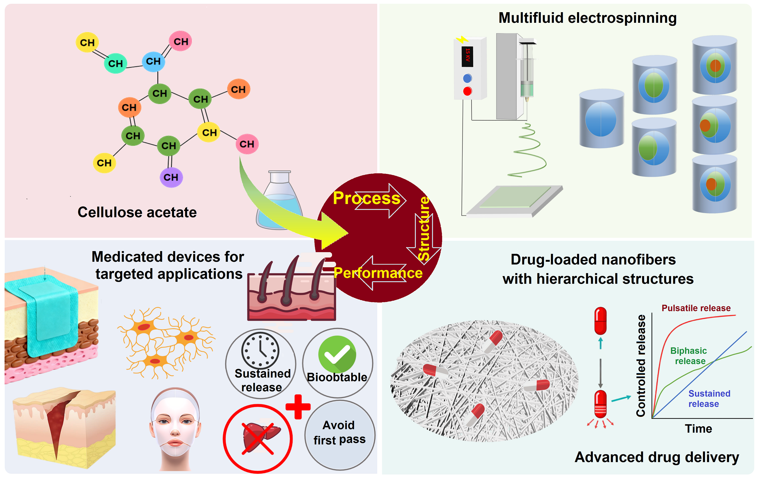
Electrospun cellulose acetate (CA) nanofibers have emerged as structurally programmable platforms for smart material systems and devices because of their tunable porosity, controllable fiber morphology, biocompatibility, compatibility, and processability using single-fluid, coaxial, side-by-side, and multifluid electrospinning. Unlike conventional drug-loaded fibrous mats, recent CA-based systems have been endowed with the ability to regulate payload distribution, diffusion pathways, interfacial wettability, and stimulus-responsive release through rational structural designs. This review summarizes the process-structure-function relationships of electrospun CA nanofibers, with emphasis on solvent engineering, fiber architecture, multiscale morphology control, and programmable release profiles. Particular attention is given to core-sheath, Janus, and trilayer core-sheath nanofibers that enable sustained, biphasic, pulsatile, and near-zero-order release profiles. The potential of these fibrous systems as skin-interfaced smart platforms for transdermal delivery, wound management, and wearable therapeutic interfaces is discussed. Finally, future directions involving green manufacturing, AI-assisted formulation design, sensing-feedback-release integration, and clinical translation are proposed.
Graphical Abstract
Keywords
Cellulose acetate, electrospun nanofibers, drug delivery, controlled release, transdermal drug delivery, nanostructure
1. Introduction
Fibers, including continuous filaments and elongated structures, are widely present in nature[1]. With the development of nanotechnology, fiber diameters have been reduced to the micro- and nanoscale. Among one-dimensional nanomaterials, nanofibers with diameters of tens to hundreds of nanometers show an exceptionally high specific surface area and porosity[2], which give them unique physicochemical properties. These features make nanofibers useful in information technology, environmental protection, food safety testing, and localized drug delivery. More importantly, when their morphology and internal structure are carefully controlled, nanofibers can also serve as structure-programmed platforms for smart material systems.
The performance of nanofibers is closely related to their building materials. Commonly used materials include polymers, carbon-based materials, composites, and semiconductors. Natural polymers are abundant, biocompatible, biodegradable, and sometimes antimicrobial. Typical examples include sodium alginate, zein, gelatin, cellulose, collagen, chitosan, hyaluronic acid, dextran, and cyclodextrins. In contrast, synthetic polymers usually show better mechanical strength, thermal stability, and chemical resistance. Frequently used synthetic polymers include polyvinylpyrrolidone (PVP), polyvinyl alcohol, polyethylene oxide, hydroxypropyl methylcellulose, ethyl cellulose, cellulose acetate (CA), polystyrene, polylactic acid, and polycaprolactone (PCL)[3,4]. Therefore, rational material selection is the first step in designing functional and smart nanofiber systems.
Several fabrication strategies have been developed to prepare polymeric nanofibers, including template synthesis, phase separation, self-assembly, sea-island bicomponent spinning, and electrospinning[5-8]. Among these methods, electrospinning is considered one of the most promising strategies because of its simple operation, high efficiency, and strong structural tunability. During electrospinning, charges are induced in a polymer solution and stretched by an electric field to form a stable jet[9]. After stretching and whipping, the jet solidifies into fibers. The fiber diameter can be adjusted from the micrometer scale to the nanometer scale by changing processing parameters[10,11]. The obtained nonwoven mats have a high surface area and interconnected pores, making them suitable for drug loading, controlled release, and skin-interfaced delivery.
Cellulose-based materials have attracted wide academic and industrial interest because of their biocompatibility, renewability, and biodegradability[12]. Cellulose nanomaterials also show a high specific surface area, high crystallinity, high mechanical strength, low density, flexibility, elasticity, and impact resistance. Among different cellulose derivatives, CA is a commonly used polymer for electrospinning because of its good biocompatibility and degradability. The introduction of acetyl groups improves the solubility and mechanical properties of CA, giving it strong adaptability in drug delivery systems. Compared with many other polymer fibers, CA nanofibers show advantages such as biocompatibility, water-insolubility, biodegradability, good mechanical performance, low cost, high binding affinity, and chemical resistance[13]. These properties make CA a suitable matrix not only for conventional drug delivery but also for structure-controlled fibrous systems.
Transdermal drug delivery systems provide a noninvasive route that can avoid first-pass metabolism and reduce the discomfort caused by injections. Therefore, they are useful for chronic disease management and pain control. However, the stratum corneum acts as a strong barrier and limits the penetration of many drugs. Because of their high surface area, tunable porosity, and adjustable surface morphology, electrospun CA nanofibers can improve drug loading, support controlled release, and enhance transdermal transport[14]. From a smart materials perspective, these advantages are not only related to the chemical properties of CA, but are also strongly affected by fiber structure, including fiber diameter, porosity, surface morphology, and internal architecture.
In the Web of Science database, literature related to CA nanofibers over the past 20 years was searched using the following terms: “TS = cellulose acetate and TS = electrospinning or nanofibers”; “TS = cellulose acetate and TS = electrospinning or nanofibers and TS = drug delivery”; and “TS= cellulose acetate and TS = electrospinning or nanofibers and TS = transdermal delivery”. The search trend is shown in Figure 1. The number of studies on “cellulose acetate and electrospinning or nanofibers” has increased continuously, indicating that this field has become an active research area. Although studies related to drug delivery are fewer, they also show a clear upward trend. In contrast, studies on transdermal delivery remain relatively limited, suggesting that this direction is still at an early stage and has broad development potential.
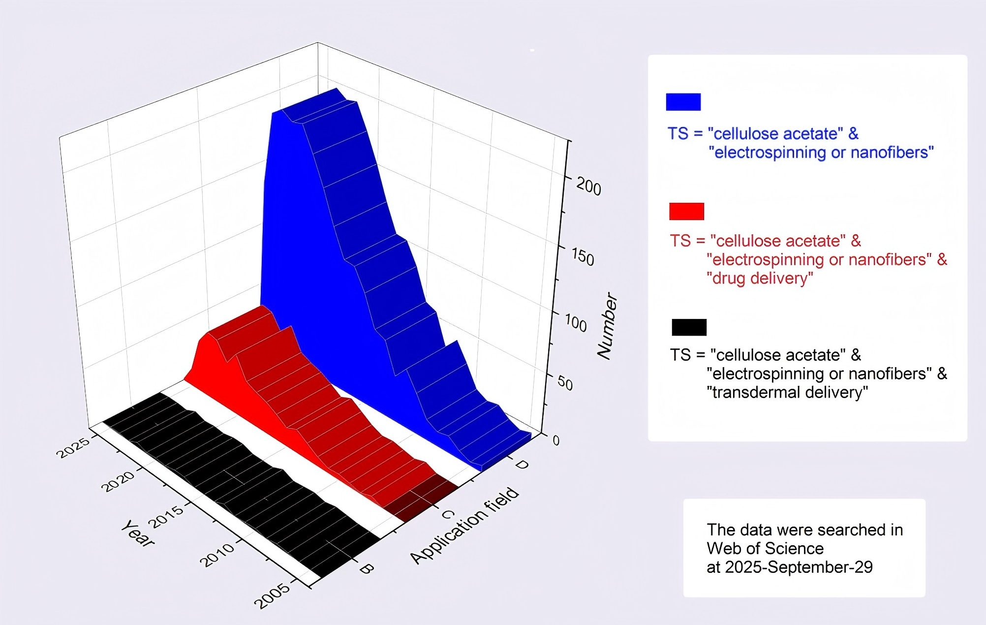
Figure 1. Trends in the number of studies retrieved via the Web of Science platform: search tips were “TS=(cellulose acetate) and TS=(electrospinning or nanofibers)”, “TS=(cellulose acetate) and TS=(electrospinning or nanofibers) and TS=(drug delivery)”, and “TS=(cellulose acetate) and TS=(electrospinning or nanofibers) and TS=(transdermal delivery)”.
As shown in Figure 1, the increasing number of publications suggests that electrospun CA nanofibers are developing from conventional drug carriers toward more advanced smart fibrous systems. Therefore, it is necessary to discuss not only their drug delivery performance, but also the relationship among processing strategy, fiber structure, and functional behavior. CA is hydrophobic, and when applied to the skin, it acts merely as a passive carrier, unable to utilize skin moisture to achieve controlled swelling or erosive drug release. CA has no penetration-enhancing effect. It is unlike certain polymers (such as PVP, poly(vinyl alcohol) (PVA), chitosan, and hyaluronic acid) that are inherently endowed with permeation enhancement or stratum corneum hydration capabilities. Therefore, CA offers no advantages over other polymers as a substrate for transdermal drug delivery. Moreover, CA solutions tend to clog the spinneret during electrospinning. Taken together, these factors have resulted in very limited research on this topic. However, CA itself exhibits excellent spinnability, high mechanical strength, and low-cost availability as an electrospinning material. Therefore, by employing multi-fluid electrospinning processes to solve the spinneret clogging problem and by constructing multi-chamber structures incorporating other polymers to modify its hydrophobicity, the drug release controllable capacity and transdermal permeation performance can be improved, thereby enabling the development of CA-based transdermal nanoscale drug delivery devices.
The potential applications of electrospun CA nanofibers are summarized in Figure 2. These applications are not limited to conventional drug delivery. They also include transdermal patches, wound dressings, antibacterial interfaces, responsive release systems, and wearable therapeutic materials. With the development of advanced electrospinning strategies, CA nanofibers can be further designed into porous, core-shell, multilayer, Janus, and other complex structures[15,16]. Similar structure-regulated designs, such as reverse-gradient core-shell nanofibers and radial polymer-distribution nanofibers, have also been reported to control drug distribution, diffusion pathways, and sustained release behavior[17].
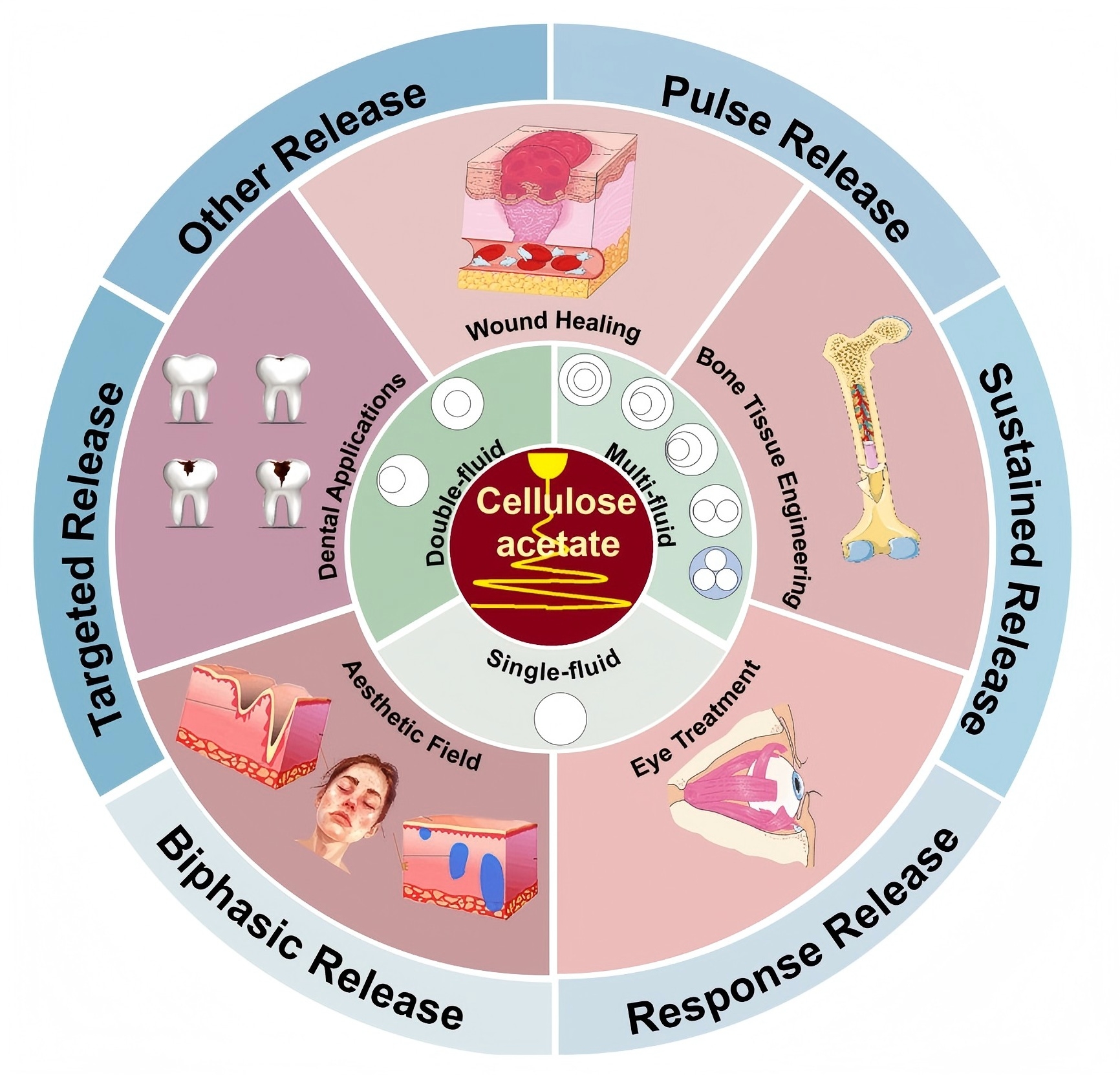
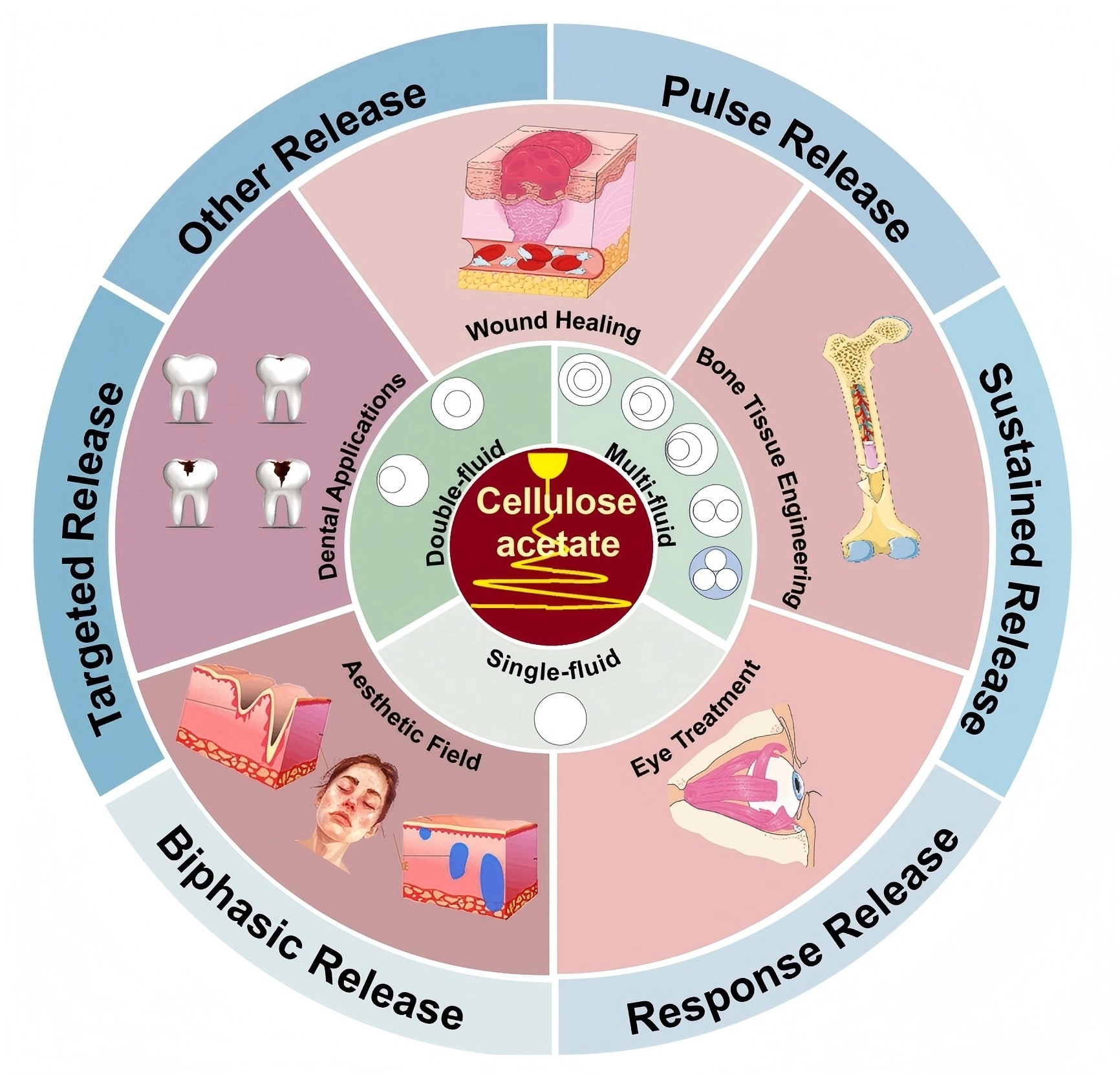
Figure 2. Electrospun CA nanofibers for programmable release and some potential applications. CA: cellulose acetate.
These structure-programmed fibers can regulate drug distribution, diffusion pathways, surface wettability, and release kinetics, providing new possibilities for smart skin-interfaced applications.
Although many studies have reported CA-based electrospun nanofibers for drug loading and release, most of them are still discussed according to drug type or application field. A deeper structure-based understanding is still needed. For example, fiber diameter, porosity, shell thickness, asymmetric structure, and spatial drug distribution can all influence release behavior and skin-interface performance. Therefore, it is important to connect process design, fiber structure, and functional output.
In this review, electrospun CA nanofibers are discussed as structure-programmed smart material platforms. First, the material features of CA and the basic principles of electrospinning are introduced. Then, different electrospinning strategies, including single-fluid, coaxial, Janus, triaxial, and multifluid electrospinning, are compared. Next, structure-regulated release behaviors and smart skin-interfaced applications are discussed. Finally, current challenges and future directions are proposed, including green manufacturing, scalable production, long-term stability, sensing-feedback-release integration, and clinical translation.
2. Basic Principles and Structural Design Strategies of Electrospinning
2.1 Technical principles
Electrospinning is an electrohydrodynamic process that uses a high-voltage electrostatic field to produce nanofibers. The basic setup consists of a high-voltage supply, a syringe pump loaded with a polymer solution, and a collector. When voltage is applied, surface charges on the pendant droplet are pulled by the field, deforming the droplet into a Taylor cone. As soon as the electrostatic force overcomes surface tension, a charged jet erupts from the cone tip. While traveling to the collector, the jet undergoes whipping and stretching instabilities, and the solvent evaporates rapidly, leaving solid nanofibers deposited on the collector.
In electrospinning, fiber morphology and diameter are governed by three primary factors: solution parameters, process parameters, and environmental parameters[13]. The solution parameters include the polymer concentration and viscosity[18], polymer molecular weight[19], surface tension[20], electrical conductivity[21-23], and solvent properties[24]; the process parameters include the voltage[25], solution flow rate[26,27], spinning distance[28], needle diameter and shape, and collector composition and surface morphology; and the environmental parameters include the temperature, humidity[29], and ambient airflow rate. Adjusting these variables within appropriate ranges can effectively tailor the morphology and size of the nanofibers.
2.2 Process type
Electrospinning is one of the popular electrohydrodynamic atomization (EHDA) methods taking advantage of the facile interactions between the working fluid and the electrostatic energy[30-32]. Based on the formats in which the working fluids are guided into the electrical field, electrospinning can be categorized into three types, i.e., needle-based, free surface, and their combinations[4,17,33-35]. The most common one is needle-based electrospinning, which can be further categorized into single-fluid, dual-fluid, and multifluid types on the basis of the simultaneous treatments of the numbers of working fluids. Furthermore, depending on the nozzle structures of the spinnerets, they can also be divided into bi-chamber core-sheath, Janus, and tri-chamber triaxial, tri-chamber Janus, and hierarchical structures based on the combinations of Janus and core-sheath[36,37], as illustrated in Figure 3.
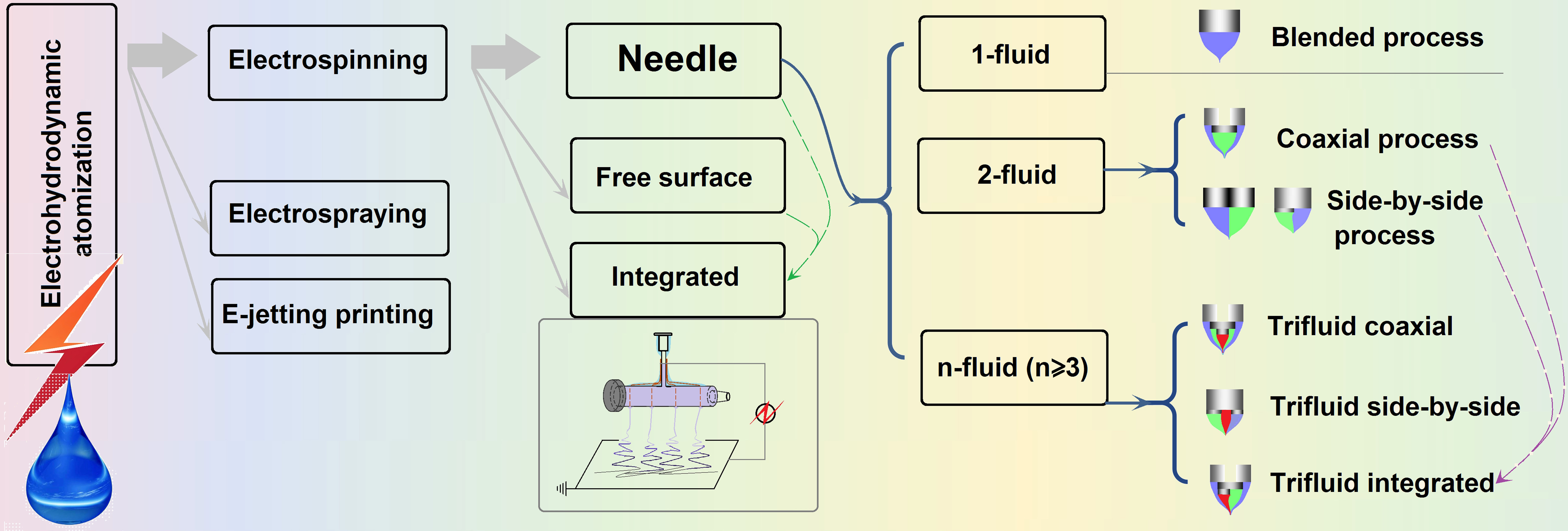
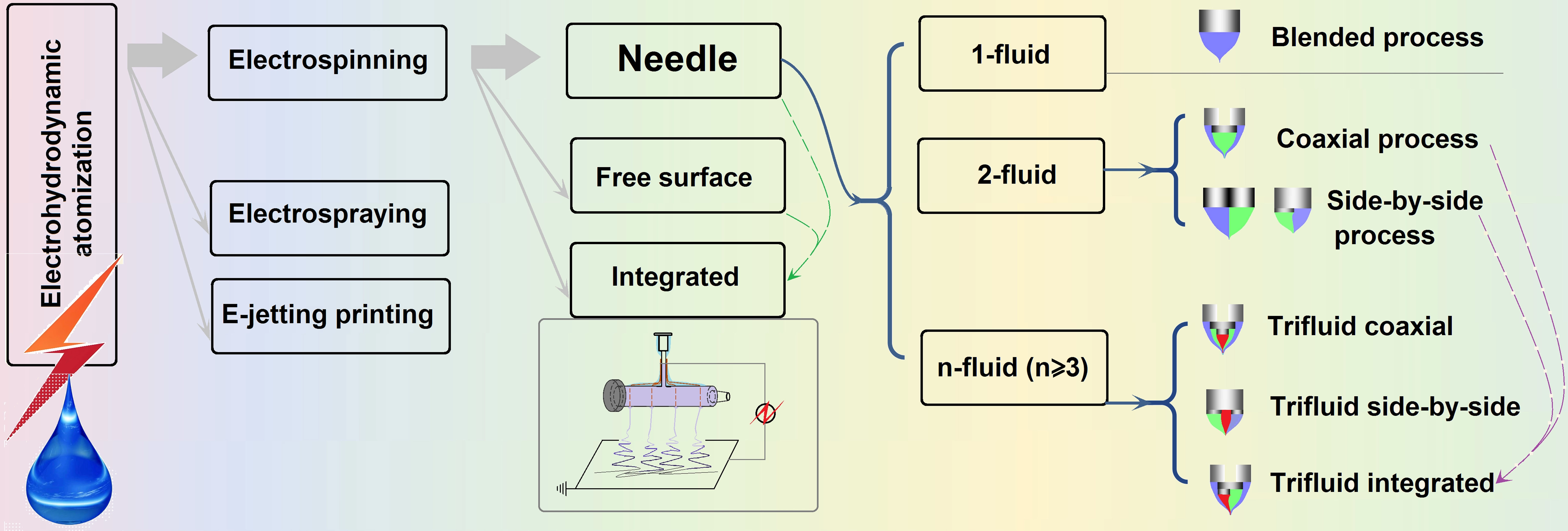
Figure 3. Categories of electrohydrodynamic atomization and electrospinning.
As the name hints, there are two types of multifluid electrospinning. One involves the use of multiple parallel spinnerets to process multiple fluids for scalable production. The other involves the simultaneous processing of multiple fluids through a structured spinneret with multiple channels that guide the fluids in a convergent manner to create multi-chamber nanostructures. Here, all the multifluid electrospinning processes refer to the latter. Unlike single-fluid blended electrospinning, multifluid electrospinning faces challenges that must be addressed to achieve robust and continuous implementation. These challenges include fluid-fluid compatibility and the associated interfacial tensions, multiple fluid-environment interactions, matching of the bending and whipping processes, spinneret fabrication difficulty, low production throughput and poor reproducibility, and the absolute flow rates of the various fluids and their suitable ratios, among others.
2.2.1 Single-fluid electrospinning
As the name implies, single-fluid electrospinning employs one solution stream. Its simplicity and ease of operation have made it a workhorse across many disciplines. Owing to their unique fiber architecture and readily tuned physicochemical properties, these materials create an ideal drug carrier, allowing a wide range of drug molecules to be dissolved directly in the polymer solution for biomedical use. Gong et al.[38] used ketoprofen (KET) as a model drug and an ethyl cellulose (EC)/PVP blend as the polymer matrix to fabricate three distinct nanocomposites: uniform, spindle-shaped, and beaded nanofibers (Figure 4a). All three architectures exhibited a biphasic KET release profile: an initial rapid burst followed by sustained delivery. Shen et al.[39] prepared diclofenac sodium (DS)-loaded Eudragit L 100-55 nanofibers by electrospinning a 5:1 (v/v) ethanol-N,N-dimethylacetamide (DMAc) solvent mixture (Figure 4b). The resulting fibers clearly displayed pH-responsive release behavior.
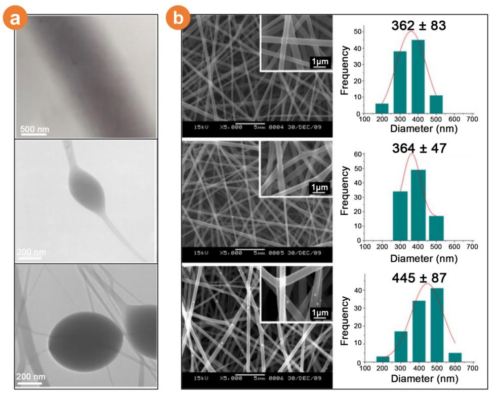
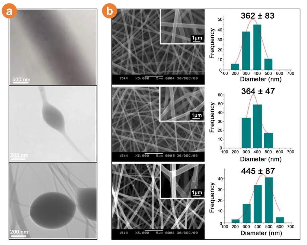
Figure 4. Micrographs and diameter distributions of electrospun products. (a) TEM images of the EHDA products, including straight nanofibers and spindle-on-a-string and bead-on-a-string structures. Reproduced from reference[38]. CC BY 4.0; (b) SEM images of Eudragit® L100-55 fibers prepared at 20% (w/v) in methanol (top), ethanol (middle), and a 5:1 ethanol/DMAc mixture (bottom), with corresponding diameter distributions. Insets show the same fibers at × 10,000 magnification. Reproduced with permission from reference[39]. Copyright © 2011 Elsevier. TEM: transmission electron microscopy; EHDA: electrohydrodynamic atomization; SEM: scanning electron microscopy; DMAc: N,N-dimethylacetamide.
Although single-fluid electrospinning is simple and easy to perform, it imposes several structural and functional limitations on the resulting nanofibers[40,41]: (a) most drugs and biomolecules show marginal compatibility with the organic solvents required for spinning; (b) release profiles are hard to fine-tune, and the rate is often too fast for optimal therapy; (c) poorly water-soluble drugs are difficult to deliver; (d) drug-encapsulation efficiency needs improvement; (e) a universal solvent that dissolves every polymer in a single blend is rarely available. These drawbacks have driven continuous innovation and motivated researchers to develop dual- and multifluid architectures.
2.2.2 Double-fluid electrospinning
Compared with conventional single-fluid methods, double-fluid electrospinning uses two fluids and represents a major advance. The two dominant architectures are coaxial and Janus (side-by-side) spinning; both offer distinct advantages and have become the foundation for more complex multifluid designs.
2.2.2.1 Coaxial electrospinning
Core-sheath nanofibers are the most widely studied complex architecture, with applications across virtually every scientific discipline[42]. Traditional coaxial spinning requires the sheath fluid to be spinnable, but Yu et al.[43] introduced a modified coaxial process that employs an unspinnable polymer solution as the core and a nonspinnable solution, or even a pure solvent, as the sheath. In 2011, the group refined the method by using a concentric spinneret with a slightly protruding core (Figure 5a)[43]. The key advantages of coaxial core-shell nanofibers include the following:
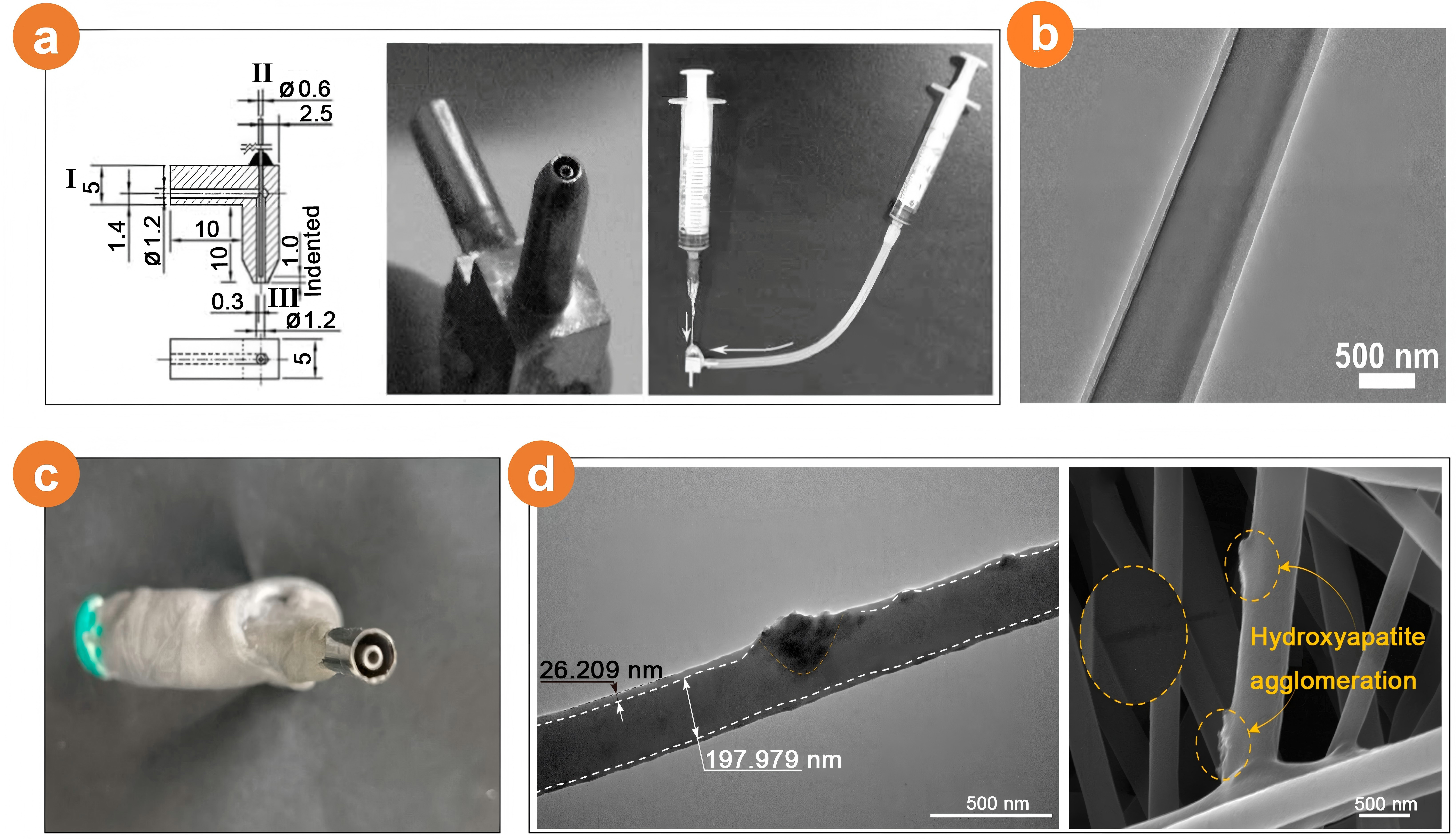
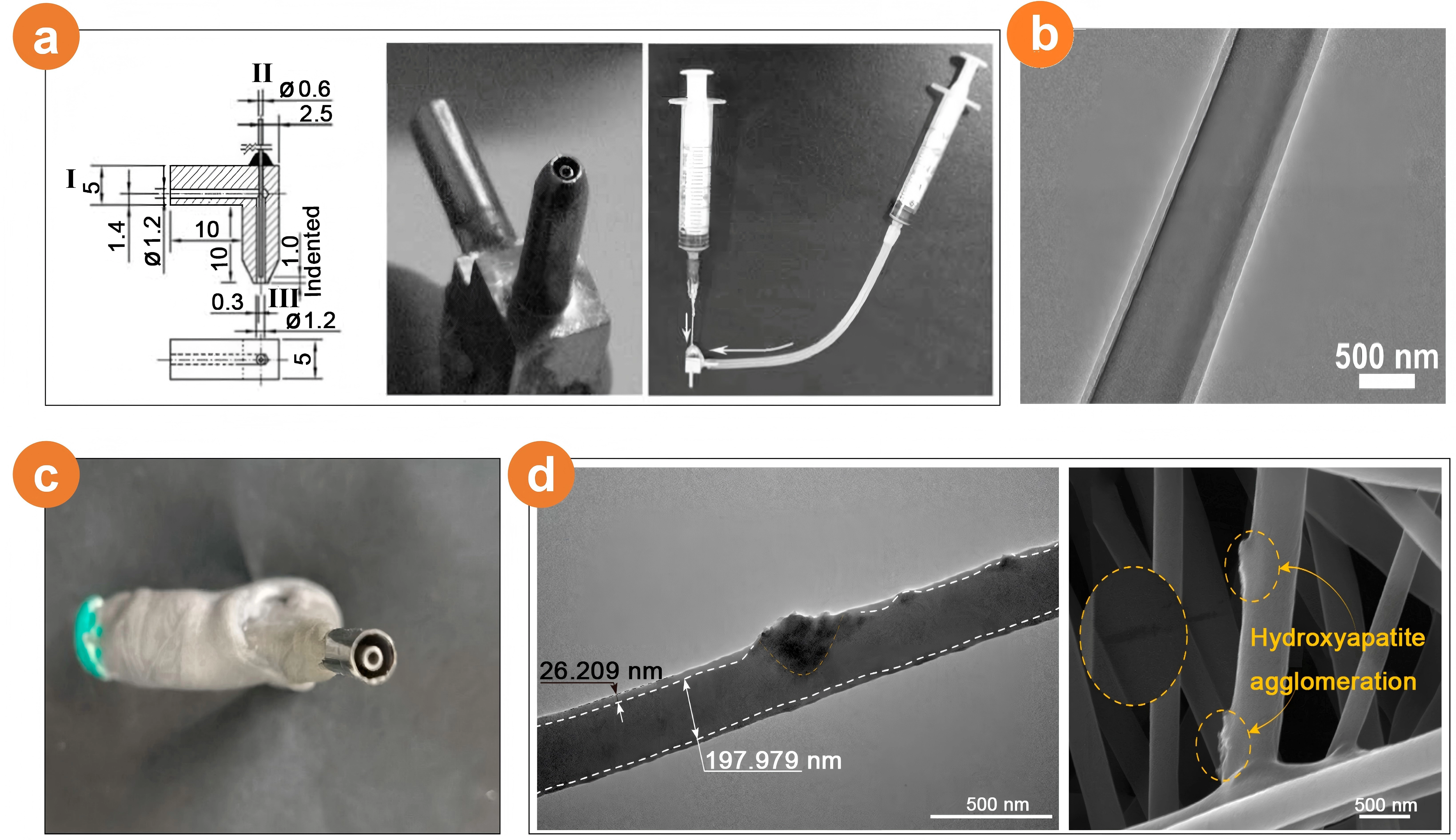
Figure 5. Concentric spinneret and core-sheath nanofibers from the coaxial electrospinning: (a) The left section is a diagram of the spinneret featuring a 1.0 mm recess of the core stainless steel capillary from the sheath capillary tip (I: sheath fluid inlet, II: core fluid inlet, III: concentric outlet). The middle section is an image of the spinneret. The right section shows the spinneret connected to two syringes. Reproduced with permission from reference[43]. Copyright © 2011 John Wiley and Sons; (b) TEM analysis revealing the core‒sheath structure. Reproduced from reference[44]. CC BY 4.0; (c) Close-up of the spinneret outlet. Reproduced from reference[46]. CC BY 4.0; (d) TEM and SEM images of core-sheath nanofibers, with dotted circles highlighting a surface crack and two HAP aggregates in the right SEM section[46]. TEM: transmission electron microscopy; SEM: scanning electron microscopy; HAP: hydroxyapatite.
Highly controllable: The diameter, morphology, and architecture can be finely tuned by adjusting the process parameters. Zhang et al.[44] reported a series of electrospun core-sheath structured PCL/polyethylene oxide (PEO)-PVP nanofibrous membranes co-loaded with ciprofloxacin hydrochloride and curcumin. Various core-to-sheath flow rate ratios can be facilely exploited to manipulate the core and sheath thicknesses. One TEM image of the nanofibers from a core-to-sheath flow rate ratio of 1:4 was exhibited in Figure 5b.
High specific surface area: A small fiber diameter yields a large surface area that enhances adsorption and reactivity. Wang et al.[45] fabricated a core-sheath nanofiber dressing loaded with astragaloside, PVP, and polylactic acid by coaxial spinning. The fibers averaged 605 ± 128 nm in diameter and exhibited a well-defined core-sheath structure.
Multifunctionality: By selecting different combinations of core-shell materials, core-shell nanofibers can perform a wide range of functions, such as sustained drug release or catalytic support. Huang et al.[46] fabricated fenofibrate/PCL@TA/hydroxyapatite (HAP)/PVP nanofibers via an improved coaxial electrospinning technique (Figure 5c). The fibers exhibited a linear morphology with diameters less than 1.0 µm. HAP showed minimal aggregation and was clearly embedded in the core region of the fibers (Figure 5d). The resulting nanofibers provided rapid release of TA and HAP alongside prolonged release of fenofibrate, achieving synergistic and sequential therapeutic effects.
Good mechanical performance: Core-shell nanofibers are usually flexible and strong, resulting in stable, reliable performance in a wide range of applications. Yang et al.[47] first synthesized silver chloride (AgCl) nanoparticles (NPs) in a water bath and then combined them with zinc oxide (ZnO) to produce AgCl/ZnO composite photocatalyst particles. Porous fibers were prepared from a CA and PCL matrix, with AgCl/ZnO deposited on the fiber surface. Using coaxial electrospinning, the team produced nanofibers with markedly better mechanical properties, offsetting the strength loss normally caused by porosity and reducing the decrease in photocatalytic efficiency that accompanies the photodegradation of AgCl/ZnO NPs.
2.2.2.2 Side-by-side electrospinning
The term “Janus” comes from Roman mythology: a god with two faces looking simultaneously into the past and the future. In materials science, Janus membranes, possessing asymmetric wettability, have attracted intense interest because of their distinct dual interfaces and tailorable bulk properties. Building on conventional membrane platforms, Janus architectures are evolving into multifunctional, “smart” materials[48]. The key attributes include the following:
Dual-sided functionality: Janus nanofibers expose two chemically or physically different surfaces that can perform separate tasks in the same environment. One side, for example, can be hydrophilic, whereas the other side is hydrophobic, enabling applications such as oil/water separation or charge separation. Huang et al.[49] fabricated Janus fibers from hydrophilic gelatin (Gel) and hydrophobic CA, loading ZnO nanoparticles into the CA layer and curcumin into the gel layer (Figure 6a). The membrane delivers two distinct antimicrobial agents, addressing both acute and chronic wound challenges.
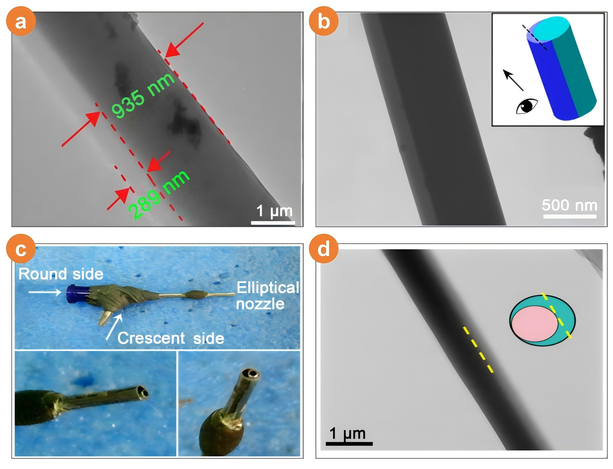
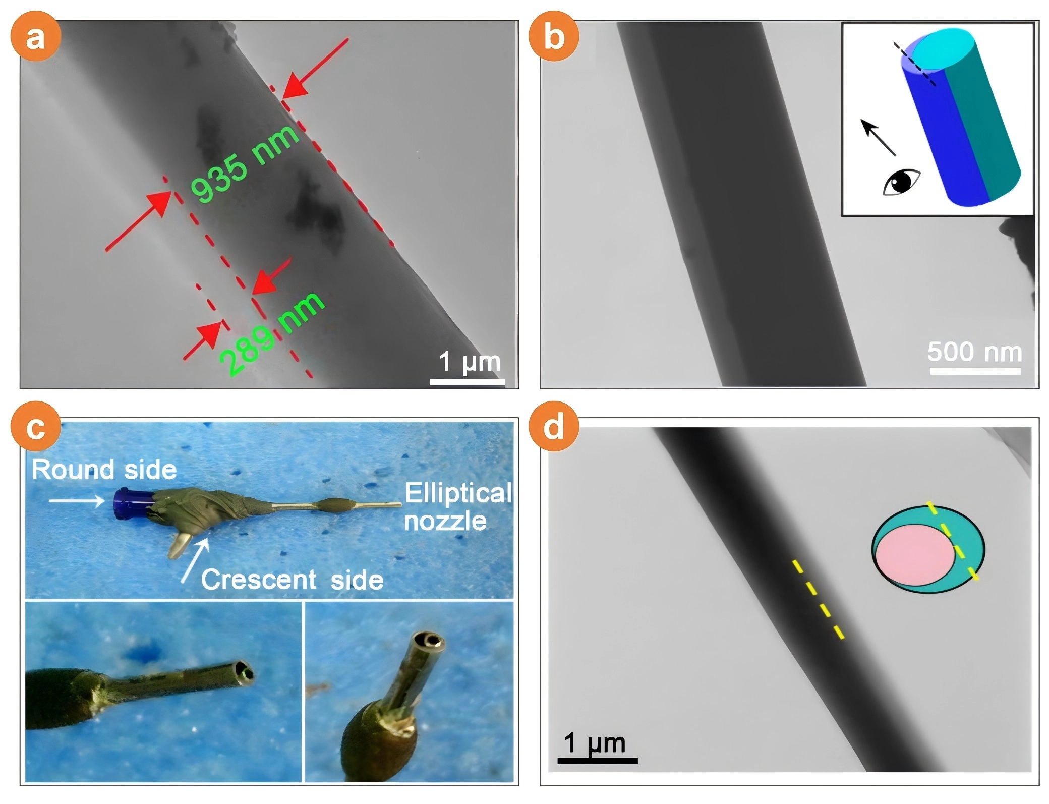
Figure 6. Side-by-side electrospinning and the electrospun Janus nanofibers: (a) TEM image of Janus nanofibers F2. Reproduced from reference[49]. CC BY 4.0; (b) TEM images of Janus nanofibers. Reproduced with permission from reference[50]. Copyright © 2018 American Chemical Society; (c) Digital images of the eccentric side-by-side spinneret captured from various angles. Reproduced from reference[51]. CC BY 4.0; (d) TEM images of the Janus nanofibers. Reproduced with permission from reference[52]. Copyright © 2024 Elsevier. TEM: transmission electron microscopy.
High efficiency: The bilateral design can increase performance. In drug delivery, each face can supply a different active agent, effectively doubling the release rate. Wang et al.[50] designed a hydrophilic Janus composite from nonspinnable PVP K-10 and spinnable PVP K-90 (Figure 6b). One face contained PVP K-90 + helicid (an herbal drug), and the opposite face contained PVP K-10 + sodium dodecyl sulfate (SDS). Water contact angle measurements revealed superior hydrophilicity relative to single-phase fibers, and the transmembrane flux increased more than tenfold.
Precise controllability: Fiber morphology, surface chemistry, and release kinetics can be tuned during fabrication. Yang et al.[51] used side-by-side electrospinning with a centerless spinneret to produce Janus fibers in which one hemisphere carried ciprofloxacin (CIP) in PVP and the opposite hemisphere carried silver nanoparticles (AgNPs) in EC (Figure 6c). The PVP-CIP hemisphere releases >90% of its drug within 30 min, whereas the EC-AgNP hemisphere provides long-lasting antibacterial activity, outperforming conventional blended fibers.
Broad application: Janus nanofibers are being explored for energy storage, catalysis, and biomedicine. Zhou et al.[52] prepared an eccentric Janus photothermal fiber (Figure 6d) with Gel loaded with the chemotherapeutic dihydromyricetin (DMY) and PCL loaded with the photothermal agent CuS. The membrane is hydrophilic and mechanically robust; in a simulated tumor microenvironment, the in vitro DMY release rate increases significantly, whereas near-infrared irradiation tests reveal an excellent photothermal response and stability. These photothermal Janus membranes are promising candidates for implantable cancer therapy.
2.2.3 Multifluid electrospinning
Multifluid electrospinning simultaneously controls three or more distinct working fluids. No matter how complex the final architecture becomes, it can always be viewed as an extension (or hybrid) of the basic core-sheath (coaxial) and side-by-side (Janus) motifs. This versatility has produced tertiary coaxial, “pig-nose”, and Janus-in-shell constructs (Figure 7a). Expanding the number of fluids, however, greatly increases processing complexity; fluid-fluid compatibility is a critical hurdle[53]. Consequently, optimizing multifluid protocols and translating the resulting fibers into practical applications remain active research goals.
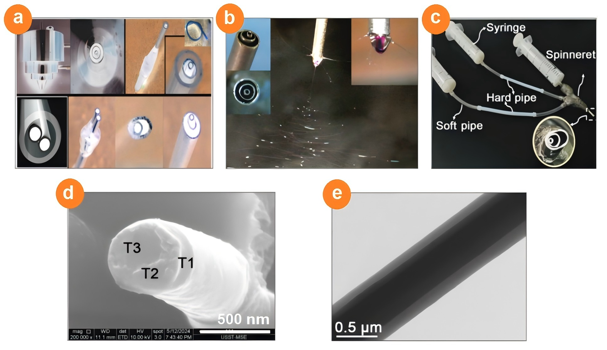
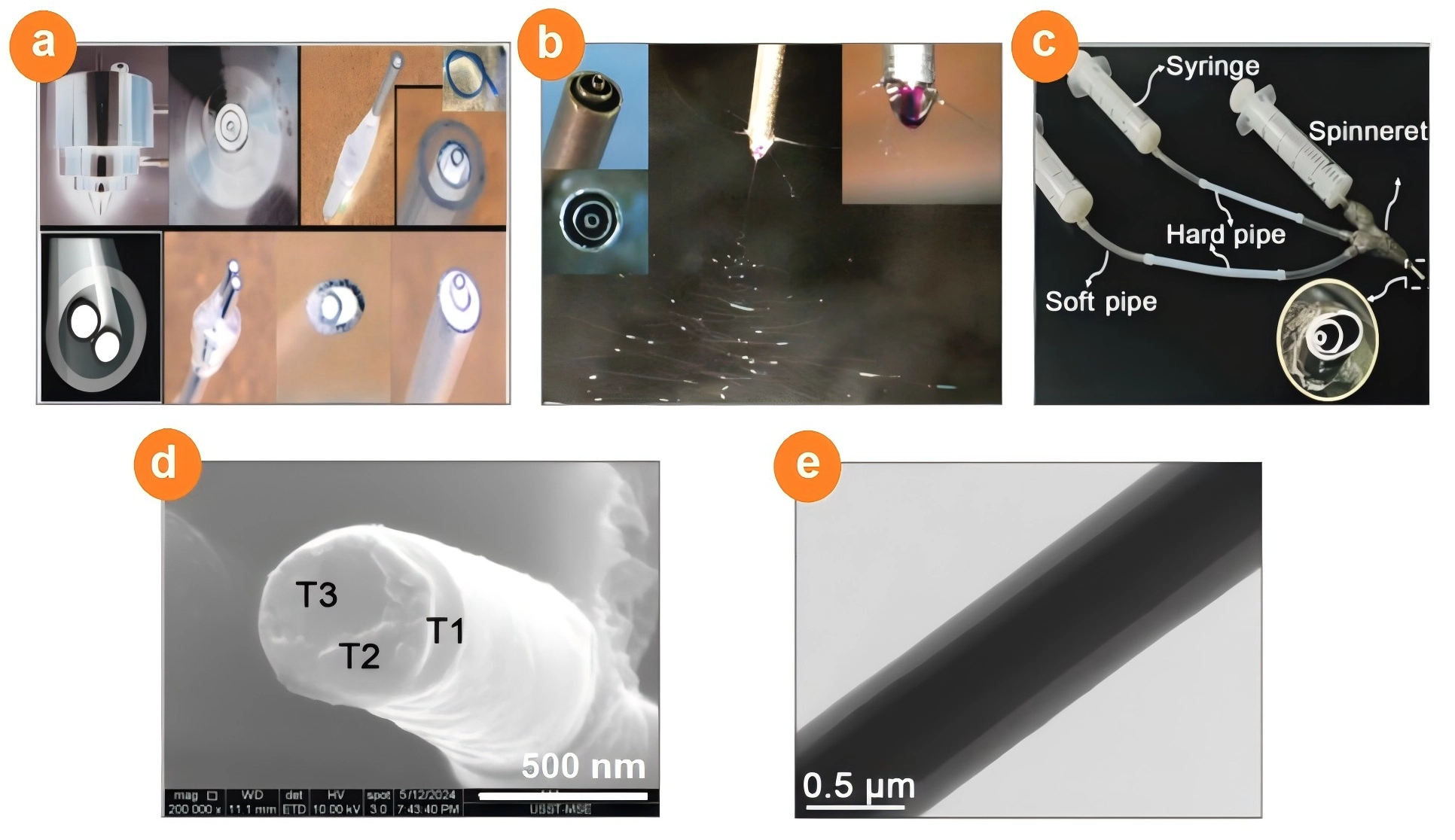
Figure 7. Trifluid electrospinning and the resultant tri-chamber nanostructures: (a) Schematic illustration of the spinneret used in multifluid electrospinning. Reproduced with permission from reference[53]. Copyright © 2019 John Wiley and Sons; (b) Images captured during the triaxial electrospinning process. The upper-left insets provide details on the structure of the custom-made spinneret, whereas the upper-right inset displays a typical compound Taylor cone. Reproduced with permission from reference[54]. Copyright © 2015 American Chemical Society; (c) Setup depicting the connection of three syringes to the spinneret, with the bottom-right inset emphasizing the trilayer eccentric co-outlet of the spinneret. Reproduced with permission from reference[55]. Copyright © 2024 Springer Nature; (d) SEM images revealing cross-sectional views of tri-chamber nanofibers. Adapted with permission from reference[56]. Copyright © 2024 Elsevier; (e) TEM image of a trilayer structure. Reproduced with permission from reference[54]. Copyright © 2015 American Chemical Society. TEM: transmission electron microscopy; SEM: scanning electron microscopy.
Yu et al.[54] used triple-coaxial electrospinning to produce three-layer nanofibers in which every layer was EC at the same concentration, while the KET concentration increased stepwise from the outer layer to the inner layer (Figure 7b). The 0.74 ± 0.06 µm fibers were straight, smooth, and clearly stratified (Figure 7e). In vitro dissolution resulted in zero-order KET release for 20 h. Zhao et al.[55] combined PCL (the primary fiber-forming polymer) with PVP (auxiliary) in a triple-fluid, eccentric side-by-side setup to generate a three-layer periodontal implant membrane in a single step (Figure 7c). The outer stripe contains ketoprofen (anti-inflammatory), the middle stripe contains metronidazole (antibacterial), and the inner stripe contains nanohydroxyapatite (nHA, pro-regeneration), yielding a single membrane that concurrently suppresses inflammation, kills bacteria, and promotes tissue repair. Xu et al.[56] fabricated a triple-section Janus barrier by side-by-side electrospinning of hydrophobic polylactic acid (PLA) and PCL. Each section contains one functional additive: beeswax (BW), quercetin (QUER), or KET, giving a crescent cross-section whose hydrophobicity is tunable via BW content (Figure 7d). The resulting fibers provide long-acting analgesia, anti-inflammatory activity, antiadhesion properties, and enhanced tendon healing.
3. Material Characteristics and Advantages of CA
3.1 Chemical properties
CA is a semi-synthetic cellulose ester obtained by acetylating cellulose, commonly using acetic anhydride as the acetylating agent and glacial acetic acid as the reaction medium[57]. CA can be synthesized under heterogeneous or homogeneous conditions. The reaction temperature, time, and reagent ratio control both the molecular weight and degree of substitution (DS, acetyl groups per anhydroglucose unit)[58]. DS is a key parameter for CA, which profoundly influences its electrospinnability, degradation rate, and skin-interface performance. DS dictates hydrogen-bonding capacity, solubility, hydrophobicity, chain flexibility, and enzyme accessibility[59]. For high-DS CA (DS ≥ 2.3), the polymer is readily soluble in classic electrospinning solvent systems such as acetone, acetone/water, N,N-dimethylformamide (DMF)/acetone, and dichloromethane (DCM)/MeOH. However, the resulting CA solution often has low viscosity and conductivity and generally requires a higher concentration or additives (salts, surfactants) to reach the entanglement threshold for bead-free nanofiber formation. In contrast, low-DS CA (DS < 1.8) is poorly soluble in the volatile solvents preferred for electrospinning and often requires dimethyl sulfoxide (DMSO), DMAc/lithium chloride (LiCl), or ionic liquids. The retained -OH groups enhance intermolecular hydrogen bonding, so the solution typically exhibits higher viscosity, a stronger gel tendency, and a narrower spinnable window[59,60].
3.2 Biocompatibility and biodegradability
CA generally shows good biocompatibility, and its suitability for skin-contact materials makes it a promising matrix for transdermal delivery. Degradation is slow but can be accelerated by lowering the DS or increasing the surface area. It is common sense that low-DS CA degrades faster, releasing fewer persistent fragments and thus avoiding the long-term foreign-body response associated with high-DS CA[61]. Nevertheless, high-DS CA is more popular owing to its superior electrospinnability and is more frequently used in the development of sustained-release drug delivery devices, stable barriers, and non-adherent wound dressings.
3.3 Regulated physicochemical properties
CA offers a high specific surface area, good breathability, and rapid moisture absorption. The fibers are soft, smooth, elastic, and antistatic, critical characteristics for comfortable transdermal patches. CA also selectively adsorbs trace organics, enabling precise drug loading and controlled release. Plasticizers, heat, or compression readily modify the CA morphology, so membranes, scaffolds, and fibers are easily molded.
Owing to their advantages of biocompatibility, biodegradability, thermal stability, hydrophilicity, large specific surface area and low cost, these materials are considered promising polymer matrices for membrane technology, drug delivery, gas sensing, biosensing, adsorption, and degradation[62,63]. Therefore, CA is widely used in cell culture[64], skin tissue engineering[65,66], filtration systems and a variety of medical and pharmaceutical products. In drug-delivery applications, CA provides sustained release that improves therapeutic outcomes, whereas in tissue engineering, it serves as a structural scaffold for cell growth[67].
4. Process Optimization of CA Nanofiber Prepared by Electrospinning
The formation of homogeneous spinning solutions is essential for electrospinning. The solubility of CA is mainly influenced by its degree of substitution. For the production of electrospun CA nanofibers, typical solvents used include acetone, chloroform, DMF, DMAc, DCM, methanol, acetone/DMF, acetone/DMAc, and acetic acid/water (H2O)[68,69]. These solvents can be used either individually or in combination. Owing to CA’s excellent electrospinnability, single-fluid electrospinning is commonly employed to directly produce nanofibers, a method that is straightforward, easily implementable, and cost-effective. Therefore, solvent selection, CA concentration, and spinneret configuration should be considered key processing variables for regulating fiber morphology, porosity, diameter distribution, and surface structure.
However, to maintain a continuous and stable electrospinning process, a relatively high concentration of CA is generally required to ensure sufficient chain entanglement and avoid capillary breakup. Nevertheless, excessively high CA concentrations may lead to spinneret clogging, thereby disrupting the electrospinning process. Among all solvent systems, acetone is the most critical component; however, dissolving CA solely in acetone often results in clogging, which hinders nanofiber formation. Therefore, incorporating cosolvents such as DMF, DMAc, or water can effectively mitigate this issue.
Both the CA concentration and the solvent significantly influence the fiber morphology. Various CA concentrations can result in fibers with different surface morphologies and various porosities[69] (Figure 8a). In recent years, various solvent systems have been explored, with acetone:DMAc = 2:1 being one of the most widely used. The fibers prepared with this ratio exhibit smooth, linear morphologies without bead defects. Recently, Yu et al.[68] introduced an improved coaxial electrospinning method for fabricating CA nanofibers, whose molecular structural depiction is shown in Figure 8b. In this approach, an organic solvent mixture of acetone:DMAc:ethanol (4:1:1 v/v) was used as the sheath solution, while the core solution consisted of CA dissolved in the same solvent system. Compared with single-fluid electrospinning, the coaxial technique yields nanofibers with smaller average diameters, narrower size distributions, improved structural uniformity, and enhanced surface smoothness.
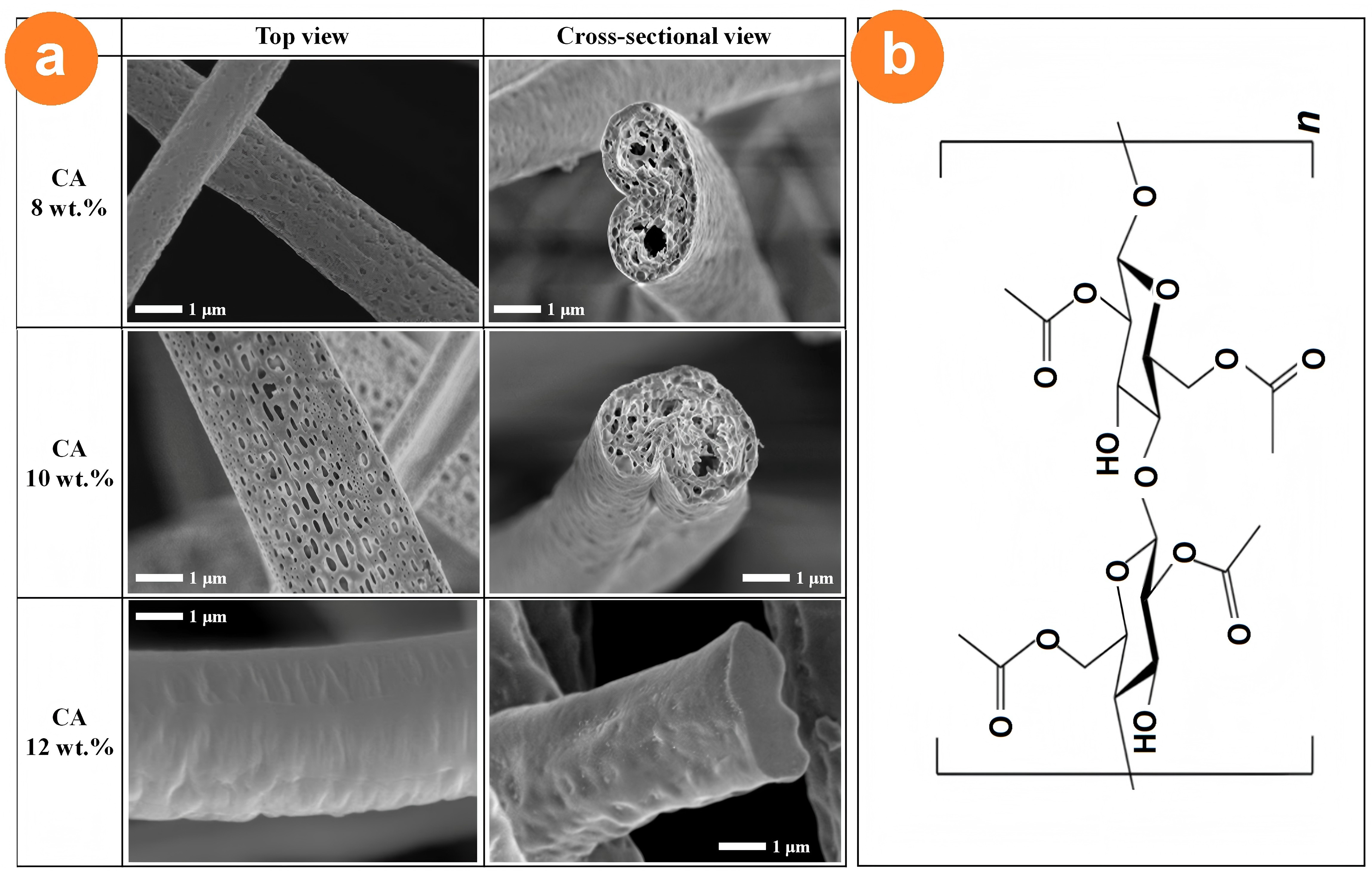
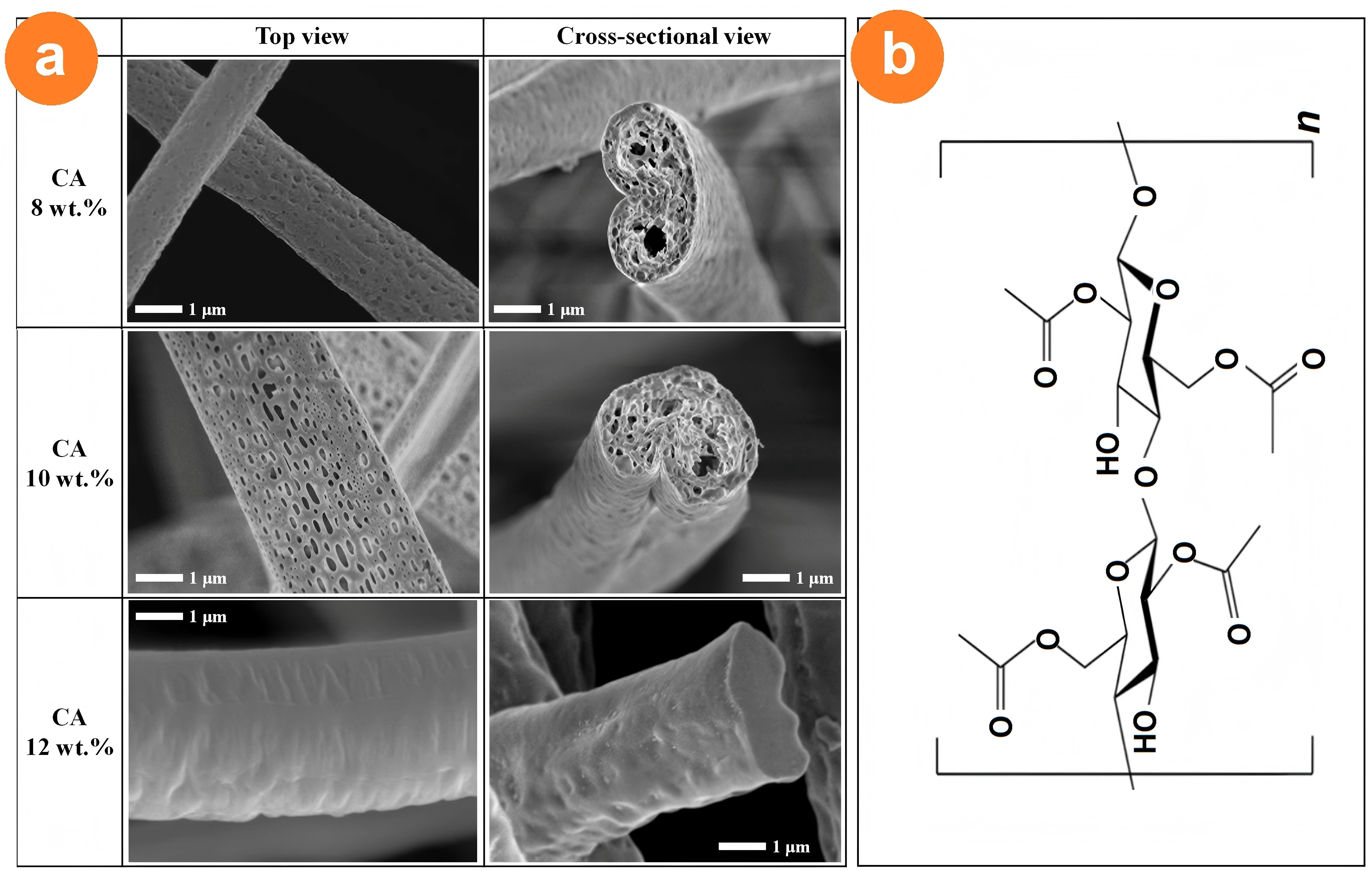
Figure 8. Various morphologies of electrospun CA nanofibers and its chemical structure: (a) Morphological features of electrospun CA nanofibers with concentrations between 8 and 12 wt.%. Reproduced from reference[69]. CC BY 4.0; (b) Structural depiction of CA. CA: cellulose acetate.
The methods for preparing electrospun CA nanofibers using different solvents are summarized in Table 1.
Table 1. Process parameters and structural characteristics of electrospun CA nanofibers prepared using different solvent systems.
| CA (Mw = 30,000) | ||||||||
| Fluids | Proportions | Concentration (wt%) | Voltage (kV) | Flow rate (ml/h) | Acceptance distance (cm) | Fiber diameter (nm) | Fiber morphology | Ref. |
| Acetone | N/A | 15,20 | 5-10 | N/A | 7 | N/A | Fiber-poor | [70] |
| Acetic acid | N/A | 13 | 10-15 | N/A | 7-8 | N/A | Fiber-poor | |
| DMAc | N/A | 15-25 | 10-15 | N/A | 6-10 | N/A | No Fibers | |
| Acetic acid: acetone | 3:1 | 13 | 15 | N/A | 10 | N/A | Fiber membrane with beading | |
| Acetone:DMAc | 1:21:3 | 15 | 10-15 | N/A | 6-10 | N/A | Beaded, no fibers | |
| 3:1 | 12.5 | 18 | N/A | N/A | N/A | Fibers with beads | ||
| 2:1 | 12 | 20 | 0.15 | 17 | 299 ± 68 | Smooth linear form, no beading | [71] | |
| DCM:Methanol | 3:1 | 7.5 | 10 | 0.4 | 15 | N/A | Porous nanofibers with more pores | [69] |
| 10 | N/A | Porous nanofibers with fewer pores | ||||||
| Acetone:DMF | 4:6 | 21 | N/A | Nanofibers with a small number of beads | ||||
| 6:4 | 19 | N/A | Smooth, relatively uniform nanofiber structure | |||||
| 8:2 | 17 | N/A | Smooth, no beading | |||||
| Acetic acid:H2O | 75:25 | 17 | 25 | 3 | 10 | 180 | Long and uniform, Fiber diameter increases with acetic acid content | [72] |
| CA (Mw =50,000) | ||||||||
| Acetone:DMF | 2:1 | 13 | 16.47 | 0.5 | 17.99 | 199 | Low and relatively uniform fiber diameter, CA spinnability increases with increasing concentration in the range 13-17 | [73] |
| 5:5 | 15 | 35 | 10 | 15 | N/A | Very fine fibers, high porosity | [74] | |
| CA (Mw = 100,000) | ||||||||
| Acetone:DMAc: Ethanol | 4:1:1 | 11 | 15 | 1.0 | N/A | N/A | linear fiber | [68] |
N/A: not available. CA: cellulose acetate; DMAc: N,N-dimethylacetamide; DMF: N,N-dimethylformamide; DCM: dichloromethane.
5. Structure-Regulated Release Profiles of Electrospun CA Nanofibers in Drug-Delivery Systems
Drug delivery systems (DDSs) are a central theme in pharmaceutical and biomedical research. Among the rapidly expanding family of micro- and nanosized platforms, fibrous scaffolds have attracted particular interest. Conventional dosage forms often suffer from poor aqueous solubility, nonspecific distribution, and erratic release, all of which limit their clinical utility. Nanocarriers can be engineered to improve drug bioavailability, regulate drug distribution, and reduce off-target effects[75]. Electrospinning uses polymer solutions or melts to fabricate nano- to microscale fibers under the action of an electric field. The resulting nonwoven mats combine a high specific surface area, interconnected porosity, and easily functionalized surfaces, making them suitable for high payload loading and controlled release.
CA, as a semisynthetic derivative of natural cellulose, has good biocompatibility, film-forming ability, and tunable degradation behavior. Fiber architectures have evolved from simple single-layer monolithic composites to sophisticated core-sheath, Janus, trichamber core-sheath, and their combined hybrid configurations. These structurally designed fibers allow drugs to be embedded as nanoparticles or amorphous solid dispersions within the fiber matrix, enabling pulsatile, sustained, biphasic, stimuli-responsive, or site-specific release[76]. CA nanofibers have already been loaded with antimicrobials, antioxidants, anti-inflammatory agents, and many other drug classes and can be delivered through different administration routes.
5.1 Sustained release profiles of electrospun CA nanofibers
Among the various drug release kinetic models (classified by the drug release rate over time), such as pulsatile, sustained or extended, delayed, zero-order, and biphasic/multiple-step release, the zero-order, biphasic, and sustained release profiles are the most popular. The zero-order model is a kinetic model in which the drug is released at a constant rate, independent of drug concentration[77,78]. The biphasic model describes release profiles consisting of two distinct sequential phases, typically an initial fast “pulsatile” release followed by a slow sustained phase, commonly fitted with two sets of kinetic parameters (fast and slow rate constants)[79,80]. Sustained release delivers a drug slowly and predictably over an extended period, reducing the dosing frequency while maintaining therapeutic plasma levels. CA is frequently exploited for developing drug delivery devices that achieve these release profiles. Electrospun CA nanofibers are ideal for these purposes because their porosity, fiber diameter, and degradation rate can be precisely tuned. The following examples illustrate how CA-based fibers provide long-acting delivery of representative anti-infective agents.
Over the past half century, various models have been proposed to elucidate drug release mechanisms from the standpoint of molecular behavior. Among them, the Korsmeyer-Peppas model is a popular semi-empirical power-law model that describes drug release from polymeric matrices, in which the diffusional exponent n distinguishes Fickian diffusion, anomalous (non-Fickian) transport, and case-II transport; it is applicable mainly to the first ~60% of the release curve[81,82]. The Baker-Lonsdale model is a theoretical equation describing diffusion-controlled drug release from spherical matrices, such as microspheres and microcapsules[83,84]. These two models are often employed to characterize the drug release behavior of electrospun CA nanofibers, and most drugs are released primarily via the typical Fickian diffusion mechanism, especially from high-DS CA matrices.
5.1.1 Sustained release of anti-infective agents
Anti-infectives: antibiotics, antivirals, antifungals, and antiparasitics, remain cornerstones of clinical therapy[85], yet antimicrobial resistance (AMR) is increasing; in 2021 alone, resistant infections caused an estimated 4.7 million deaths worldwide. New drugs and smarter delivery systems are urgently needed[86]. Electrospun CA nanofibers, with high surface areas, adjustable porosities, and sustained-release capabilities, are emerging as powerful platforms for local antibacterial therapy. The representative studies are summarized below.
Daptomycin (Dap)-orthopedic coatings. Dap is a cyclic lipopeptide antibiotic that has excellent efficacy against gram-positive bacterial infections[87,88]. Faria’s group[89] developed a Dap-loaded CA fiber coating for stainless-steel (SS) implants. A 22 kV, 1 mL h-1, 10 cm tip-to-collector setup yielded uniform 4.1 ± 1.5 µm fibers from dichloromethane/acetone (9:1). An electrosprayed chitosan interlayer improved adhesion, whereas Dap was encapsulated in PMMA-Eudragit microcapsules dispersed within the CA matrix. In simulated body fluid, the coating released 3 × 10⁻³ mg mL-1 within 2 h and maintained bactericidal levels against gram-positive pathogens. The system combines biocompatibility, strong metal adhesion, and sustained antibiotic release, offering a simple strategy for infection-free implants.
Azithromycin (AZM)-ocular inserts. AZM, a macrolide antibiotic, is widely used to treat ocular infections, but it suffers from issues such as low bioavailability and the need for frequent dosing[90]. Taghe et al.[91] fabricated AZM-loaded CA and PCL nanofiber eye inserts (18 kV, 0.5-1.0 mL h-1, 15 cm). The mean fiber diameters were 160 nm (CA) and 190 nm (PCL). The CA fibers followed first-order kinetics and provided smooth, burst-free release, whereas the PCL fibers showed an initial burst followed by sustained diffusion. In rabbits, both formulations maintained therapeutic AZM levels for >6 days; CA achieved a higher Cmax and longer ocular residence. Both scaffolds inhibited Staphylococcus aureus (S. aureus) and Escherichia coli (E. coli) in vitro and were nonirritating (Figure 9a), confirming their suitability as long-acting topical antiinfective inserts.
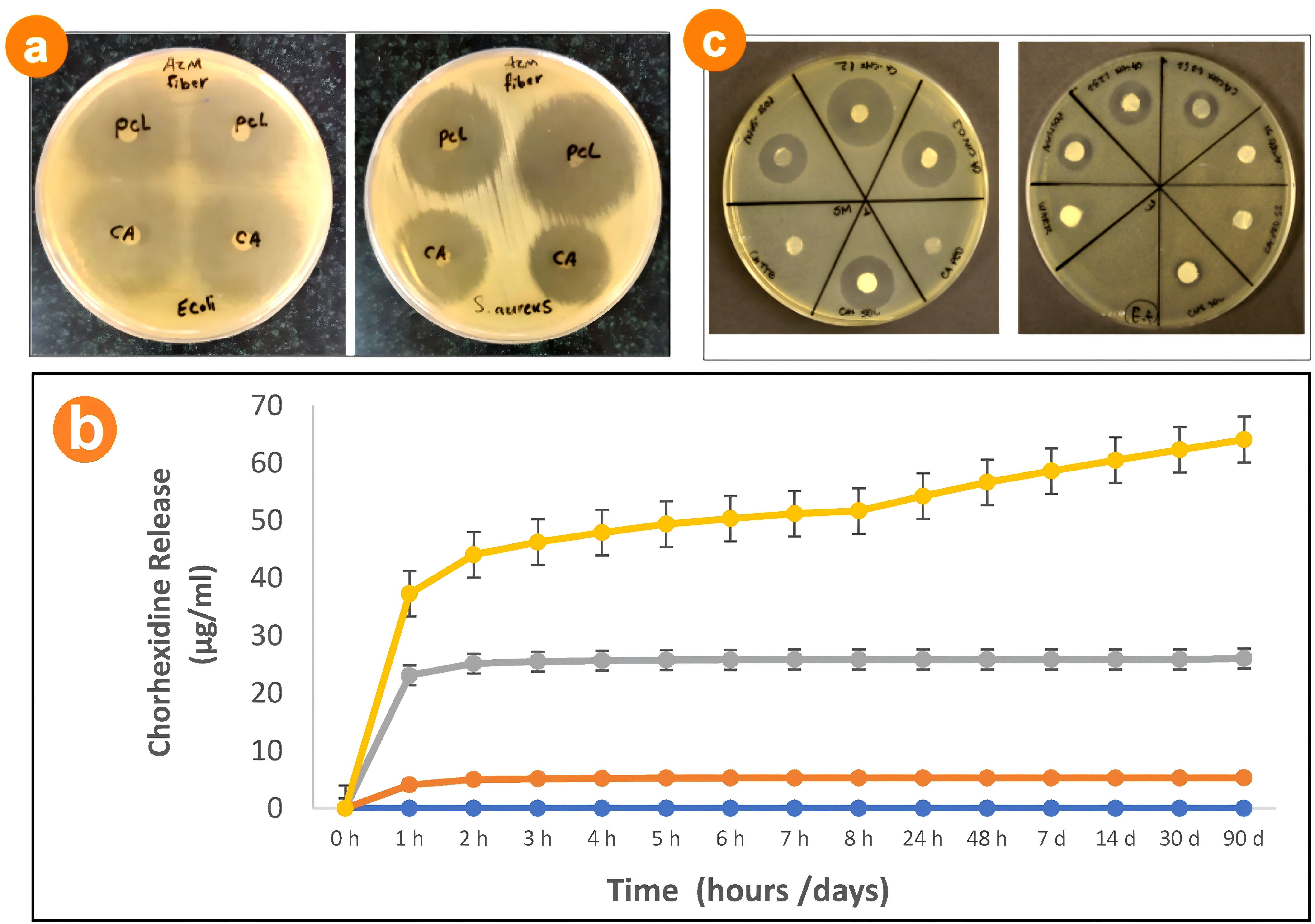
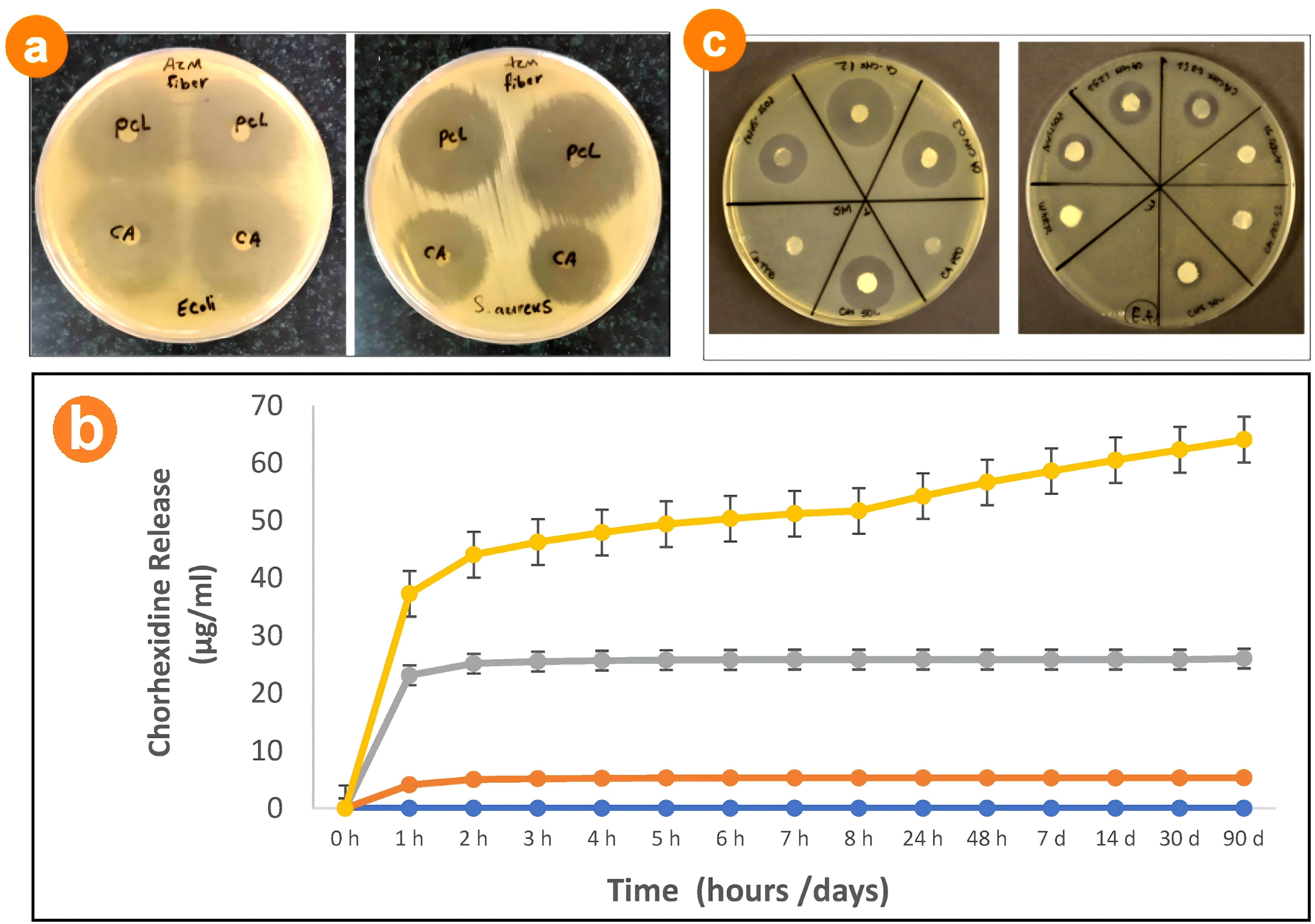
Figure 9. Antibacterial effects of medicated CA nanofibrous films: (a) Inhibition growth zones of AZM-CA and AZM-PCL nanofibers against E. coli (gram-negative) and S. aureus (gram-positive). Reproduced from reference[91]. CC BY 4.0; (b) Sustained release of chlorhexidine from the nanofibrous CA membranes. Reproduced from reference[96]. CC BY 4.0; (c) Inhibition halos observed when the experimental mats were applied against S. mutans and E. faecalis. Reproduced from reference[96]. CC BY 4.0. CA: cellulose acetate; E. coli: escherichia coli; AZM: azithromycin; S. aureus: staphylococcus aureus; PCL: polycaprolactone.
Tetracycline hydrochloride (TC): wound dressings. As a broad-spectrum antibiotic, the low bioavailability of TC limits its clinical application[92]. Khan et al.[93] recovered CA from discarded cigarette filters (CFCAs) and electrospun it with TC (20 kV, 1 mL h-1, 15 cm). The fibers (190-800 nm) formed a hydrophilic, cross-linked network. Seventy-seven percent of the antibiotic was released within 4 h, creating 23-42 mm inhibition zones against S. aureus and E. coli. 3-(4,5-Dimethylthiazol-2-yl)-2,5-diphenyltetrazolium bromide (MTT) assays revealed >97% cell viability, underscoring the potential of recycled CA/TC mats as low-cost, antibacterial wound dressings.
Chlorhexidine(CHX): dental membrane. CHX is commonly used in oral anti-infective therapy, but it may cause adverse reactions such as tooth discoloration[94,95]. de Carvalho et al.[96] prepared flexible CA membranes loaded with 0.3-1.2 wt% CHX (14 kV, 14 cm, 12 h slow spinning). The 640 nm hydrophilic mats absorbed >600% of the water. An initial 56% burst was followed by sustained release for 90 days (Figure 9b). Disc diffusion tests revealed robust activity against Streptococcus mutans and Enterococcus faecalis (inhibition zones 23-42 mm) (Figure 9c); the 1.2 wt % group outperformed the positive control (p < 0.05). The excellent cytocompatibility confirmed that electrospun CA-CHX membranes are promising local drug reservoirs for periodontal therapy.
5.1.2 Sustained release of anticancer drugs
Anticancer agents interrupt uncontrolled cell proliferation, but multidrug resistance and off-target toxicity often limit their clinical value. Electrospun CA nanofibers are now being explored as drug carriers to manipulate drug release time periods[97] or to improve the therapeutic index of cytotoxic drugs[98-101].
Stoyanova et al.[97] reported a new kind of composite nanofibers containing CA, polyethylene glycol, and a natural biologically active compound, quercetin, which were prepared using single-fluid blended electrospinning. The scanning electron microscope (SEM) images are shown in Figure 10a. Although these nanofibers have beads and spindles among them, the sustained release of quercetin can be kept continuously for 360 min with an amount of 85.3% is released (Figure 10b). It is verified that the CA nanofibers exhibited not only high antioxidant activity, but also a high cytotoxic effect against both HeLa tumor cells and SH-4 melanoma skin cells.
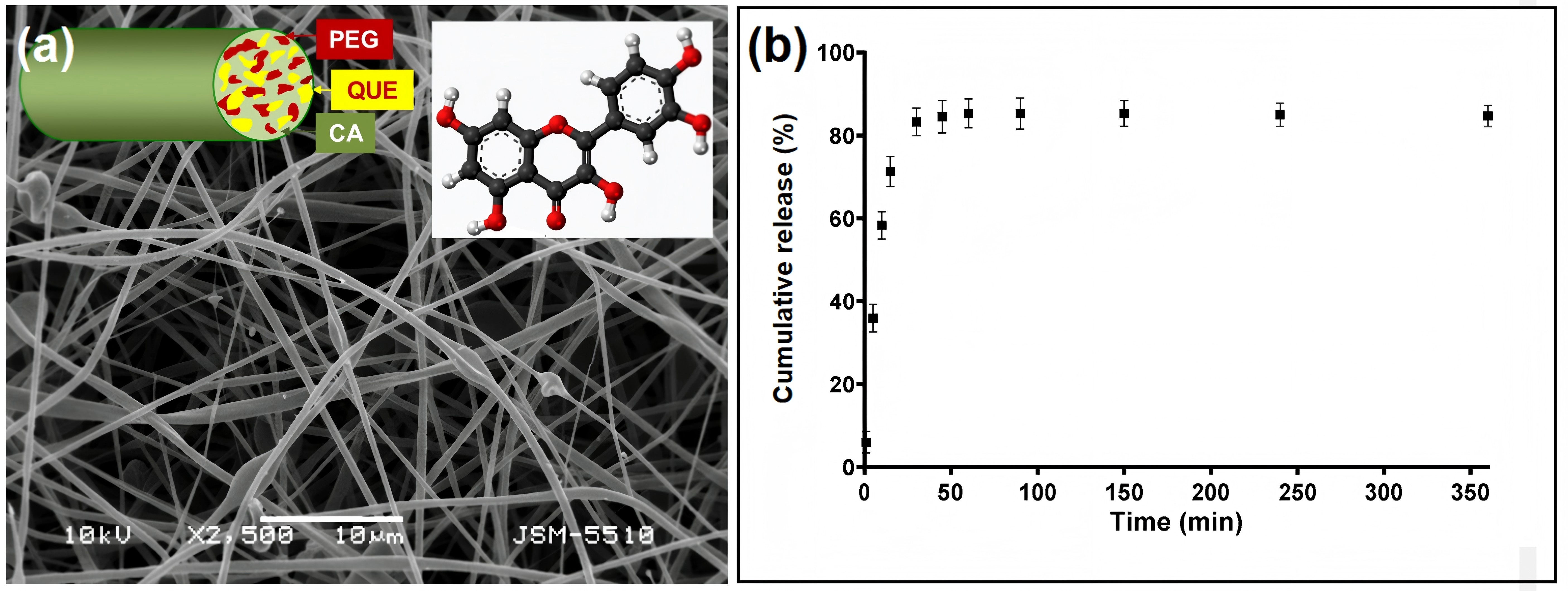
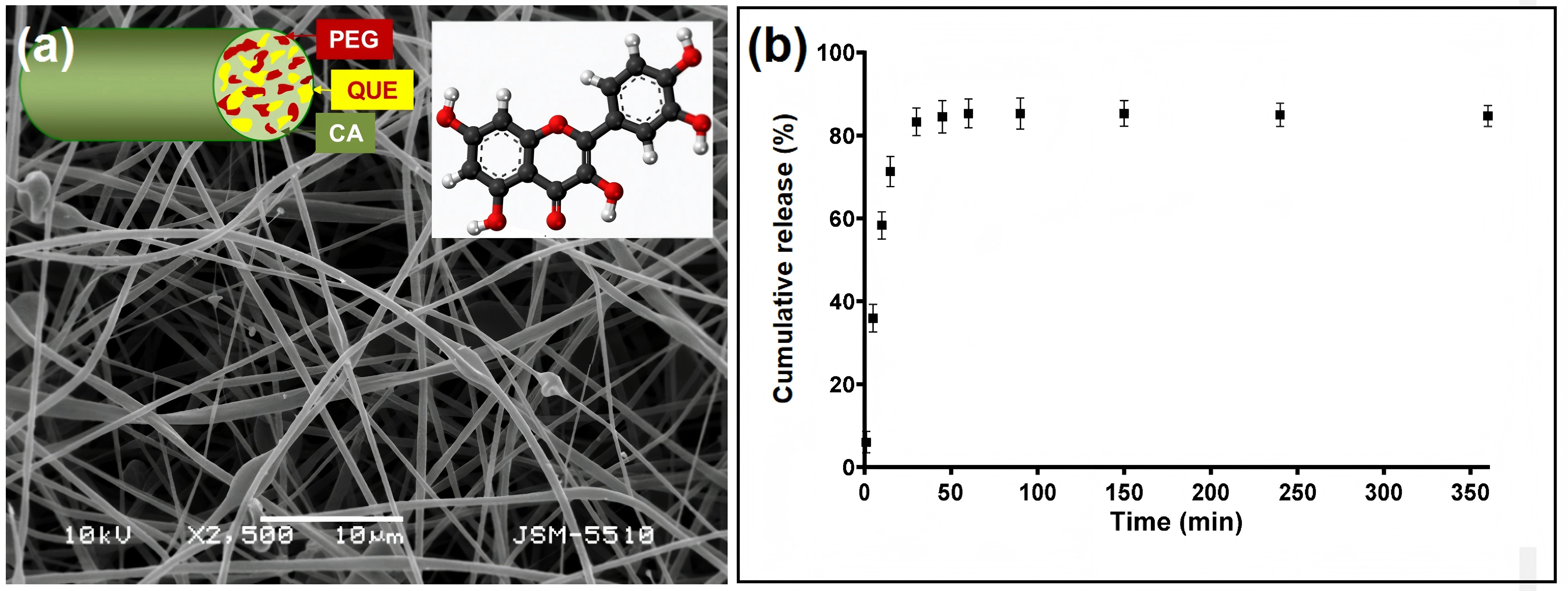
Figure 10. Anticancer drug-loaded nanofibers and the drug sustained release profile: (a) SEM images of the quercetin-loaded CA/PEG nanofibers. Reproduced from reference[97]. CC BY 4.0; (b) The in vitro sustained release profiles of the composite nanofibers. Reproduced from reference[97]. CC BY 4.0. PEG: polyethylene glycol; SEM: scanning electron microscopy; CA: cellulose acetate; QUE: quercetin.
Doxorubicin, a broad-spectrum agent used against breast, lung, and other malignancies, is hindered by drug resistance and systemic toxicity[98]. To enhance delivery, Alisani et al.[99] incorporated multiwalled carbon nanotubes (MWCNTs) and DOX into CA/PCL-diol/polytetramethylene ether-glycol polyurethane (PCL-Diol/PTMG-PU) fibers. Under optimized conditions (20 kV, 14 cm working distance, 0.5 mL h-1), flexible 230-410 nm fibers were obtained that adsorbed up to 1.5 g DOX per gram of mat. At pH 7.4, release followed non-Fickian (Korsmeyer-Peppas) kinetics for 120 h. In LNCaP prostate cancer cells, the fibers induced 59.9% total killing (43.7% apoptosis), whereas L929 fibroblasts were largely unaffected (>90% viability), demonstrating selective anticancer activity.
Sorafenib is a multikinase inhibitor approved for liver and kidney cancer. It has limited clinical use because of its poor water solubility and low oral bioavailability[100]. Alemomen et al.[101] encapsulated sorafenib (SF) in PCL/CA nanofibers (≈ 281 nm) prepared by electrospinning. A full-factorial design yielded defect-free fibers with 7.5% drug loading and 98% encapsulation efficiency. CA increased the hydrophilicity and increased the Young’s modulus to 75 MPa. X-ray diffraction confirmed the molecular dispersion of SF. In vitro, 51% of the payload was released within 24 h, following the Baker-Lonsdale (Fickian diffusion) model. In the infected mice, the fibers markedly reduced lesion size and hepatic/splenic parasite burden, performing comparably to intravenous Glucantime® and outperforming oral SF, thus validating the benefit of local sustained release.
5.1.3 Sustained release of antioxidants
Antioxidants neutralize excess free radicals, curb oxidative stress, and slow the tissue damage that underlies many chronic diseases. Because endogenous defenses are often insufficient, exogenous antioxidant supplementation has become a routine preventive and therapeutic strategy. Therefore, antioxidants are frequently encapsulated in advanced biomedical drug delivery devices, where long-term sustained-release profiles are highly desired to exert their functional effects. Many antioxidants, such as ferulic acid and curcumin, have poor water solubility[102-107]. Electrospun nanofibers have been shown not only to improve the solubility of these compounds but also to provide predictable, prolonged release, enabling their use in precision medicine.
Ferulic acid is a natural phenolic antioxidant with potent radical-scavenging and anti-inflammatory activity, but its poor aqueous solubility and low oral bioavailability limit its clinical use[102,103]. To overcome this, Anand et al.[104] encapsulated ferulic acid (FA) in silk sericin (SS)/PCL/CA nanofibers. Optimized electrospinning (10-25 kV, 0.8 mL h-1, 10 cm) produced smooth, 168-171 nm fibers whose tensile strength reached 14.7 MPa after CA incorporation. The hydrophilic CA/SS phase provided wettability, while the PCL core prolonged release: ≈ 59% of the payload was liberated within 12 h (Figure 11a), followed by a slow diffusion-controlled tail. The mats were noncytotoxic (> 97% viability), inhibited bacterial biofilms, and accelerated wound closure in diabetic rats (≈ 100% re-epithelialization within 14 days), demonstrating that antioxidation, antibacterial action, and sustained FA delivery can be combined in a single dressing.
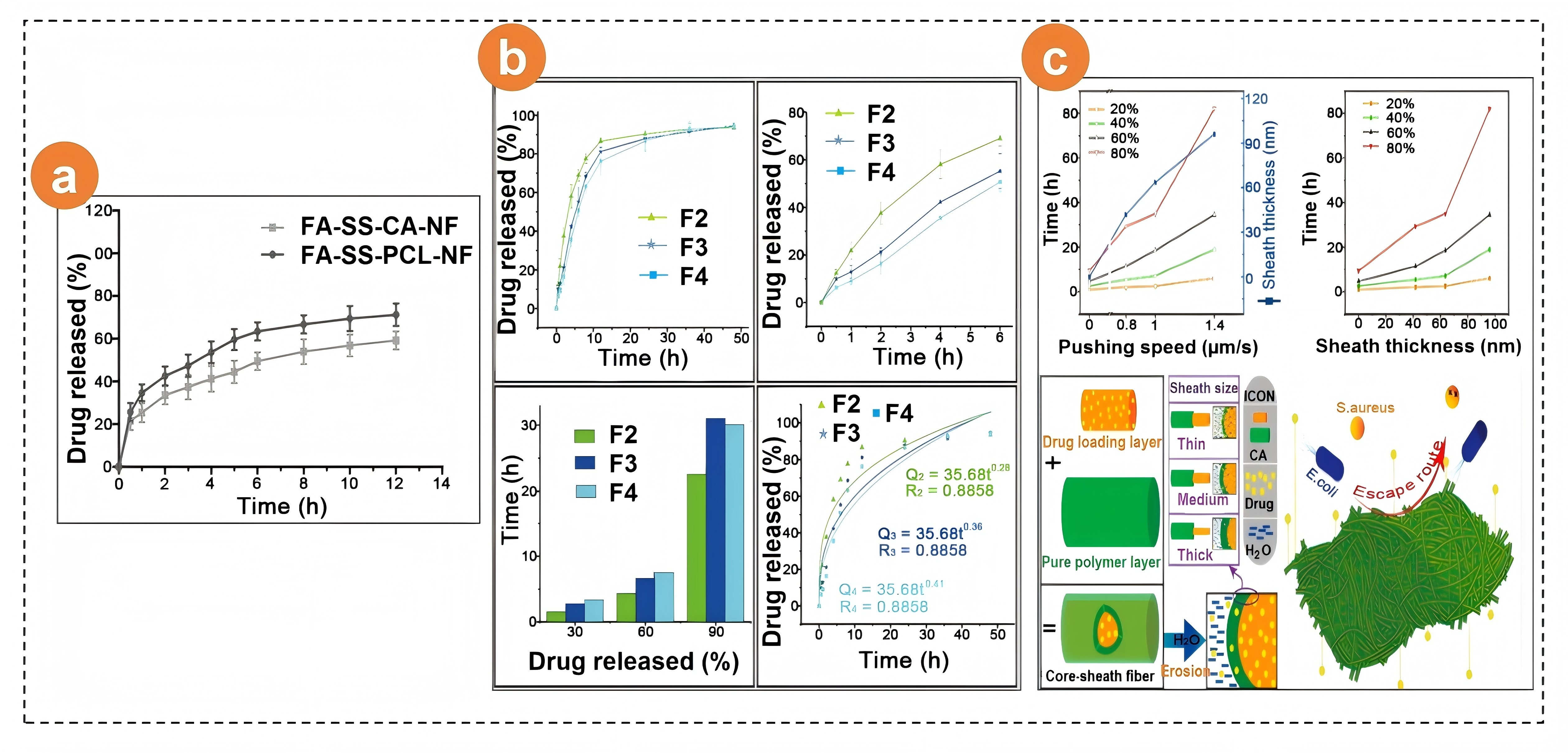
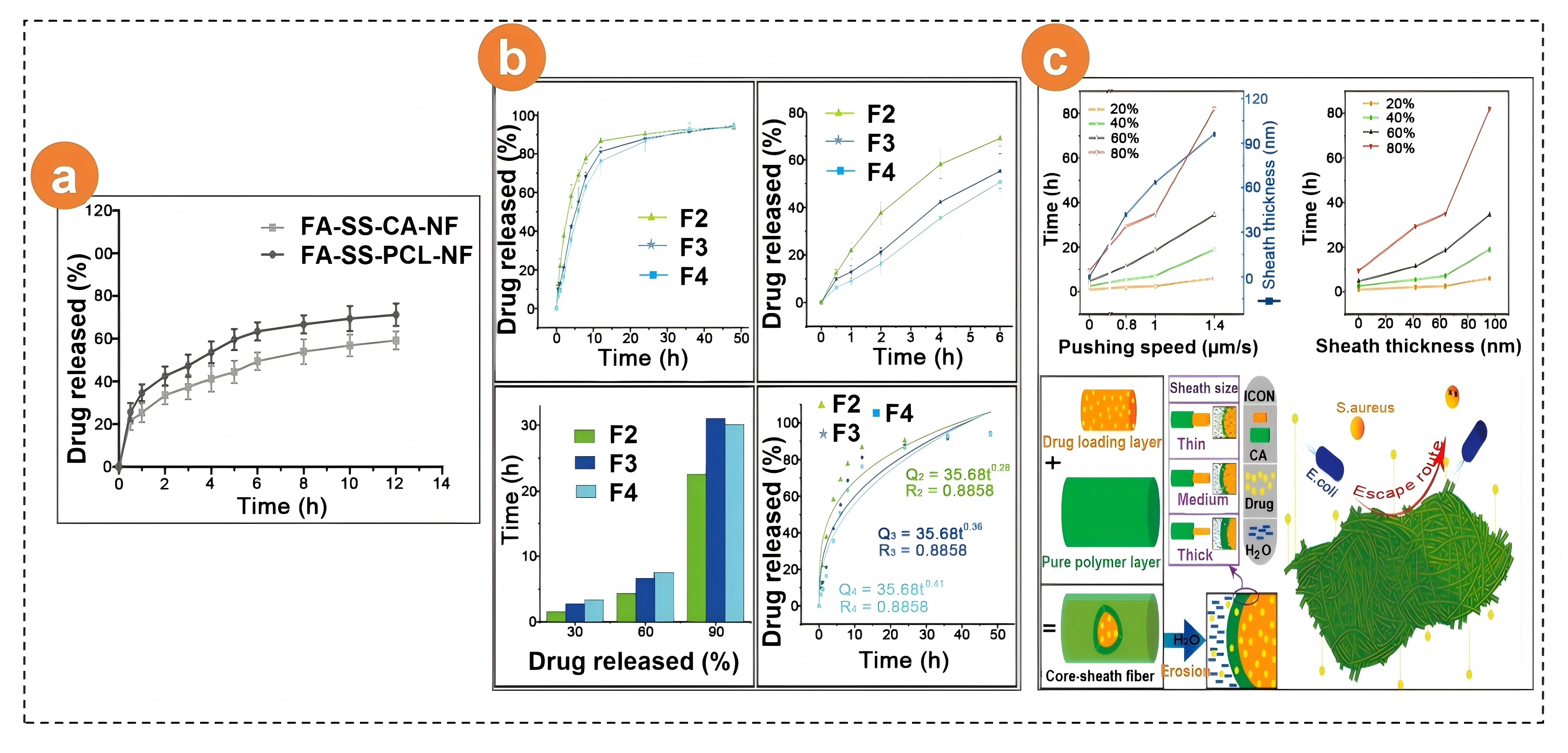
Figure 11. Sustained release of antioxidants from electrospun CA nanofibers: (a) In vitro drug release of the FA-SS-PCL-NF and FA-SS-CA-NF nanofibrous formulations in PBS at pH 7.4 (mean ± S.D.). Reproduced from reference[104]. CC BY 4.0; (b) Drug dissolution profiles of fibers F2-F4 for the entire 48 h period; Dissolution profiles of F2-F4 in the previous 6 h; The time required for the Cur release of fibers F2-F3 to reach 30, 60, and 90%; Drug release analysis of fibers F2-F4 through the Pappas model. Reproduced with permission from reference[106]. Copyright © 2024 American Chemical Society; (c) Relationships between the pushing speed of the middle pump (flow rate of the middle fluid), the sheath thickness of the core-sheath T1-T3 fibers, and the release time of curcumin. Mechanism of drug release and antibacterial activity for manipulating the sheath thickness of core-sheath fibers. Reproduced from reference[107]. CC BY 4.0. CA: cellulose acetate; FA: ferulic acid; PCL: polycaprolactone; PBS: phosphate-buffered saline; NF: nanofibers; SS: stainless-steel.
Curcumin, the active constituent of turmeric, has antibacterial, anti-inflammatory, antioxidant, and wound-healing activities, but its poor water solubility and low bioavailability restrict its clinical efficacy[49,107]. Research groups have optimized curcumin delivery with electrospun CA. Chen et al.[106] prepared core-sheath fibers with ZnO nanoparticles and Cur in the core and a CA sheath that acted as a diffusion barrier. The sheath extended Cur release to 36 h (Figure 11b) and coreleased Zn2+ ions, synergistically enhancing fibroblast proliferation while suppressing bacterial growth. Liu et al.[107] used tri-axial electrospinning to vary the CA shell thickness from 7~41 nm by adjusting the middle-fluid flow rate; a 20 nm sheath provided near-zero-order Cur release for ≈ 60 h and maximized antibacterial activity against both gram-positive and gram-negative strains (Figure 11c). In both studies, curcumin was converted from a crystalline to an amorphous form during spinning, dispersed uniformly within CA fibers, and retained full antioxidant and antibacterial activity. These results confirm that microstructural control of CA nanofibers via advanced electrospinning is an effective strategy for the sustained delivery of hydrophobic antioxidants and for enhancing their therapeutic performance.
5.1.4 Sustained release of natural medicines
Anthocyanins are water-soluble natural pigments with strong antioxidant and radical-scavenging activities, but their poor stability in terms of temperature, light, and pH limits their use[105]. Liu et al.[108] prepared anthocyanin-loaded CA ultrafine fibers (CA@Anth UFs) by electrospinning to improve stability and provide controlled release. The optimized conditions (20 kV, 1.0 mL h-1, 16 cm) yielded smooth, bead-free fibers with uniform diameters. At 7 wt% loading, the encapsulation efficiency peaked at 49%. CA markedly protected the pigment; 95% cumulative release occurred in simulated gastric fluid within 240 min, while 65% antioxidant activity was retained, demonstrating the potential for natural drug delivery and green packaging.
Resveratrol (RES), a polyphenolic phytoalexin found in red wine, peanuts, grape skins, and blueberries, has antioxidant, anti-inflammatory, anticancer, and cardioprotective effects[109]. Its clinical utility is hindered by poor aqueous solubility, low oral bioavailability (< 1%), and chemical instability[110]. Chen et al.[111] used coaxial electrospinning to create core-shell CA/PVP fibers with a reverse drug gradient: core fluid (high drug/low CA) and shell fluid (low drug/high CA). This architecture suppressed burst release (only 18.9% in 1 h) and prolonged 90% release to 19.5 h while minimizing tailing, offering a new strategy for efficient natural drug delivery via spatial control.
5.1.5 Sustained release of other drugs
Pramipexole, a dopamine receptor agonist approved for Parkinson’s disease, also accelerates diabetic wound healing via antioxidant pathways[112-114]. Tan et al.[115] electrospun CA nanofibers containing 3, 5, or 10% pramipexole. All formulations yielded uniform, bead-free fibers (CA/3%: 828 ± 109 nm; pure CA: 874 ± 185 nm). The 3% dressing released 64 ± 4% within 24 h and 96 ± 5% by 144 h via a diffusion-controlled mechanism. It absorbs ~31% of the water, has a tensile strength of 2.0 ± 0.4 MPa, and has a contact angle of 68°, enabling exudate management and mechanical protection. In diabetic rats, the CA/3% dressing achieved 94% wound closure at 14 days and reduced oxidative stress (downregulated GPx) and inflammation (inhibited nuclear factor kappa-B (NF-κB)).
Probucol, a cholesterol-lowering drug with antioxidant and anti-inflammatory activity[116], was incorporated into CA nanofibers by Abbaszadeh et al.[117] The 1% probucol formulation produced bead-free, randomly oriented fibers (1,059 ± 21 nm vs. 783 ± 34 nm for pure CA; p < 0.05). The release was 41 ± 4% at 24 h and nearly complete at 168 h (diffusion-controlled). The dressing absorbed 22 ± 3% of the water, had a tensile strength of 2.7 ± 0.3 MPa, and had a contact angle of 65°. In diabetic rats, healing reached 91 ± 4% at 14 days, with an epithelial thickness of 45 ± 2 µm versus 62 ± 4% and 27 ± 4 µm for pure CA (p < 0.05), which was associated with reduced oxidative stress and sustained probucol release.
Acetaminophen (AAP, paracetamol), a widely used analgesic/antipyretic, suffers from poor water solubility[118]. Wang et al.[119] employed triaxial electrospinning to produce three-layer CA fibers: an outer blank CA shell (10-15 nm) and two inner gradient drug layers. This architecture reduced burst release (30% required 8.1 h) and prolonged 90% release to 43.2 h with a gentle tail. In vivo studies revealed stable plasma levels (twin peaks at 6 and 24 h). CA ensured fiber integrity, while electrospinning achieved an amorphous AAP dispersion and a precise multilayer geometry, markedly enhancing the sustained-release performance.
5.2 Biphasic release
Biphasic systems deliver an initial rapid dose to achieve therapeutic levels quickly, followed by sustained release to maintain efficacy, reducing dosing frequency and improving patient adherence. In electrospun CA-based systems, biphasic release is usually achieved by combining a rapidly dissolving phase with a slower CA-containing diffusion barrier or by constructing hybrid microsphere/fiber, blended, or layered architectures.
Berberine hydrochloride (BH): transdermal/mucosal patch. Berberine, an isoquinoline alkaloid from Coptis chinensis, exhibits broad-spectrum antibacterial activity, but its oral bioavailability is < 1%, and parenteral use is toxic, necessitating frequent dosing[120]. Zhou et al.[121] combined electrospraying and electrospinning (electrohydrodynamic atomization (EHDA)) to create a hybrid microsphere/nanofiber system: BH was loaded into CA microspheres embedded within a PVP nanofiber mesh (Figure 12a). PVP fibers eroded rapidly, releasing 42% of the drug within 0.5 h (burst phase); CA microspheres then released the remaining 56% by Fickian diffusion over 60 h (sustained phase), eliminating tailings (Figure 12b). The construct offers a straightforward route for high-performance biphasic delivery across the skin or mucosa.
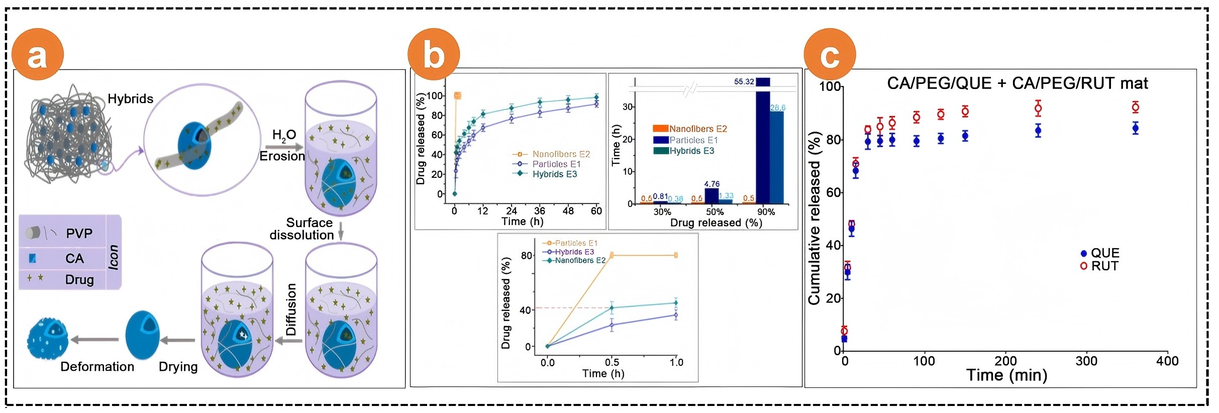
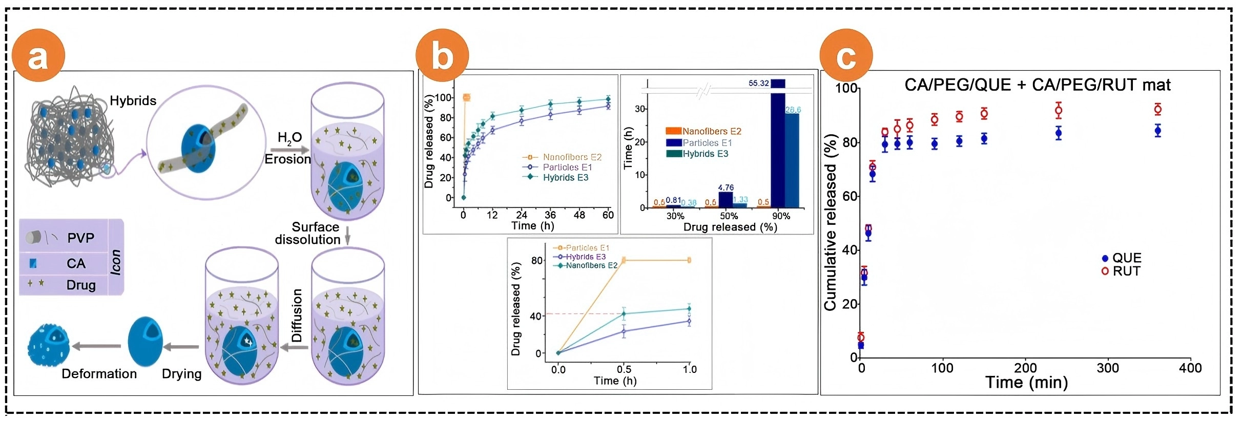
Figure 12. Biphasic release profiles from the CA-based nanofibers: (a) Mechanism of biphasic release of BH from electrospun PVP nanofibers and electrosprayed CA microparticles. Reproduced from reference[121]. CC BY 4.0; (b) In vitro dissolution test results: the percentage (%) of cumulative drug release throughout the entire experimental period and within the first hour and estimated times for specific BH release percentages (30%, 50%, and 90%) from three EHDA products. Reproduced from reference[121]. CC BY 4.0; (c) Release profiles of QUE and RUT from CA/PEG/QUE+CA/PEG/RUT mats. The data are presented as average values ± standard deviations from three measurements: acetate buffer-to-Tween 80 volume ratio (99.2/0.8 v/v), ionic strength (0.1), pH 5.5, and 37 °C. Reproduced from reference[128]. CC BY 4.0. CA: cellulose acetate; QUE: quercetin; RUT: rutin; PEG: polyethylene glycol; PVP: poly(vinyl alcohol); EHDA: electrohydrodynamic atomization.
Timolol maleate (TIM): ocular patch. Timolol is a first-line therapy for glaucoma, but eye drops provide < 5 min of corneal residence, require twice-daily instillation, and can irritate patients[122,123]. Mirzaeei S et al.[124] electrospun PCL/CA (1:1) nanofibers (104-176 nm) containing 2% TIM. The drug was amorphous and stable. Release was biphasic: 82% burst within 12 h (rapid intraocular pressure (IOP) reduction) followed by sustained release for > 72 h (93% total), fitting Korsmeyer-Peppas kinetics (n = 0.43, anomalous diffusion). The mucoadhesion time was 133 ± 21 s, and the mechanical strength withstood blinking. A single patch placed in a rabbit glaucoma model lowered IOP ≈ 5 mmHg for 6 days, superior to twice-daily drops, without corneal irritation.
Rutin (RUT) & quercetin (QUE): flavonoid platform. Rutin and quercetin are antioxidant/anti-inflammatory flavonoids hampered by poor solubility, extensive first-pass metabolism, and low oral bioavailability[125-127]. Stryanova et al.[128] electrospun CA/polyethylene glycol (PEG) blends (1:1-3:1) loaded with 5% RUT + 5% QUE. The fibers (366-780 nm) contained both drugs in amorphous form; PEG lowered the contact angle to 0°, increasing the wettability. Release was biphasic: 94% RUT and 85% QUE within 6 h (fast antioxidant burst), followed by a low-level sustained tail (Figure 12c). The membrane combines radical scavenging and anticancer activity and can be tailored for transdermal or implant use by adjusting the CA/PEG ratio or adopting layered spinning.
5.3 Pulsatile release
Pulsatile delivery mimics circadian or stimulus-triggered physiology by liberating drugs rapidly at a preset time or upon a specific cue (pH, temperature, enzyme). This boosts efficacy, reduces side effects, and improves adherence. For CA-based electrospun nanofibers, pulsatile release can be realized by localizing drug molecules or drug crystals on the fiber surface, thereby creating a fast-release domain outside the slower polymer diffusion matrix.
Ciprofloxacin (CIP): surface-crystal pulsatile system. CIP, a second-generation fluoroquinolone, is active orally, IV, otically, and ophthalmically, but its low aqueous solubility limits its bioavailability. Zhou et al.[129] used a pure CIP solution as the sheath fluid in coaxial electrospinning to deposit crystalline CIP nanoparticles onto CA nanofibers, forming polymer@drug nanohybrids (NHs) that were linear and bead-free. In vitro dissolution resulted in a rapid “pulse” of surface CIP followed by slow diffusion. Compared with single-fluid CA-CIP mats containing 16.7% drug loading, NHs with only 3.9% drug loading exhibited superior antimicrobial activity against S. aureus and E. coli, demonstrating the advantage of pulsatile presentation.
5.4 Other CA-based nanofibrous films for wound repair
In addition to classical sustained, biphasic, and pulsatile profiles, CA-based nanofibers can also regulate therapeutic performance through matrix composition, fiber diameter, surface wettability, scaffold integrity, and drug-polymer interactions[130-137]. Ullah et al.[130] report a kind of electrospun Manuka honey (MH) loaded nanofibrous mats. The four types of nanofibers: CA, CAH1, CAH2, and CAH3, (with MH loadings relative to CA of 0%,10%, 15%, and 20%, respectively) showed improved antibacterial performance, which was demonstrated by the diameters of the zones of inhibition against both S. aureus and E. coli. Elbhnsawi et al.[133] similarly reported that the nano-chitosan/eucalyptus oil/cellulose acetate fibers exhibited enhanced antibacterial activity for potential wound-healing applications, as indicated by the inhibition zones against S. aureus in Figure 13a.
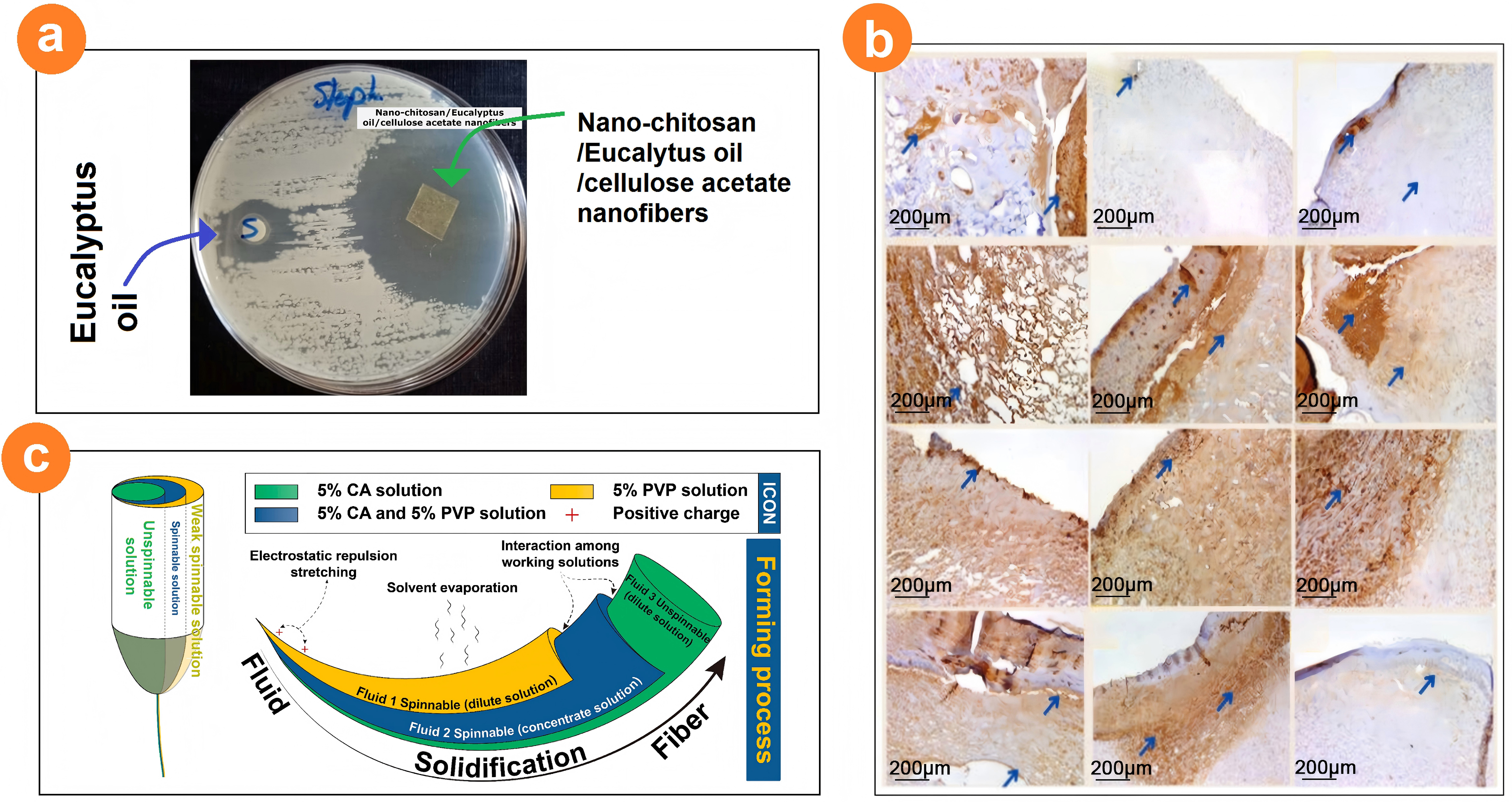
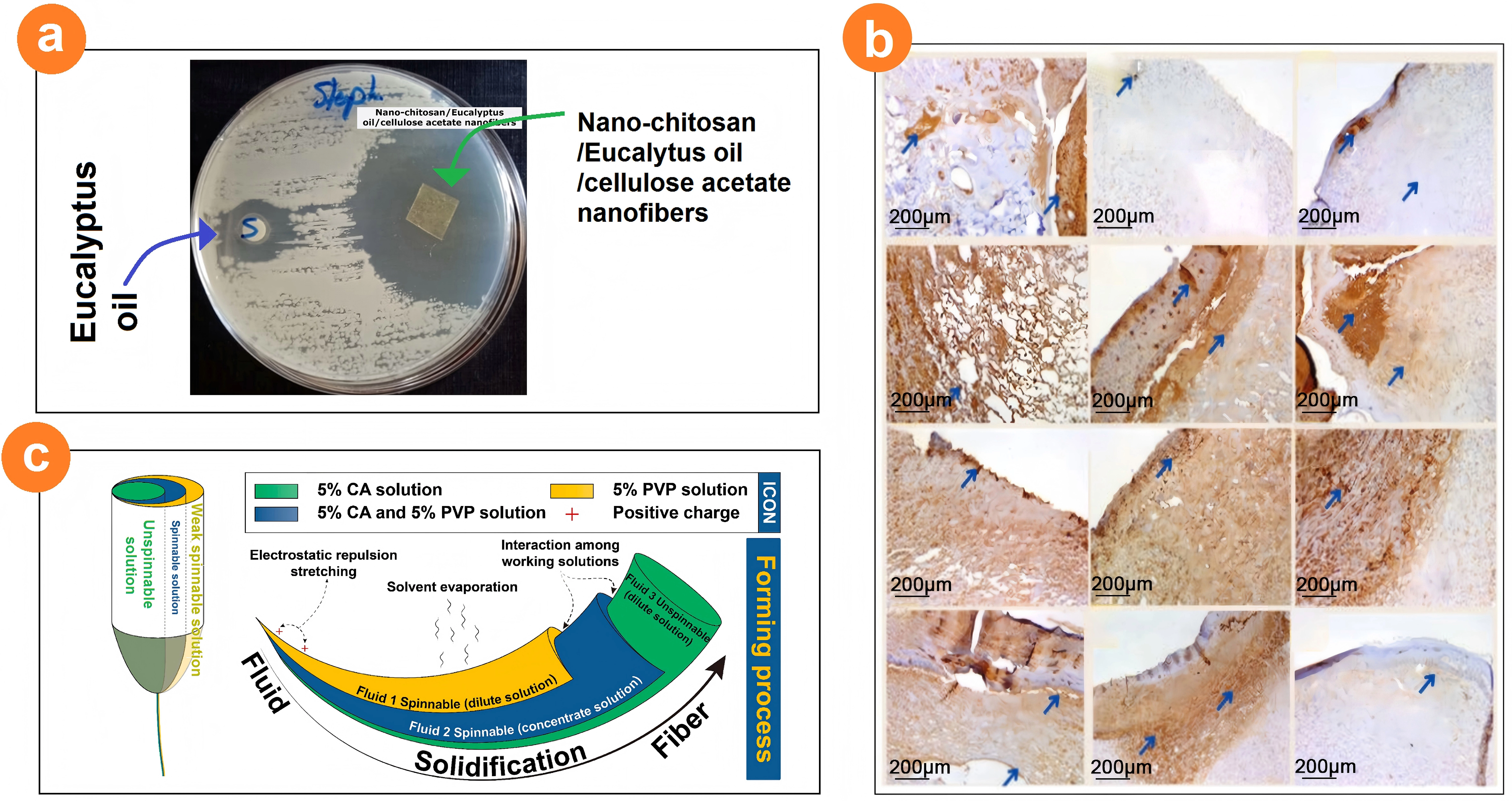
Figure 13. Electrospun CA-based nanofibrous films for wound healing: (a) The inhibition zones against S. aureus by CA nanofibers. Reproduced from reference[133]. CC BY 4.0; (b) Photomicrograph of wound tissue sections stained for the angiogenesis factor CD31 on days 4, 8 and 12. Reproduced from reference[133]. CC BY 4.0; (c) Tri-chamber Janus structure from the tri-fluid side-by-side electrospinning for loading various active ingredients in various polymeric matrices for a joint wound healing application. Reproduced with permission from reference[137]. Copyright © 2025 Elsevier. CA: cellulose acetate.
Eucalyptus essential oils (EEOs) are volatile antioxidants. EEO possesses analgesic, anti-inflammatory, and broad-spectrum antimicrobial activity, but its volatility and water insolubility limit its clinical use[132]. Elbnsawi NA. et al.[133] coelectrospun chitosan + EEO + CA to create 98 nm nanofibers that exhibited 80% cell viability, strong antibacterial activity, and upregulated transforming growth factor-β (TGF-β) and type I/III collagen, promoting safe and efficient wound repair (Figure 13b).
Besides routine active ingredients such as honey and plant oils, many plant extracts have anti-inflammatory, antioxidant, and antimicrobial properties due to the polyphenols and flavonoids they contain[134-136]. These extracts are frequently reported to be encapsulated into polymers as wound dressings. However, these nanofibers are mainly monolithic medicated nanocomposites from single-fluid blended electrospinning. As progress is made in of the related scientific fields and materials conversion techniques, many new strategies have been developed for developing new types of wound dressings.
Wang et al.[137] most recently reported a kind of tri-chamber composite nanofibrous films. In the films, CA was exploited as the backbone support and PVP was exploited to improve the hydrophilicity of the composite films. Meanwhile, two active ingredients, i.e., the naturally sourced berberine hydrochloride (BHC) and aloin (a popular component for cosmetics), were co-loaded into the structural films. Diagrams of trifluid side-by-side electrospinning and the resultant tri-chamber Janus structures are shown in Figure 13c. Owing to the specific structure and polymers selected, BHC and aloin presented different release processes with a two-stage release profile for BHC and sustained release for aloin. The combination of different drug release profiles should be a powerful platform in synergistic treatment. The fibrous membrane loaded with BHC and aloin exhibited good antibacterial performance, and it was especially effective against S. aureus. The processing parameters, fiber structures, loaded drugs, and release modes of representative electrospun CA-based drug delivery systems[52,91,93,96,104,106-108,111,115,117,119,128,133,138-148] are summarized in Table 2.
Table 2. Processing parameters, fiber structures, and release modes of drug-loaded electrospun CA-based nanofibers.
| Process | Polymer | Solvent | Drug | Diameter (nm) | Fiber structure | Drug release mode | Ref. |
| Single fluid | 10% CA-(10%/20%) AZM | DCM: acetone: Methanol = 3:2:1 | AZM | 160 | There are a few beads | delayed release | [91] |
| Single fluid | 17% CFCA-TC | Acetic acid: acetone = 3:1 | TC | 220-800 | Circular fibers with a smooth surface cross section | delayed release | [93] |
| Single fluid | (15% CS-EEO)-CA | Acetone: DMAc = 2:1 | EEO | 98 | The fibers are evenly distributed | Other releases | [133] |
| Single fluid | 6% CA-0.2% PEO-1% TTE-1.2% CHX | DMF | CHX | 613 ± 122 | The surface of the fiber is smooth, cylindrical, without beads, and evenly distributed | delayed release | [96] |
| Single fluid | 17% CA & 1.8% Budesonide | Acetic acid | Budesonide | 200 ± 100 | A beads-on-string morphology, short nanofibers | Sustained release | [138] |
| Single fluid | 10% (CA-CuO/AgNP) | Acetone: DMF = 2:1 | CuO/AgNP | 136 | The fiber is in a regular shape without beads | Other releases | [139] |
| Single fluid | 15% PCL-5% CA-7% RP | DCM: DMF = 7:3 | R.prinoides | 332 ± 74 | The nanofibers show a smooth fiber surface, no beads, and uniform distribution | Biphasic release | [140] |
| Single fluid | 12% CA-5% PCL-0.5% IBU | Acetone: DMF = 2:1; DCM:DMF = 7:3 | IBU | N/A | N/A | delayed release | [141] |
| Single fluid | 2% FA-8% SS-12% CA | Acetone | FA | 168 ± 26 | Nanofibers show small, smooth and flexible interconnecting structure fibers | delayed release | [104] |
| Single fluid | 16% CA-7% anthocyanin | DMAc: acetone: anhydrous methanol = 1:1:1 | Anthocyanidin | 320 ± 51 | The fiber diameter is small, relatively uniform and smooth | delayed release | [108] |
| Single fluid | 18% CA-1% Probucol | DMF: acetone = 3:2 | Probucol | 1059 ± 21 | A fibrous structure with randomly oriented fibers | delayed release | [117] |
| Single fluid | 18% CA-3% Pramipesol | DMF: acetone = 3:2 | Pramipesol | 828 ± 109 | It consists of randomly oriented fibers and a smooth surface | delayed release | [115] |
| Single fluid | (13% CA:15% PCL = 90:10)-1.0% Cur | Acetic acid: acetone = 3:1; Chloroform:methanol = 3:1 | Cur | N/A | A random network of fibers without beads | delayed release | [142] |
| Single fluid | 10% (CA : PVP = 7:3) -10% caffeine | Acetone: DMAc = 2:1 | Caffeine | 471.3 ± 0.1 | It is characterized by smooth, beadless and uniform cylindrical fibers | delayed release | [143] |
| Double electrostatic spinning | 18% CA-5% EUSOL+10% PVA-5% Cur | Acetone: DMF = 2:1 | EUSOL, Cur | 390 ± 50 | The fiber is characterized by a highly smooth and beadless morphology | Biphasic release | [144] |
| Double electrospinning | CA/PEG/QUE + CA/PEG/RUT | Acetone: water = 4:1 | QUE, RUT | 375 ± 230 | N/A | Biphasic release | [128] |
| Two levels in parallel | Crescent layer: 12% CA-2% CurCircular layer: 14% Gel | TFEA; HFIP | Cur | 1070 ± 20 | The surface of the nanofiber is smooth and there are no obvious beads | Biphasic release | [52] |
| Two levels of coaxiality | Sheath: 10% CA-5% PVP-5% RESCore layer: 5% CA-5% PVP-10% RES | Acetone: DMAc: ethanol = 4:1:1 | RES | 530 ± 160 | Linear nanofibers without beads have an obvious core-shell structure | delayed release | [111] |
| Two levels of coaxiality | CA-PCL loads FLA-HS | N/A | FLA | 400~600 | Uniform distribution, spherical particles | delayed release | [145] |
| Two levels of coaxiality | Sheath: 3% CA-3% ZnOCore layer: 15% CA-2% Cur | Acetone: DMF: ethanol = 4:1:1 | ZnO,Cur | 650 ± 240 | The fiber is cylindrical, without beads, and has a clear core sheath structure | delayed release | [106] |
| Two levels of coaxiality | Sheath: 10% PVP-10% RA-10% honeyCore: 5% CA-1% borneol | Absolute ethyl alcohol; Acetone: DMAc: = 2:1 | RA | 831 ± 150 nm | The surface of the fiber is smooth without beads, and the surface of the fiber has a porous structure. The overall core-shell structure of the fiber is clear | Biphasic release | [146] |
| Three-stage electrospinning | Top layer: 9% PVA and 3% CA crosslinkMiddle layer: 16%CA load 200 ppm AgNPsBase: 10%PCL | Distilled water; Acetone: DMF= 3:1; Chloroform:DMSO = 3:1 | AgNPs | N/A | N/A | Biphasic release | [147] |
| Three levels of coaxiality | Sheath: acetone: DMAc: ethanol = 4:1:1Middle layer: 11% CA-0.5% TiO2Core layer: 11% CA-2% Cur | Acetone: DMAc: ethanol = 4:1:1 | Cur,TiO2 | 720±170 | The core of the nanofiber is coated with a large number of TiO2 nanoparticles | delayed release | [148] |
| Three levels of coaxiality | Sheath: 2% CAMiddle layer: 11% CA-1% AAPCore layer: 11% CA-4% AAP | Acetone: DMAc: ethanol = 4:1:1 | AAP | 440 ± 80 | Linear nanofibers with a core-shell structure and a shell thickness of approximately 50-90 nm | delayed release | [119] |
| Three levels of coaxiality | Sheath: acetone: DMF: ethanol = 4:1:1Middle layer: 12% CACore layer: 12% CA-2% Cur | Acetone: DMF: ethanol = 4:1:1 | Cur | 570 ± 200 | Excellent cylindrical morphology and inner core sheath structure were obtained | zero order release | [107] |
N/A: not available. CA: cellulose acetate; DMAc: N,N-dimethylacetamide; DMF: N,N-dimethylformamide; DCM: dichloromethane; AZM: azithromycin; CFCA: CA from discarded cigarette filter; TC: tetracycline hydrochloride; EEO: eucalyptus essential oil; CHX: chlorhexidine; PEO: polyethylene oxide; AgNP: silver nanoparticles; PCL: polycaprolactone; SS: stainless-steel; FA: ferulic acid; PVP: polyvinylpyrrolidone; PVA: poly(vinyl alcohol); PEG: polyethylene glycol; QUE: quercetin; RUT: rutin; FLA: fluocinolone acetonide; ZnO: zinc oxide; PCL: polycaprolactone; AAP: acetaminophen; Gel: gelatin; CS: chitosan; TTE: tea tree extract; RP: rhamnus prinoides; IBU: ibuprofen; EUSOL: Edinburgh University solution of lime; RES: resveratrol; HS: hyalurosomes; TFEA: 2,2,2-trifluoroethanol; HFIP: hexafluoroisopropanol.
6. Transdermal Delivery with Electrospun CA Nanofibers
Electrospun CA mats combine nanoscale diameters (50-500 nm), open porosity, excellent biocompatibility, and mechanical flexibility, making them promising platforms for next-generation transdermal drug delivery systems (TDSs). The following section critically reviews recent advances under four complementary headings: high drug loading, controlled release, biosafety, and enhanced skin permeation.
6.1 Structural features for high drug loading
In fibrous drug delivery systems, drug loading is determined not only by polymer-drug compatibility, but also by fiber diameter, porosity, surface roughness, and the physical state of the drug within the CA matrix.
CA nanofibers possess an extremely high specific surface area and a porous structure, providing ample loading space for drugs, especially poorly soluble ones. Drugs can be uniformly dispersed within the fibers via methods such as blend spinning, emulsion spinning, or surface adsorption, often existing in an amorphous state. This significantly enhances drug solubility and bioavailability. Purnamasari[149] investigated the effect of graphene oxide (GO) incorporation into CA-based nanofibers on the transdermal delivery of naproxen (NA). Through GO-CA co-blended electrospinning (Figure 14a), the authors observed that GO addition significantly reduced the average fiber diameter (from 802 nm to 447 nm), produced uniform fiber morphology without bead-like structures (Figure 14b), and enhanced conductivity and mechanical strength, thereby accelerating drug release rates. Furthermore, the drug release kinetics conformed to the Korsmeyer-Peppas model (R2 > 0.98), indicating a diffusion-controlled release mechanism. Shafi et al.[150] developed a transdermal fiber patch for progesterone (PGS) delivery to address limitations in menopausal hormone replacement therapy. The authors combined EC and CA with PVP and incorporated polysorbate 80 (PS80) as a permeation enhancer. PS80 significantly increased PGS release (98% from wet fibers vs. 87% from dry fibers) while increasing fiber diameter [CA fibers decreased from 0.5 μm to 0.7 μm (Figure 14c)]. Fourier transform infrared (FTIR) analysis confirmed hydrogen bonding interactions between the drug and polymer, ensuring drug stability. The patch demonstrated sustained release characteristics in vitro, offering novel insights for personalized therapy. Additionally, the authors[151] investigated PS80’s enhancement of PGS diffusion from PVP fibers. Rheological analysis revealed that PS80 reduced solution surface tension, optimizing fiber formation. SEM images revealed that PS80 incorporation reduced fiber diameter from 960 nm to 760 nm while increasing specific surface area. PS80 enhanced the PGS release rate nearly fivefold, achieving 89% transdermal release in Franz diffusion cells. Kinetic models (e.g., Makoid-Banakar) exhibited excellent fitting (root mean square error (RMSE) < 7), highlighting PS80’s potential in clinical patch design. Shi et al.[152] addressed artemisinin (ART) recrystallization by preparing core-shell fibers ([ART/CA]/PVP) via coaxial electrospinning, where CA served as the core carrier and PVP as the shell. X-ray diffraction (XRD) and differential scanning calorimetry (DSC) analyses confirmed amorphous ART distribution within the fibers, effectively preventing crystallization. In vitro release experiments demonstrated sustained delivery (over 90% released within 6 days) and enhanced transdermal permeability from the core-shell fibers. This strategy offers an effective approach to improving the bioavailability of hydrophobic drugs. Wu et al.[153] evaluated the delivery performance of naproxen ester prodrugs (e.g., methyl ester, ethyl ester) in CA fiber pads. The electrospun fibers, with an average diameter of 100-500 nm, exhibited a high specific surface area that promoted drug dissolution. FTIR and DSC results revealed good compatibility between the prodrugs and CA, achieving a loading capacity of 20 wt%. In vitro release studies demonstrated sustained release for up to 6 days in physiological saline, with the ester prodrugs enhancing skin permeability. The authors emphasize that this fiber system can replace traditional patches, reducing first-pass hepatic metabolism.
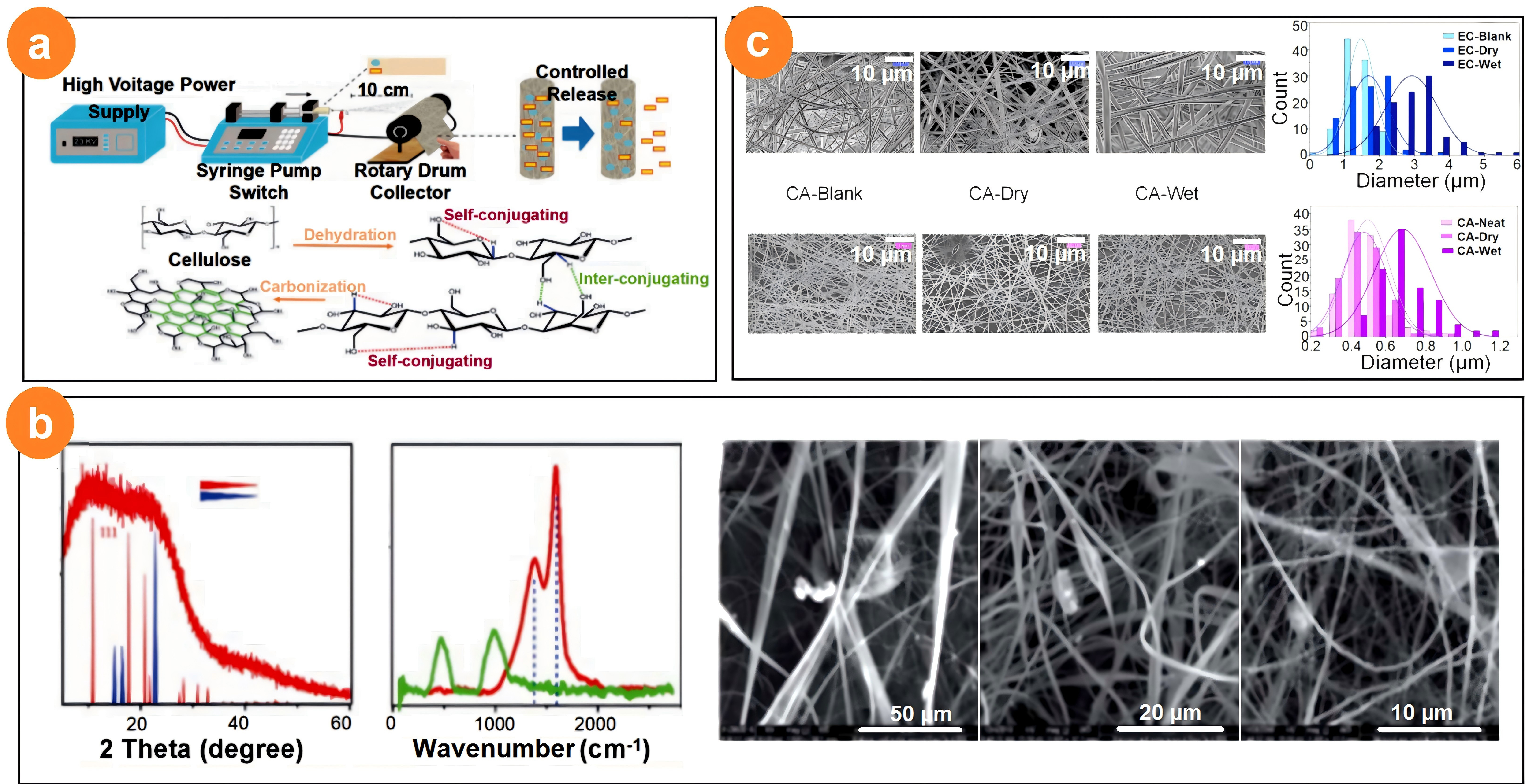
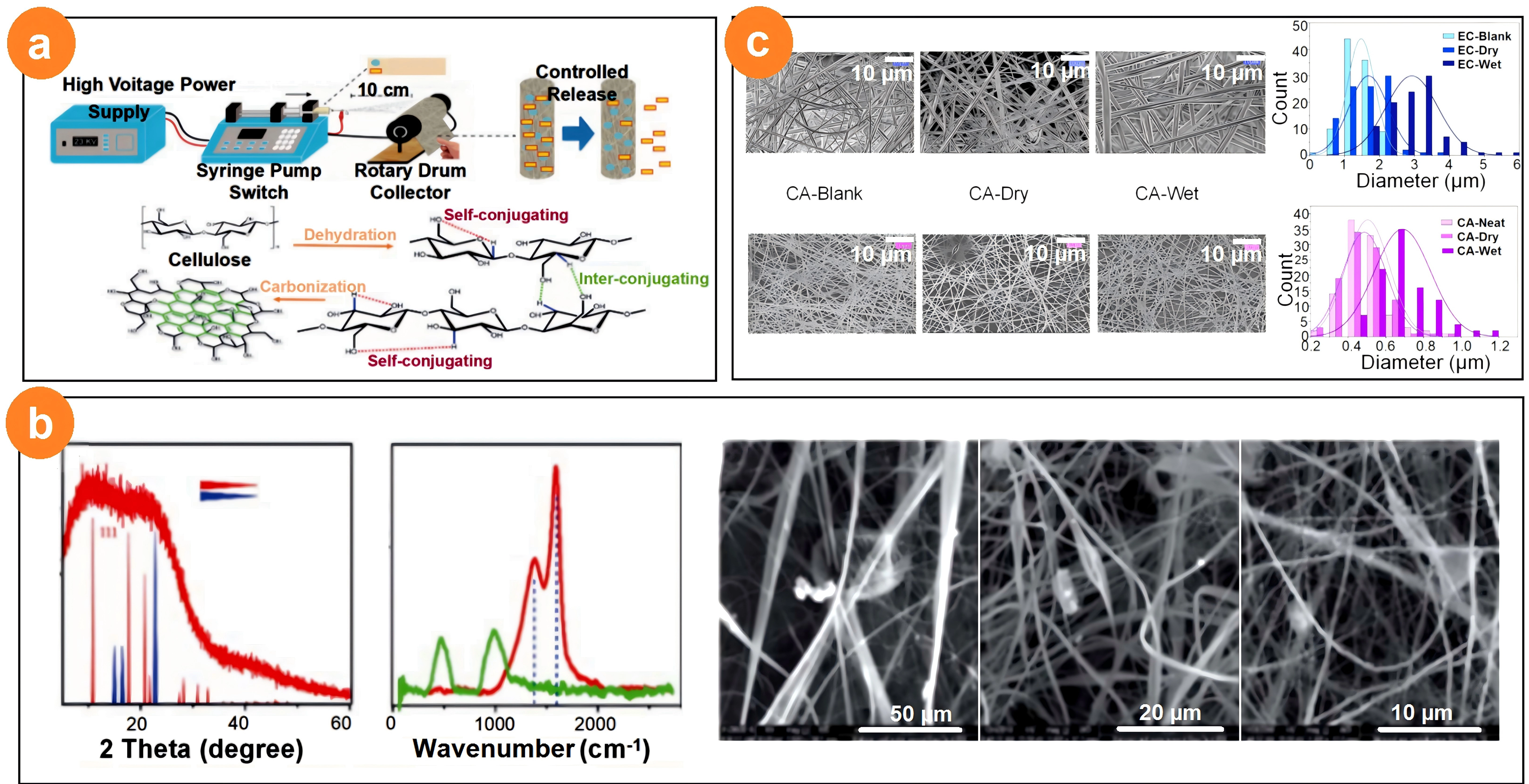
Figure 14. Electrospun CA-based nanofibers for a high drug loading and encapsulation efficiency: (a) Schematic illustration of the preparation of CA-GO-NA nanofibers. Naproxen was embedded into the nanofibers and used as a mechanism for the synthesis of graphene oxide from cellulose. Reproduced from reference[149]. CC BY 4.0; (b) XRD, FTIR and SEM images of CA, CA-GO, and CA-GO-NA nanofibers. Reproduced from reference[149]. CC BY 4.0; (c) SEM images of cellulose-PVP fibers with different diameter distributions with a 10 µm scale bar. n = 100 for each sample. Reproduced from reference[150]. CC BY 4.0. CA: cellulose acetate; PVP: poly(vinyl alcohol); GO: graphene oxide; NA: naproxen; XRD: X-ray diffraction; FTIR: fourier transform infrared; SEM: scanning electron microscope.
These studies demonstrate that CA nanofibers effectively enhance the loading capacity and release performance of poorly soluble drugs. Through supramolecular solubilization and crystal engineering, CA fibers can improve the apparent solubility, physical stability, and release performance of poorly soluble drugs, establishing a high-concentration gradient foundation for subsequent sustained release.
6.2 Structure-regulated controlled-release performance
As the dominant drug-loaded polymeric carrier, CA enables controlled drug release to be interpreted as a structure-dependent process involving diffusion pathways, pore connectivity, fiber swelling, drug crystallinity, and polymer-drug interactions.
The pore structure, diameter, composition, and post-treatment processes of CA nanofibers can precisely regulate drug release behavior, enabling multistage kinetics ranging from rapid release to sustained release over several weeks. CA fibers can be independently programmed across molecular, nanoscale, and micrometer scales to achieve a three-stage kinetic profile: “early burst release-plateau sustained release-late pulse release”. Ghorani et al.[154] successfully loaded the water-soluble molecule L-tryptophan onto CA nanofibers by developing a novel ternary solvent system (acetone:DMAc:methanol, 2:1:2). Fiber diameter (520-1,010 nm) and morphology were controlled via electrospinning parameters, thereby influencing release kinetics. The release profiles followed pseudo-second-order kinetics), with fibers averaging 720 nm in diameter exhibiting the highest initial desorption rate (2.80 mg g-1 min-1) and rate constant (0.0166 mg g-1 min-1)), attributed to their lack of bead-like defects and high specific surface area. The authors emphasized that the uniform fiber structure shortened drug diffusion pathways, thereby optimizing release controllability. Shafi et al.[150] found that adding the surfactant PS80 significantly enhanced PGS release (EC fibers from 87% to 98%, CA fibers from 82% to 96%) by improving drug permeability through micelle formation. Release kinetics were described by the Makoid-Banakar and Peppas-Sahlin models, with EC fibers exhibiting the lowest RMSE (1.9), indicating accurate prediction of release behavior. The authors noted that pore size and the polymer hydrophilicity-hydrophobicity balance are key factors regulating release rates. Prakash et al.[155] embedded GO/TiO2/curcumin nanocomposites into CA fibers to achieve pH-responsive curcumin release. Release profiles at pH 5, 7.4, and 8.2 demonstrated sustainability: cumulative release rates of 87%, 54%, and 48% over 12 days, respectively. This pH dependency was attributed to carboxyl group ionization and fiber swelling changes, with acidic conditions (pH 5) promoting faster release, mimicking dynamic pH shifts during wound healing. The authors emphasize that the fiber degradation rate (79 ± 3% porosity) synergizes with the release behavior, ensuring sustained drug efficacy at the wound site. Kontogiannopoulos et al.[156] achieved high encapsulation efficiency (74-95%) and controlled release by loading a Polygonum cuspidatum extract/vanillin (A/S) mixture onto electrospun fibers of CA, poly(lactic-co-glycolic acid) (PLLA), and poly(lactic-co-glycolic acid-co-glycerol) (PLGA). The release profiles exhibited biphasic characteristics: an initial burst release (30-40% within 2 hours) attributed to surface-adsorbed drug, followed by a slow release (50-90% within 72 hours) governed by diffusion within the fibers. Kinetic analysis aligned with Fickian diffusion mechanisms (n ≈ 0.5), indicating that release was primarily concentration gradient-driven. The authors note that polymer crystallinity (e.g., the semi-crystalline nature of PLLA) retards release, whereas the amorphous structure of PLGA accelerates drug diffusion. Shi et al.[157] compared four patch designs (Drug-in-fiber, Drug-in-adhesive, etc.), finding that patches based on CA/PVP electrospun fibers (Drug-in-fiber) effectively suppressed ibuprofen recrystallization, ensuring drug release in an amorphous state. In vitro release studies showed that the Drug-in-adhesive/fiber design delivered the highest cumulative release (2496.0 μg cm-2) within 24 hours, combining the reservoir function of fibers with the rapid diffusion advantage of the adhesive layer. Adhesion testing confirmed that the design maintains excellent mechanical properties (peel strength: 5.13 N mm-1), demonstrating that the fiber carrier sustains controlled release without compromising patch usability.
These studies achieved controlled drug release via electrospinning, with performance dependent on fiber morphology regulation, polymer-drug interactions, and external stimulus responsiveness. Common advantages include high encapsulation efficiency, sustained release profiles, and precise prediction through mathematical models (e.g., pseudo-second-order kinetics, Makoid-Banakar model). Future work may focus on in vivo validation and clinical translation to further advance the application of fiber-based delivery systems.
6.3 Skin-compatible structures and biosafety
From a structural perspective, membrane softness, breathability, pore interconnectivity, surface wettability, and extracellular matrix (ECM)-like fibrous morphology are key factors affecting skin compatibility and cell response. As a natural polysaccharide derivative with an acetylation degree of 39-42%, CA endows fibers with a controllable degradation rate (t½ ≈ 7-30 days) and extremely low immunogenicity. This confers excellent biodegradability and skin compatibility. Its nanofiber membranes are soft and breathable, capable of mimicking extracellular matrix structures to support cell adhesion and proliferation, making them suitable for long-term dressings and wound dressings. Prakash et al.[155] developed curcumin-loaded CA/GO/TiO2 composite nanofibers for wound healing applications. The hemolysis rate of CA/GO/TiO2 curcumin nanofibers was below 5% (4.2% after 4 h), meeting the requirements for blood compatibility in biomaterials. Following co-culture with fibroblasts (NIH3T3), the composite nanofibers demonstrated no cytotoxicity and significantly accelerated fibroblast migration, achieving approximately 96% wound closure within 24 hours. This indicates their effective promotion of the skin wound healing process. These findings indicate that the CA-based nanofiber scaffold exhibits excellent biocompatibility and provides a suitable microenvironment for cell growth and migration. Suwantong et al.[158] explored the potential of curcumin-loaded CA nanofibers as transdermal drug delivery carriers. After co-culturing with normal human dermal fibroblasts (NHDF), the CA nanofiber membrane exhibited high cell viability (~79-107%) even at high extract concentrations (10 mg/mL). Following drug loading, curcumin’s chemical integrity was preserved during electrospinning, and its radical scavenging capacity (antioxidant activity) was retained, facilitating its anti-inflammatory and antioxidant roles in wound healing. In vitro release studies using acetic acid buffer (pH 5.5) mimicked the acidic environment of the skin surface, suggesting the drug delivery system’s adaptability to the skin environment. ELhabal et al.[145] designed hyaluronic acid-CA/PCL nanofibers loaded with fluocinolone acetonide (FLA) for treating rheumatoid arthritis. In vitro release studies conducted in phosphate-buffered saline (pH 7.4) mimicking physiological conditions demonstrated sustained FLA release from the nanofibers for over 72 hours, indicating their potential as a transdermal patch for prolonged therapy. Joint inflammation treated with FLA nanofiber patches showed significantly reduced cellular infiltration, improved cartilage damage, and restored cellular architecture closer to normal. Furthermore, these nanofibers markedly downregulated pro-inflammatory factors (tumor necrosis factor alpha (TNF-α), NF-κB, mechanistic target of rapamycin (mTOR)) while upregulating anti-inflammatory factors (IL-10), molecularly demonstrating excellent biocompatibility and anti-inflammatory effects that effectively modulate the immune microenvironment at the lesion site. Adepu et al.[159] investigated the effect of micro-patterning on the release of diclofenac sodium from CA nanofibers. While the study focused on controlling drug release, its transdermal experiments, using Franz diffusion cells and nylon membranes with specific pore sizes (0.3 μm) to simulate the skin barrier, demonstrated that the developed nanofiber membranes are structurally suitable for transdermal drug delivery systems. The hydrophobic surface of the nanofiber membrane effectively mitigated the burst release of hydrophilic drugs. This controlled release characteristic helps maintain stable local drug concentrations, thereby reducing potential skin irritation and indirectly demonstrating its suitability for cutaneous applications. Suwantong et al.[160] evaluated CA nanofibers loaded with asiaticoside (AC) or its crude extract (CACE). CA nanofibers loaded with pure asiaticoside (PAC) showed no significant impact on the viability of NHDF even at higher concentrations (10 mg/mL), maintaining survival rates of ~86-98%, demonstrating excellent biocompatibility. Release studies conducted in buffers at pH 5.5 (simulating the skin surface) and pH 7.4 (simulating the wound microenvironment) demonstrated the adaptability of this drug delivery system to different physiological conditions, enabling its use as both a transdermal patch and a wound dressing.
The above studies collectively confirm that electrospun CA nanofibers exhibit excellent biocompatibility, manifested by low cytotoxicity, low hemolysis, and the ability to support cell proliferation and migration. Concurrently, these nanofiber systems demonstrate excellent adaptability to skin environments. This includes evaluation using release media simulating skin pH and in vitro/in vivo experiments confirming their efficacy in promoting wound healing or alleviating skin inflammation without inducing significant adverse reactions. These properties position CA nanofibers as highly promising candidate materials for wound dressings and transdermal drug delivery systems.
6.4 Structural strategies for enhanced skin permeation
In transdermal systems, fiber architecture can enhance skin contact, maintain local drug concentration gradients, suppress recrystallization, and prolong drug residence at the skin interface. The high specific surface area and porous structure of CA nanofibers promote close contact between drugs and skin. When synergized with technologies such as surfactants, microneedles, and iontophoresis, they can significantly enhance transdermal delivery efficiency, particularly for macromolecular or poorly soluble drugs. Prakash et al.[155] developed CA nanofibers loaded with GO, TiO2, and curcumin for wound dressing applications. These composite nanofibers exhibit high porosity (79-94%) and excellent swelling properties, effectively absorbing wound exudate while maintaining a moist healing environment to facilitate sustained drug release. Furthermore, GO incorporation enhances the nanofibers’ mechanical strength and antimicrobial activity, while hydrogen bonding interactions between GO and CA further improve drug loading efficiency and release stability. Shi et al.[152] prepared core-shell fibers with CA as the core and PVP as the shell via coaxial electrospinning for loading ART. This structure effectively suppressed drug recrystallization, maintaining its amorphous state to enhance solubility and transdermal permeability. The PVP shell exhibits good hydrophilicity and erosion properties, enabling controlled drug release rates and preventing burst release. In vitro permeation studies demonstrated sustained and stable drug release behavior on a porcine skin model, with significantly higher transdermal flux compared to conventional composite fibers. Elhabal et al.[145] designed hyaluronic acid-modified nanofibers loaded with FLA for rheumatoid arthritis treatment. This approach achieved dual-controlled drug release by encapsulating the drug within hyaluronic acid-mediated nanovesicles, which were further embedded into the nanofibers. In vitro release studies demonstrated sustained delivery across a pH range of 5-8.2, with significantly increased drug retention in the skin and minimal systemic absorption, thereby reducing systemic side effects. Furthermore, the nanofibers’ high specific surface area and skin-mimetic structure enhanced local drug penetration and retention. Kontogiannopoulos et al.[156] investigated electrospun fiber mats loaded with lithospermic acid and its derivatives (CA, PLLA, PLGA). Hydrogen bonding between lithospermic acid and the polymer matrix enabled its uniform dispersion in an amorphous state, improving drug release efficiency and stability. The fibers’ high specific surface area and tunable pore structure significantly enhanced transdermal drug permeability. Different polymers, such as PLGA 75:25, further modulated release behavior by adjusting degradation rates. This study provides an effective carrier strategy for the topical delivery of comfrey-derived compounds.
Electrospun CA nanofibers offer a comprehensive platform for transdermal drug delivery: a high payload, tunable release, excellent biocompatibility, and superior skin permeation. By integrating supramolecular solubilization, multiscale controlled release, active biocompatibility, and barrier-crossing permeability, they overcome the loading and release limitations of conventional patches and show clear clinical potential for anti-inflammatory therapy, hormone regulation, oncology, antioxidant care, and neurodegenerative diseases. Future work will target green, large-scale production (solvent-free melt spinning, supercritical CO2-assisted processes), smart multiresponsive systems (reactive oxygen species (ROS)/pH/enzyme logic gates), and personalized 3D-printed patches to accelerate the translation and commercialization of CA nanofibers in precision transdermal medicine.
Although CA electrospun nanofiber patches offer the above-mentioned advantages for tunable release kinetics[161,162], their therapeutic efficacy remains constrained by the stratum corneum (SC, the 10~50 μm “bricks-and-mortar” layer of corneocytes embedded in intercellular lipid lamellae that accounts for the majority of skin barrier function and restricts passive permeation to small (< 500 Da), moderately lipophilic molecules)[163,164]. Physical enhancement strategies are therefore frequently combined with CA nanofibrous patches. Among them, microneedle (MN) and iontophoresis (IP) are the two popular ones. MN pretreatment creates transient microchannels that bypass the SC, enabling rapid onset and delivery of hydrophilic/macromolecular payloads from the nanofiber matrix, and is synergistic with dissolvable MN-nanofiber hybrid designs. Its drawbacks include insertional pain perception risk, manufacturing complexity, potential infection at micropores, and limited drug loading capacity per array[165]. IP, which drives charged species through the skin via electromigration and electro-osmosis under low-level current, offers non-invasive, programmable flux control and reduced lag time, advantageous for CA patches delivering ionizable drugs, but is restricted to charged species, requires power sources and electrodes, and can cause tingling, erythema, or current-density-dependent irritation at the electrode sites[164,165]. Notably, combined MN + IP systems show synergistic enhancement, since MN-created microchannels markedly lower skin resistance and allow the applied current to propel drug deeper into the dermis[165]. Overall, MN-CA nanofibrous hybrids favor macromolecule delivery with rapid bolus release, whereas CA + IP combinations suit chronic, on-demand dosing of small ionizable drugs; both, however, add device complexity and cost compared with passive CA patches alone[164,165].
7. Conclusions
With their unique three-dimensional network structure and high specific surface area, electrospun CA nanofibers provide exceptional drug loading capacity. They not only efficiently encapsulate hydrophilic or hydrophobic drugs but also disperse poorly soluble drugs in an amorphous state, significantly increasing their dissolution rates and bioavailability. By precisely controlling the fiber diameter and porosity and employing core-shell structures, these nanofibers enable precise drug release control, ranging from rapid to sustained delivery lasting weeks. They can also respond to specific pathological environments for intelligent, on-demand drug release. The excellent biocompatibility and safety of these materials have been validated via in vitro cell experiments and in vivo models, supporting their potential biomedical safety. Furthermore, the porous structure of CA nanofibers synergistically enhances transdermal drug delivery efficiency when CA nanofibers are combined with chemical permeation enhancers or physical techniques (e.g., microneedles). This provides an efficient solution for the transdermal delivery of macromolecular drugs (e.g., insulin), fully demonstrating their immense application potential in transdermal drug delivery. Nevertheless, the application of CA nanofibers in transdermal delivery still faces several challenges. For example, the current product portfolio remains limited, and production scales are constrained, making it difficult to meet the demands of large-scale clinical applications. Furthermore, issues such as solvent selection and recovery during spinning, as well as fiber uniformity and stability, require further resolution.
8. Perspectives
Electrospun CA nanofibers have unique advantages in the field of drug delivery, yet they also face numerous challenges. The excellent biocompatibility and strong degradability of these materials allow them to integrate well with human tissues, minimizing immune reactions and side effects. After completing drug release, they can gradually degrade within the body, eliminating the need for secondary surgical removal. Furthermore, their nanofibrous structure offers a high specific surface area and porosity. In transdermal drug delivery, this enables sustained drug release, prolonging the therapeutic effect. This is particularly significant for scenarios requiring long-term, stable drug administration, such as chronic disease management and wound healing.
In particular, in the context of nanoscience, electrospinning, and electrospraying[166] have been differentiated, and a series of multiple fluid processes have been developed. These new processes include coaxial, side-by-side, trifluid coaxial, trifluid side-by-side, and trifluid combined processes of coaxial and side-by-side processes. These techniques have greatly expanded the ability of electrospinning to create various inner complex multichamber nanostructures and outer configurations in a straightforward and highly controllable manner, which is very difficult or even impossible for other top-down or bottom-up nanofabrication methods. These processes and their structural products provide a broad platform for developing many CA-based medicated nanofibers for transdermal drug delivery applications.
On the basis of current research progress and developmental needs, future studies on electrospun CA nanofibers for transdermal drug delivery should focus on deep integration and synergistic breakthroughs across four core directions: green sustainable manufacturing, smart responsive systems, multifunctional synergistic therapies, and clinical translation with personalized medicine. By optimizing raw materials and solvent systems, developing continuous and automated production equipment, and increasing solvent recovery rates, it is possible to reduce costs, improve production efficiency, and overcome bottlenecks in large-scale manufacturing. Building upon this foundation, designing smart nanofibers capable of precisely responding to pathological signals (such as pH, enzymes, or ROS) can enable on-demand drug release. Further integration of therapeutic, diagnostic, and monitoring functions can lead to the development of smart patches with “sensing-feedback-release” capabilities[167]. Ultimately, by strengthening validation in large animal models and clinical studies while exploring personalized formulation strategies tailored to patient variability (e.g., skin characteristics, metabolic differences), this technology will transition from the laboratory to clinical application, providing a safe and efficient transdermal delivery strategy for precision medicine[168,169].
In terms of wound healing applications, CA nanofibrous dressings are promising, but their favorable reputation rests largely on in vitro data from non-sterile, laboratory-processed materials. For clinical translation, the critical path is: (i) controlling and validating residual solvent to pharmacopeial/ICH limits; (ii) irritation/sensitization testing of the final, sterilized, additive-containing formulation on compromised-skin models; and (iii) a sterilization strategy chosen jointly with the formulation, with full post-sterilization re-characterization[170-172]. Regulatory-grade biological evaluation (ISO 10993 series) of the finished, sterilized product, not the as-spun mat, is the single most important gap to close.
Acknowledgements
Ms. Wenwen Zhang is gratefully acknowledged for her assistance with data collection and for providing valuable information.
Authors contribution
Zhu J, Wang B: Writing-original draft.
Du L: Writing-review & editing.
Yu DG, Zhu Y: Writing-review & editing, supervision, methodology, project administration.
All authors have read and approved the final version of the manuscript.
Conflicts of interest
Deng-Guang Yu is an Editorial Board member of Smart Materials and Devices. The other authors declare no conflicts of interest.
Ethical approval
Not applicable.
Consent to participate
Not applicable.
Consent for publication
Not applicable.
Availability of data and materials
Not applicable.
Funding
This work was supported by the National Natural Science Foundation of China (Grant No. 52371146) and the Shanghai Industrial Collaboration Project (Grant No. XTCX-KJ-2023-44).
Copyright
© The Author(s) 2026.
References
-
1. Xue J, Wu T, Dai Y, Xia Y. Electrospinning and electrospun nanofibers: Methods, materials, and applications. Chem Rev. 2019;119(8):5298-5415.[DOI]
-
2. Xia Y, Yang P, Sun Y, Wu Y, Mayers B, Gates B, et al. One-dimensional nanostructures: Synthesis, characterization, and applications. Adv Mater. 2003;15(5):353-389.[DOI]
-
3. Bayat F, Hashtrodylar Y, Karimi H, Mehryab F, Haeri A. A precise look at electrospinning parameters in fabricating the polymeric nanofibers: A review on synthetic and natural polymers. J Pharm Investig. 2024;54(6):699-750.[DOI]
-
5. Che C, Lv Y, Wu X, Dong P, Liang N, Gao H, et al. A dual-template strategy assisted synthesis of porous coal-based carbon nanofibers for supercapacitors. Diam Relat Mater. 2023;137:110140.[DOI]
-
7. Wan Y, Hu D, Xiong G, Li D, Guo R, Luo H. Directional fluid induced self-assembly of oriented bacterial cellulose nanofibers for potential biomimetic tissue engineering scaffolds. Mater Chem Phys. 2015;149:7-11.[DOI]
-
8. Kamiyama M, Soeda T, Nagajima S, Tanaka K. Development and application of high-strength polyester nanofibers. Polym J. 2012;44(10):987-994.[DOI]
-
9. Abdulhussain R, Adebisi A, Conway BR, Asare-Addo K. Electrospun nanofibers: Exploring process parameters, polymer selection, and recent applications in pharmaceuticals and drug delivery. J Drug Deliv Sci Technol. 2023;90:105156.[DOI]
-
11. Han WH, Wang QY, Long Y, Xin M, Long YZ, Hao CC. Personalized customization of in-plane thermal conductive networks by a novel electrospinning method. Compos Part B Eng. 2025;290:111971.[DOI]
-
12. Kerwald J, de Moura Junior CF, Freitas ED, de Moraes Segundo JDDP, Vieira RS, Beppu MM. Cellulose-based electrospun nanofibers: A review. Cellulose. 2022;29(1):25-54.[DOI]
-
14. Taepaiboon P, Rungsardthong U, Supaphol P. Vitamin-loaded electrospun cellulose acetate nanofiber mats as transdermal and dermal therapeutic agents of vitamin A acid and vitamin E. Eur J Pharm Biopharm. 2007;67(2):387-397.[DOI]
-
15. Xing J, Feng J, Wang Q, Yu DG, Annie Bligh SWA. Recent advances in electrospun nanofiber-based implantable drug delivery systems for breast cancer therapy. RSC Pharm. 2026;3(2):276-304.[DOI]
-
16. Sun L, Zhao Y, Caomeng X, Yu DG, Liu P. Core-sheath nanofibers from a modified coaxial electrospinning for transdermal delivery of finasteride. ChemistryOpen. 2026;15(6):e202500564.[DOI]
-
17. Yu DG, He C, Feng J, Liu Y, Annie Bligh SW. New generations of electrospun Janus nanofibers for drug delivery. J Pharm Sci. 2026;115(8):104307.[DOI]
-
18. Bosworth LA, Downes S. Acetone, a sustainable solvent for electrospinning poly(ε-caprolactone) fibres: Effect of varying parameters and solution concentrations on fibre diameter. J Polym Environ. 2012;20(3):879-886.[DOI]
-
19. Gupta P, Elkins C, Long TE, Wilkes GL. Electrospinning of linear homopolymers of poly(methyl methacrylate): Exploring relationships between fiber formation, viscosity, molecular weight and concentration in a good solvent. Polymer. 2005;46(13):4799-4810.[DOI]
-
20. Chen P, Zhou Q, Chen G, Wang Y, Lv J. Numerical simulation and experimental research of electrospun polyacrylonitrile Taylor cone based on multiphysics coupling. E-Polymers. 2023;23(1):20228106.[DOI]
-
21. Sun B, Long YZ, Zhang HD, Li MM, Duvail JL, Jiang XY, et al. Advances in three-dimensional nanofibrous macrostructures via electrospinning. Prog Polym Sci. 2014;39(5):862-890.[DOI]
-
22. Khan JS, Akram R, Shahid K, Rafique S, Hussain M, ur Rehman A. High electrical conductivity and low thermal conductivity in Bi2Te3 mixed PPy/PVP composite nanofibers by electrospinning. Mater Lett. 2023;351:134989.[DOI]
-
23. AL-SAMET MAMM, Akbulut MŞ, Burgaz E. Fabrication of novel Cu-embedded core-shell carbon nanofibers with high electrical conductivity via coaxial electrospinning. Mater Today Commun. 2024;41:111012.[DOI]
-
24. Jarusuwannapoom T, Hongrojjanawiwat W, Jitjaicham S, Wannatong L, Nithitanakul M, Pattamaprom C, et al. Effect of solvents on electro-spinnability of polystyrene solutions and morphological appearance of resulting electrospun polystyrene fibers. Eur Polym J. 2005;41(3):409-421.[DOI]
-
26. Zargham S, Bazgir S, Tavakoli A, Rashidi AS, Damerchely R. The effect of flow rate on morphology and deposition area of electrospun nylon 6 nanofiber. J Eng Fibers Fabr. 2012;7(4):155892501200700414.[DOI]
-
27. Chowdhury M, Stylios GK. Analysis of the effect of experimental parameters on the morphology of electrospun polyethylene oxide nanofibres and on their thermal properties. J Text Inst. 2012;103(2):124-138.[DOI]
-
28. Matabola KP, Moutloali RM. The influence of electrospinning parameters on the morphology and diameter of poly(vinyledene fluoride) nanofibers-effect of sodium chloride. J Mater Sci. 2013;48(16):5475-5482.[DOI]
-
29. De Vrieze S, Van Camp T, Nelvig A, Hagström B, Westbroek P, De Clerck K. The effect of temperature and humidity on electrospinning. J Mater Sci. 2009;44(5):1357-1362.[DOI]
-
30. He W, Lv H, Feng J, Yi W, Wang H, Yu DG, et al. Electrospun Janus nanofibrous mats as multifunctional fish preservation gaskets. Macromol Mater Eng. 2026;311(9):e70345.[DOI]
-
32. Zhao W, Ma Y, Zhou F, Feng S, Zhang L, Chen Y, et al. An electrospun Janus membrane for integrated radiative cooling, photothermal conversion and phase change energy storage for adaptive solar regulation. Acta Phys Chim Sin. 2026;100425.[DOI]
-
34. Gupta D, Soni S. Electrospun nanofibers in wound management: A comprehensive review of fabrication, functionality, and future directions. Polym Adv Technol. 2026;37(8):e70711.[DOI]
-
36. Sun FL, Zhao MY, Li Y, Li ZY, Li XJ, Wang N, et al. Research progress of electrospinning in food field: A review. Food Hydrocoll. 2025;158:110474.[DOI]
-
38. Gong W, Yang W, Zhou J, Zhang S, Yu DG, Liu P. Engineered beads-on-a-string nanocomposites for an improved drug fast-sustained bi-stage release. Nanocomposites. 2024;10(1):228-241.[DOI]
-
40. Goh YF, Shakir I, Hussain R. Electrospun fibers for tissue engineering, drug delivery, and wound dressing. J Mater Sci. 2013;48(8):3027-3054.[DOI]
-
45. Wang H, Wang L, Liu Z, Luo Y, Kang Z, Che X. Astragaloside/PVP/PLA nanofiber functional dressing prepared by coaxial electrostatic spinning technology for promoting diabetic wound healing. Eur Polym J. 2024;210:112950.[DOI]
-
48. Duan Y, Zhao X, Sun M, Hao H. Research advances in the synthesis, application, assembly, and calculation of Janus materials. Ind Eng Chem Res. 2021;60(3):1071-1095.[DOI]
-
53. Yu DG, Wang M, Li X, Liu X, Zhu LM, Annie Bligh SW. Multifluid electrospinning for the generation of complex nanostructures. Wiley Interdiscip Rev Nanomed Nanobiotechnol. 2020;12(3):e1601.[DOI]
-
55. Zhao P, Zhou K, Xia Y, Qian C, Yu DG, Xie Y, et al. Electrospun trilayer eccentric Janus nanofibers for a combined treatment of periodontitis. Adv Fiber Mater. 2024;6(4):1053-1073.[DOI]
-
56. Xu L, Li Q, Wang H, Liu H, Yu DG, Bligh SWA, et al. Electrospun multi-functional medicated tri-section Janus nanofibers for an improved anti-adhesion tendon repair. Chem Eng J. 2024;492:152359.[DOI]
-
58. Puls J, Wilson SA, Hölter D. Degradation of cellulose acetate-based materials: A review. J Polym Environ. 2011;19(1):152-165.[DOI]
-
59. Samadian H, Zamiri S, Ehterami A, Farzamfar S, Vaez A, Khastar H, et al. Electrospun cellulose acetate/gelatin nanofibrous wound dressing containing berberine for diabetic foot ulcer healing: In vitro and in vivo studies. Sci Rep. 2020;10:8312.[DOI]
-
61. de Freitas RRM, Senna AM, Botaro VR. Influence of degree of substitution on thermal dynamic mechanical and physicochemical properties of cellulose acetate. Ind Crops Prod. 2017;109:452-458.[DOI]
-
62. Yuan W, Wu K, Liu N, Zhang Y, Wang H. Cellulose acetate fibers with improved mechanical strength prepared with aqueous NMMO as solvent. Cellulose. 2018;25(11):6395-6404.[DOI]
-
63. Sultana N, Zainal A. Cellulose acetate electrospun nanofibrous membrane: Fabrication, characterization, drug loading and antibacterial properties. Bull Mater Sci. 2016;39(2):337-343.[DOI]
-
64. Wan Y, Lin Z, Gan D, Cui T, Wan M, Yao F, et al. Effect of graphene oxide incorporation into electrospun cellulose acetate scaffolds on breast cancer cell culture. Fibers Polym. 2019;20(8):1577-1585.[DOI]
-
67. Huang H, Dean D. 3-D printed porous cellulose acetate tissue scaffolds for additive manufacturing. Addit Manuf. 2020;31:100927.[DOI]
-
68. Yu DG, Yu JH, Chen L, Williams GR, Wang X. Modified coaxial electrospinning for the preparation of high-quality ketoprofen-loaded cellulose acetate nanofibers. Carbohydr Polym. 2012;90(2):1016-1023.[DOI]
-
70. Liu H, Hsieh YL. Ultrafine fibrous cellulose membranes from electrospinning of cellulose acetate. J Polym Sci B Polym Phys. 2002;40(18):2119-2129.[DOI]
-
71. Matos RJR, Chaparro CIP, Silva JC, Almeida Valente M, Borges JP, Soares PIP. Electrospun composite cellulose acetate/iron oxide nanoparticles non-woven membranes for magnetic hyperthermia applications. Carbohydr Polym. 2018;198:9-16.[DOI]
-
72. Han SO, Youk JH, Min KD, Kang YO, Park WH. Electrospinning of cellulose acetate nanofibers using a mixed solvent of acetic acid/water: Effects of solvent composition on the fiber diameter. Mater Lett. 2008;62(4-5):759-762.[DOI]
-
73. Sayed NM, Noby H, Thu K, El-Shazly AH. High molecular weight cellulose acetate membrane electrospinning: Parameters experimental optimization. Mater Today Proc. 2023.[DOI]
-
74. Attari N, Hausler R. Morphological investigation of Cellulose Acetate nanofibrous membranes. In: Proceedings of the 4th International Conference of Recent Trends in Environmental Science and Engineering (RTESE’20); 2020 Nov 12-14; Virtual Conference. Ottawa: Avestia Publishing; 2020. p. 124.[DOI]
-
77. Fan LT, Singh SK. Controlled release. 1st ed. Berlin: Springer;1989:[DOI]
-
78. Langer R, Peppas N. Chemical and physical structure of polymers as carriers for controlled release of bioactive agents: A review. J Macromol Sci C. 1983;23(1):61-126.[DOI]
-
79. Kikkinides ES, Charalambopoulou GC, Stubos AK, Kanellopoulos NK, Varelas CG, Steiner CA. A two-phase model for controlled drug release from biphasic polymer hydrogels. J Control Release. 1998;51(2-3):313-325.[DOI]
-
80. Dash S, Murthy PN, Nath L, Chowdhury P. Kinetic modeling on drug release from controlled drug delivery systems. Acta Pol Pharm. 2010;67(3):217-223.[PubMed]
-
81. Korsmeyer RW, Gurny R, Doelker E, Buri P, Peppas NA. Mechanisms of solute release from porous hydrophilic polymers. Int J Pharm. 1983;15(1):25-35.[DOI]
-
82. Ritger PL, Peppas NA. A simple equation for description of solute release II. Fickian and anomalous release from swellable devices. J Control Release. 1987;5(1):37-42.[DOI]
-
83. Costa P, Sousa Lobo JM. Modeling and comparison of dissolution profiles. Eur J Pharm Sci. 2001;13(2):123-133.[DOI]
-
84. Koizumi T, Panomsuk SP. Release of medicaments from spherical matrices containing drug in suspension: Theoretical aspects. Int J Pharm. 1995;116(1):45-49.[DOI]
-
85. Suckling CJ, Hunter IS, Scott FJ. Multitargeted anti-infective drugs: Resilience to resistance in the antimicrobial resistance era. Future Drug Discov. 2022;4(1):FDD73.[DOI]
-
86. Naghavi M, Vollset SE, Ikuta KS, Swetschinski LR, Gray AP, Wool EE, et al. Global burden of bacterial antimicrobial resistance 1990-2021: A systematic analysis with forecasts to 2050. Lancet. 2024;404(10459):1199-1226.[DOI]
-
88. Taylor SD. Synthesis, mechanism of action, and SAR studies on the cyclic lipopeptide antibiotic daptomycin. Can J Chem. 2024;102(7):414-424.[DOI]
-
89. Faria J, Dionísio B, Soares Í, Baptista AC, Marques A, Gonçalves L, et al. Cellulose acetate fibres loaded with daptomycin for metal implant coatings. Carbohydr Polym. 2022;276:118733.[DOI]
-
90. Kagkelaris KA, Makri OE, Georgakopoulos CD, Panayiotakopoulos GD. An eye for azithromycin: Review of the literature. Ophthalmol Eye Dis. 2018;10:2515841418783622.[DOI]
-
91. Taghe S, Mirzaeei S, Pakdaman N, Kazemi A, Nokhodchi A. Macrolide-loaded nanofibrous inserts with polycaprolactone and cellulose acetate base for sustained ocular delivery: Pharmacokinetic study in Rabbit’s eye. Int J Pharm. 2024;665:124699.[DOI]
-
92. Adir J, Barr WH. Dose-dependent bioavailability of tetracycline in man. J Pharmacokinet Biopharm. 1978;6(2):99-110.[DOI]
-
93. Khan MR, Liao S, Farooq A, Naeem MA, Wasim M, Wei Q. Regeneration and modification of cellulose acetate from cigarette waste: Biomedical potential by encapsulation of tetracycline hydrochloride. Int J Biol Macromol. 2023;250:126266.[DOI]
-
95. Oshvandi K, Lotfi RF, Azizi A, Tapak L, Larki-Harchegani A. Comparison of the effect of licorice and chlorhexidine mouthwash on the oral health of intubated patients in the intensive care unit. BMC Oral Health. 2024;24(1):682.[DOI]
-
100. Shukla SK, Goyal M, Kanabar DD, Ayehunie S, Deore B, Sanhueza CA, et al. Exploring the enhanced stability and therapeutic efficacy of Sorafenib-Cyclodextrin inclusion complex. J Mol Liq. 2024;401:124701.[DOI]
-
101. Alemomen M, Taymouri S, Saberi S, Varshosaz J. Preparation, optimization, and in vitro-in vivo evaluation of sorafenib-loaded polycaprolactone and cellulose acetate nanofibers for the treatment of cutaneous leishmaniasis. Drug Deliv Transl Res. 2023;13(3):862-882.[DOI]
-
102. Zhao Z, Moghadasian MH. Chemistry, natural sources, dietary intake and pharmacokinetic properties of ferulic acid: A review. Food Chem. 2008;109(4):691-702.[DOI]
-
103. Chen Y, Teng W, Wang J, Wang Y, Zhang Y, Cao J. The intestinal delivery systems of ferulic acid: Absorption, metabolism, influencing factors, and potential applications. Food Front. 2024;5(3):1126-1144.[DOI]
-
109. Dong M, Yang J, Fang F, Li Y, Li C. Effects of different preparation processes on the properties of resveratrol-wheat/rice starch complexes. Food Meas Charact. 2024;18(7):5243-5253.[DOI]
-
112. Jain A, Pandey G, Kolipaka T, Sharma A, Arya DK, Rajinikanth PS, et al. Electrospun poly (vinyl alcohol)/zein-based nanofibrous dressings co-loaded with pramipexole and hesperidin for diabetic wound management: In silico, in vitro, and in vivo assessments. Int J Biol Macromol. 2026;340(Pt 1):150092.
-
115. Tan C, Yuan Z, Xu F, Xie X. Electrospun cellulose acetate wound dressings loaded with Pramipexole for diabetic wound healing: An in vitro and in vivo study. Cellulose. 2022;29(6):3407-3422.[DOI]
-
117. Abbaszadeh M, Meybodi SM, Zarei A, Khorasgani EM, Heravi HM, Kasaiyan N. Cellulose acetate nanofibrous wound dressings loaded with 1% probucol alleviate oxidative stress and promote diabetic wound healing: An in vitro and in vivo study. Cellulose. 2022;29(9):5359-5374.[DOI]
-
118. Bagheri M, Shekaari H, Mokhtarpour M, Alavi A, Dorosti S, Sillanpää M, et al. Effect of novel surfactant protic ethanolamine based ionic liquids on the aqueous solubility and thermodynamic properties of acetaminophen. J Mol Liq. 2024;406:124999.[DOI]
-
120. Zhou HY, Tong JN, Ren LJ, Hao PY, Zheng HJ, Guo XM, et al. Preparation and performance of chitosan/cyclodextrin-g-glutamic acid thermosensitive hydrogel. J Drug Deliv Sci Technol. 2022;74:103504.[DOI]
-
122. Kral Ö, Turan Ayhan E, Ilbasmis-Tamer S, Tirnaksiz FF. Timolol maleate in situ ophthalmic mucoadhesive-thermosensitive gel: Development and characterization. Turk J Pharm Sci. 2024;21(4):348-354.[DOI]
-
123. Steger JS, Durai I, Odayappan A, Raman R, Sruthi T, Song AJ, et al. An evaluation of the efficacy and safety of timolol maleate 0.5% microdrops administered with the nanodropper. Ophthalmology. 2024;131(9):1045-1055.[DOI]
-
124. Mirzaeei S, Faryadras FB, Mehrandish S, Rezaei L, Daneshgar F, Karami A. Development and evaluation of polycaprolactone-based electrospun nanofibers containing timolol maleate as a sustained-release device for treatment of glaucoma: in vivo evaluation in equine eye. Res Pharm Sci. 2022;17(5):468-481.
-
125. Li Y. The utilization of Rutin-loaded lipid nanoparticles as promise carriers to enhance the therapeutic efficacy in ovarian cancer: A computational and experimental study. Inorg Chem Commun. 2025;172:113516.[DOI]
-
126. Almeleebia TM, Goyal N, Akhter MH, Alalmaie A, Al-Harbi AI, Khalilullah H, et al. β-cyclodextrin/PVP-stabilized nanocrystal gel for dual release of rutin and thymoquinone for wound healing. J Clust Sci. 2024;36(1):13.[DOI]
-
129. Zhou K, Wang M, Zhou Y, Sun M, Xie Y, Yu DG. Comparisons of antibacterial performances between electrospun polymer@drug nanohybrids with drug-polymer nanocomposites. Adv Compos Hybrid Mater. 2022;5(2):907-919.[DOI]
-
130. Ullah A, Ullah S, Khan MQ, Hashmi M, Nam PD, Kato Y, et al. Manuka honey incorporated cellulose acetate nanofibrous mats: Fabrication and in vitro evaluation as a potential wound dressing. Int J Biol Macromol. 2020;155:479-489.[DOI]
-
131. Ramanathan G, Seleenmary Sobhanadhas LS, Sekar Jeyakumar GF, Devi V, Sivagnanam UT, Fardim P. Fabrication of biohybrid cellulose acetate-collagen bilayer matrices as nanofibrous spongy dressing material for wound-healing application. Biomacromolecules. 2020;21(6):2512-2524.
-
132. Rehman R, Hussain M, Muzaffar R, Bano A, Ahmed M, Sadia H. Eucalyptus essential oil based nanoemulsions: Preparation and biological activities. Chem Biodivers. 2024;21(7):e202400406.[DOI]
-
134. Atawodi S, Liman M, Ottu J, Iliemene U. Total polyphenols, flavonoids and antioxidant properties of different parts of tamarindus indica linn of Nigerian origin. Annu Res Rev Biol. 2014;4:4273-4283.[DOI]
-
135. El-Gazzar NS. Tamarind genus chemical composition and biological activities. Nat Prod Res. 2025;39(4):935-947.[DOI]
-
137. Wang M, Huang C, Chen Y, Ji Y, Yu DG, Bligh SWA. Medicated tri-layer fibers based on cellulose acetate and polyvinylpyrrolidone for enhanced antibacterial and wound healing properties. Carbohydr Polym. 2025;348:122856.[DOI]
-
138. Shkolnikov-Lozober H, Vasilyev G, Hadad S, Vilensky R, Avrahami R, Arinstein A, et al. Localized delivery of budesonide via mucoadhesive short nanofibers for Eosinophilic esophagitis (EoE) therapy. Int J Pharm. 2026;704:127393.[DOI]
-
139. Kumar G, Khan FG, Abro MI, Aftab U, Jatoi AW. Development of cellulose acetate/CuO/AgNP nanofibers based effective antimicrobial wound dressing. Compos Commun. 2023;39:101550.[DOI]
-
140. Adamu BF, Gao J, Yuan X, Tan S, Zhao H, Jhatial AK. Rhamnus prinoides leaf extract loaded polycaprolactone-cellulose acetate nanofibrous scaffold as potential wound dressing: An in vitro study. Int J Biol Macromol. 2024;279:134934.[DOI]
-
142. Suteris NN, Yasin A, Misnon II, Roslan R, Zulkifli FH, Rahim MHA, et al. Curcumin loaded waste biomass resourced cellulosic nanofiber cloth as a potential scaffold for regenerative medicine: An in-vitro assessment. Int J Biol Macromol. 2022;198:147-156.[DOI]
-
143. Tipduangta P, Watcharathirawongs W, Waritdecha P, Sirithunyalug B, Leelapornpisid P, Chaiyana W, et al. Electrospun cellulose acetate/polyvinylpyrrolidone fiber mats as potential cosmetic under-eye masks for caffeine delivery. J Drug Deliv Sci Technol. 2023;86:104732.[DOI]
-
144. Fatima H, Khan B, Ansari AQ, Mahar FK, Mehdi M. Fabrication and characterization of cellulose acetate-EUSOL/polyvinyl alcohol-curcumin electro spun wound dressing for diabetic foot ulcer. Adv Tradit Med. 2024;24(3):777-788.[DOI]
-
145. ELhabal SF, El-Nabarawi MA, Hassanin SO, Hassan FE, Abbas SS, Gebril SM, et al. Transdermal fluocinolone acetonide loaded decorated hyalurosomes cellulose acetate/polycaprolactone nanofibers mitigated Freund’s adjuvant-induced rheumatoid arthritis in rats. J Pharm Investig. 2025;55(1):113-132.[DOI]
-
146. Zeng X, Guan Z, Yusufu R, Li S, Wu Q, Feng Y, et al. Multi-drug loaded electrospinning nanofibers promote healing with less scar formation by remodeling wound microenvironment via modulating TGF-β1/Smads/EN1 signal. Chem Eng J. 2024;499:156154.[DOI]
-
148. Deng W, Liu Y, He C, Xiong X, Zhang R, Yan T, et al. Synergistic improvements of properties of cellulose acetate based curcumin@TiO2 nanofibers via triaxial electrospinning. Chem Eng J. 2025;506:160117.[DOI]
-
150. Shafi O, Swer M, Chan UH, Kassam A, Mirzarakhimov M, Huang JY, et al. Surfactant-modified progesterone-loaded PVP/cellulose fibres for future drug delivery applications in menopause. Macromol Mater Eng. 2025;310(10):e00023.[DOI]
-
151. Shafi O, Edirisinghe M, Brako F. Polysorbate enhanced progesterone loaded drug diffusion from macromolecular fibrous patches for applications in obstetrics and gynaecology. J Drug Deliv Sci Technol. 2023;79:104062.[DOI]
-
152. Shi Y, Zhang J, Xu S, Dong A. Electrospinning of artemisinin-loaded core-shell fibers for inhibiting drug re-crystallization. J Biomater Sci Polym Ed. 2013;24(5):551-564.[DOI]
-
155. Prakash J, Venkataprasanna KS, Bharath G, Banat F, Niranjan R, Venkatasubbu GD. In-vitro evaluation of electrospun cellulose acetate nanofiber containing Graphene oxide/TiO2/Curcumin for wound healing application. Colloids Surf A Physicochem Eng Asp. 2021;627:127166.[DOI]
-
158. Suwantong O, Opanasopit P, Ruktanonchai U, Supaphol P. Electrospun cellulose acetate fiber mats containing curcumin and release characteristic of the herbal substance. Polymer. 2007;48(26):7546-7557.[DOI]
-
159. Adepu S, Gaydhane MK, Kakunuri M, Sharma CS, Khandelwal M, Eichhorn SJ. Effect of micropatterning induced surface hydrophobicity on drug release from electrospun cellulose acetate nanofibers. Appl Surf Sci. 2017;426:755-762.[DOI]
-
160. Suwantong O, Ruktanonchai U, Supaphol P. Electrospun cellulose acetate fiber mats containing asiaticoside or Centella asiatica crude extract and the release characteristics of asiaticoside. Polymer. 2008;49(19):4239-4247.[DOI]
-
162. Kumar L, Verma S, Joshi K, Utreja P, Sharma S. Nanofiber as a novel vehicle for transdermal delivery of therapeutic agents: Challenges and opportunities. Future J Pharm Sci. 2021;7(1):175.[DOI]
-
163. Parhi R, Suresh P, Patnaik S. Physical means of stratum corneum barrier manipulation to enhance transdermal drug delivery. Curr Drug Deliv. 2015;12(2):122-138.[DOI]
-
164. Alkilani A, McCrudden MT, Donnelly R. Transdermal drug delivery: Innovative pharmaceutical developments based on disruption of the barrier properties of the stratum corneum. Pharmaceutics. 2015;7(4):438-470.[DOI]
-
165. Vora D, Garimella HT, German CL, Banga AK. Microneedle and iontophoresis mediated delivery of methotrexate into and across healthy and psoriatic skin. Int J Pharm. 2022;618:121693.[DOI]
-
166. Chen S, Yun C, Wu X, Ding X. One-step eccentric side-by-side electrospraying of Janus particle coatings on cotton in an ethanol-water system for multifunctional finishing. Prog Org Coat. 2026;217:110255.[DOI]
-
168. Sarma KN, Thalluri C, Mandhadi JR. Nanofibers in drug delivery systems: A comprehensive scientific review of recent approaches. Int J Pharm Investigation. 2024;14(3):633-646.[DOI]
-
169. Hiwrale A, Bharati S, Pingale P, Rajput A. Nanofibers: A current era in drug delivery system. Heliyon. 2023;9(9):e18917.[DOI]
-
171. Havlickova K, Kuzelova Kostakova E, Lisnenko M, Hauzerova S, Stuchlik M, Vrchovecka S, et al. The impacts of the sterilization method and the electrospinning conditions of nanofibrous biodegradable layers on their degradation and hemocompatibility behavior. Polymers. 2024;16(8):1029.
-
172. Vijayakumar V, Samal SK, Mohanty S, Nayak SK. Recent advancements in biopolymer and metal nanoparticle-based materials in diabetic wound healing management. Int J Biol Macromol. 2019;122:137-148.[DOI]
Copyright
© The Author(s) 2026. This is an Open Access article licensed under a Creative Commons Attribution 4.0 International License (https://creativecommons.org/licenses/by/4.0/), which permits unrestricted use, sharing, adaptation, distribution and reproduction in any medium or format, for any purpose, even commercially, as long as you give appropriate credit to the original author(s) and the source, provide a link to the Creative Commons license, and indicate if changes were made.
Publisher’s Note
Science Exploration remains a neutral stance on jurisdictional claims in published
maps
and institutional affiliations. The views expressed in this article are solely those
of
the author(s) and do not reflect the opinions of the Editors or the publisher.
Share And Cite


Science Exploration Style
Zhu J, Wang B, Du L, Yu DG, Zhu Y. Electrospun medicated cellulose acetate nanofibers: Architectures, controlled release and transdermal drug delivery applications. Smart Mater Devices. 2027;3:202635. https://doi.org/10.70401/smd.2026.0059
Tips
Copy completed.
Submit a Manuscript
Author Instructions
Cite this Article
Article Metrics
0
View
0
Download
Cited
Article Updates

- Abstract
- Keywords
- 1. Introduction
- 2. Basic Principles and Structural Design Strategies of Electrospinning
- 3. Material Characteristics and Advantages of CA
- 4. Process Optimization of CA Nanofiber Prepared by Electrospinning
- 5. Structure-Regulated Release Profiles of Electrospun CA Nanofibers in Drug-Delivery Systems
- 6. Transdermal Delivery with Electrospun CA Nanofibers
- 7. Conclusions
- 8. Perspectives
- Acknowledgements
- Authors contribution
- Conflicts of interest
- Ethical approval
- Consent to participate
- Consent for publication
- Availability of data and materials
- Funding
- References
- Copyright
 Get citation
Science Exploration Style

Zhu J, Wang B, Du L, Yu DG, Zhu Y. Electrospun medicated cellulose acetate nanofibers: Architectures, controlled release and transdermal drug delivery applications. Smart Mater Devices. 2027;3:202635. https://doi.org/10.70401/smd.2026.0059
copy
Share Link
copy