
Cracking the Spatial Omics Data Bottleneck: From Images to Biological Insight
Published On: 10 Nov 2026
Views: 121
Subscribe
Speaker(s)
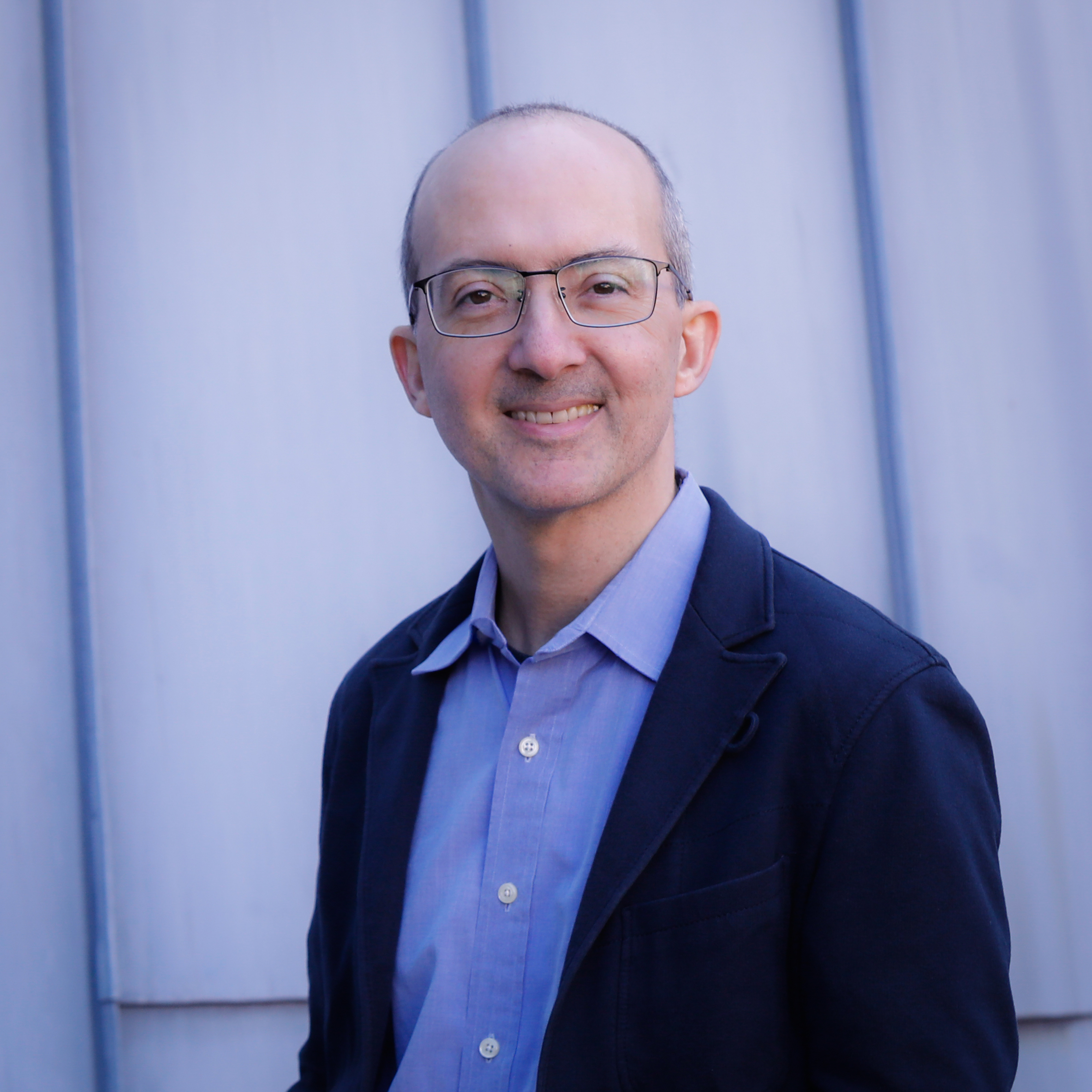
Prof. Brent R. Stockwell
Department of Biological Sciences, Department of Chemistry, Department of Pathology and Cell Biology, Columbia University, New York, NY, USA.
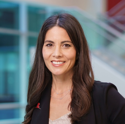
Dr. Jasmine Plummer
Center for Spatial Omics, St. Jude Children's Research Hospital, Memphis, TN, USA.
Introduction
How can researchers turn the vast - and rapidly growing - datasets generated by spatial omics into reliable biological insight? In this episode of EXO Chats, Prof. Brent Stockwell is joined by Dr. Jasmine Plummer, Director of the Center for Spatial Omics at St. Jude Children's Research Hospital, to explore this question through the EXO article, Computational workflows and data infrastructure for spatial omics analysis.
Spatial omics technologies can map thousands of genes or hundreds of proteins directly within tissues, revealing not only what cells are present, but also where they are located and how they interact with their surroundings. Yet extracting meaningful answers from these experiments remains a major challenge. Dr. Plummer discusses the need for shared data standards and purpose-built computational workflows across imaging- and sequencing-based platforms, as well as the critical importance of accurate cell segmentation and multimodal data integration.
The conversation also considers how spatial analysis can advance the study of cellular neighborhoods, ligand-receptor interactions, and precision biomarkers for patient stratification. Looking ahead, Dr. Plummer shares her vision for AI-enabled approaches that connect genomics, pathology, anatomy, and cell biology ' ultimately making spatial omics as routine and informative as a standard histological stain.
Related article published in EXO: Computational workflows and data infrastructure for spatial omics analysis
Full Transcript
Brent Stockwell:
A single spatial omics experiment can map biomolecules in a tissue section. Converting the data into biological insight, though, can take months, and the field has not agreed on how that should be done. My guest today has written the clearest account of what happens after the data come out of the machine.
Welcome to EXO Chats, the podcast of EXO - Beyond the Cell. The journal concerns itself with everything that lies outside the cell boundary: the extracellular matrix, secreted and circulating signals, the tumor microenvironment, and the spatial organization of tissues. Our subject today is that last one: The question of where a cell sits and what surrounds it has become answerable now at molecular resolution within the last few years, and instruments can generate data at a volume that some analysis methods have not fully caught up with yet.
I'm Brent Stockwell, and I'm the Editor-in-Chief of EXO - Beyond the Cell. My guest today is Dr. Jasmine Plummer, Director of the Center for Spatial Omics, an Associate Member in the Department of Developmental Neurobiology, in the Department of Molecular and Cellular Biology and a member of the Cancer Center at St. Jude Children's Research Hospital. Her lab runs spatial transcriptomics and spatial proteomics at scale, and she's been an advocate for high-quality measurement standards. She's described her ambition for spatial omics as making it as routine as a standard histological stain. And the paper under discussion today is Computational workflows and data infrastructure for spatial omics analysis published in EXO this year. Jasmine, thank you for joining me.
Jasmine Plummer:
Thank you for having me, Brent, and thank you to EXO, one for having me, but also for allowing us a platform to discuss the nitty-gritty of really what I think the big bottleneck is for the field. I don't think we spent enough time, and I think this is for all fields right now, but we're generating so much data. What are we going to do with that? And I think that's the bottleneck. I think most people, if you're honest with yourself and you run a lab, most of our time is still stuck in the analysis of the data. And most of our struggles with getting papers out or trying to formulate what we think happened in an experiment have everything to do with data analysis. I think we're really good at generating things in a wet lab now. We're suffering tremendously. And I think that's where the hope of AI is coming from, is the idea that this data struggle that we have might help us. So, thank you for having me and allowing me to talk about this.
The biggest problem in our field that really stimulated these ongoing discussions in our group - and Brent came and had a discussion with me in my office - is what are the things that are really difficult. I have the coolest biological questions, and so does Brent. But I think we can't answer those biological questions until we really focus on how we're going to analyze the data in a kind of streamlined way.
Brent Stockwell:
I totally agree. That's the future of biology, and we're going to get into it here for our listeners. So, let's just go back to the beginning a little bit, though, for listeners who have never run a spatial omics experiment. Can you just give a little introduction to, let's say, spatial transcriptomics, what are we measuring, and how is it different from what people used to do?
Jasmine Plummer:
So, I think actually spatial biology and spatial omics started with what everybody used to do. So, it's an easier thing to explain than genomics and sequencing, because most people maybe started in a lab where you stained with one antibody, or you tried to look at in situ markers, or maybe you are interested in RNA and you looked at in situ hybridization probes.
So that has not changed. We just changed it at scale. So instead of imagining the amount of data now we're generating, instead of one antibody that you may have looked at for over a year in a small cell structure, we now can plex over 100 antibodies in an experiment. So one slide, 100 antibodies - and that's just kind of genomics people have attacked it. One, the imagers got faster; two, the analysis, so acquiring it got faster; and three, we took molecular tools that we snuck from sequencing. So in sequencing, in single-cell, we kind of use these barcodes and that's how we get to scale now.
So if you think about that in situ hybridization experiment, we were using one or two probes, moving them along, putting them on a slide, and imaging them. Now we use barcodes the same way we did in single-cell. So we take those barcodes and now we attach them to every single gene in the human transcriptome or every single gene in the mouse transcriptome etc. - bumblebees, there are some bumblebee experiments. So that allows us to profile 14,000 genes now by imaging - and imagine what that data looks like.
We also do it in sequencing-based and we'll get into why I think the computation is difficult too. So the other way we do that is just take kind of a barcoded slide, slap the tissue on it, and every place we see a cell, we now make a library and can put that on. What that means now is, instead of a bulk RNA-seq experiment where you would have mushed up that tissue and put it in one tube and ascertained the brain of wild type with or without drug - now you're taking the entire brain, slicing it on a slide, and everywhere there's a cell, you're now making an individual library. So one can imagine, off of one slide, you could have 300,000 libraries, because now there's 300,000 cells there.
And that kind of scales where we start with sequencing, which is scalable because we're used to sequencing a lot of things. But the imaging one really is - we have to physically still take a picture of every single one of those genes and barcodes. We do that quickly so we can stitch them together, do that faster, but we wash them off in order to get to 14,000. You're cycling things on and off, on and off, on and off.
So, I think it's an easier way to think about where you started. Most people did start with stains, but the plexing and the ability for people to think about that one. I remember in my PhD, I was making GFP reporters, looking at three different colors, and that was like my PhD, which tells you how old I am. But given how long that used to take us, now imagine the amount of data if we put 100 antibodies on, right? So that's really one slide, 100 antibodies - that's where we're landing these days.
Brent Stockwell:
That's a perfect segue into my next question. But I'll just point out that I used to load sequencing gels manually as a graduate student. So that tells you something.
So, okay, so you mention in the paper that there's no unified analysis pipeline across these omics platforms and that a lot of these analysis methods we have were adapted from these kinds of bulk single-cell sequencing methods. And so how much is that really the issue now that there aren't as many specially designed analysis platforms? Is that what's needed?
Jasmine Plummer:
Absolutely. So I start with one simple analogy for most. You said you loaded sequencing gel - for everything that happened in sequencing I've come, I did sequencing for a very long time - we had one file, right? So we had one file where we took kind of a conversion of what an A, C, T, and a G look like right? So there was a floor for an A, C, T, and a G which is what Brent loaded on a gel, meaning we knew what an A looked like, we knew what a C looked like, we knew what a T looked like, we knew what a G looked like, and we stitched those all together and now they come out as a FASTQ file.
So no matter what modality you're doing in genomics, everything - cut-and-tag, single-cell, multi-ome, long reads, short reads, epigenomics - they all came out as the FASTQ file. The same is true in the sequencing-based, but now we have to match it to imaging. So you get a FASTQ file out of that, but it now has to match to a TIFF or a JPEG, an image file. So that's the first problem, but that's an easier problem to solve, because we've been in genomics, as Brent said, he loaded sequencing gels. We've been in it for a long time. We can do really well at like registering an image to a sequence-based method.
But remembering that file is completely different on an imager. So now you have every single person and company that's made their own machine, and every machine has their own files and no file is the same. So when you look off some of these in situ hybridization, they're taking Z-stacks, they have decoding - there's about 300 files that come off any one single capture. So, one slide, 300 files, and nobody's made a steadfast rule about what that file looks like, right? So, now you have images with decoding manuals, with coordinates.
And then it becomes even more complicated in antibodies because antibodies just have layers, right? So, they're literally just taking layers and each layer gets identified. I love that you allowed us to have a whole section in the paper about this. Like none of the files - it's like you're getting an Excel file, a PowerPoint, a PDF, you're getting some weird ImageJ / QuPath file. None of those are in the same format. And so no one's really agreed as to put it into the same version of a FASTQ and then continue on.
So we were kind of hinting at that in the paper - that some of the ways that we're doing that is, if you are working in kind of an R-based - R is more single-cell - R-based format, put it into a Seurat object. The image-based ones are just super heavy. So we tend to put that, we're hinting at that in the paper, in an AnnData object, and that can kind of move.
But that idea that the modalities will never match is the first problem. The second problem is exactly the second part of your question: we have single-cell genomicists and genomicists deciding how to analyze an image, and those two things don't coincide, right? Meaning there's so much information being lost if all we do are forcing sequencing data into what the image is telling us.
So I give one great example of T cells, you'll love this one. So in T cells, most of the major differences we're seeing in single-cell data are like T cells get exhausted, they get some transcriptional state pattern, because that's all we could see, right? All we can see is gene expression patterns that change based on a cell type we can't see. We just know they have a marker for. But when you look in a tumor, it's not a T cell that's exhausted. It's like, did the T-cell even get in, right? That's the very first problem to solve, right? You can be exhausted and outside the tumor, you would be exhausted inside the tumor, but did you get in, right?
And so, are there more exhausted T cells in the tumor? Are there more exhausted T cells outside that can't get in? So, those levels of questions, if you're only a genomicist thinking about bulk data or single-cell data that you've been looking for this marker of all these transcriptional states, and you're not thinking like an imager, right? Most imaging people think about what the structure is, where the cells are, why they went in, what cell lies around each other, and those are kind of ways by which the field needs to meet.
Brent Stockwell:
It reminds me of the old saying when you're buying a home, the three most important rules are location, location, location. So that's kind of updated for this spatial omics area.
So let me ask, you brought in this idea of different kinds of modalities. So, you talk in the paper about spatial sequencing-based spatial transcriptomics, imaging-based spatial transcriptomics, and then imaging-based multiplex spatial proteomics. So, if someone's new to this field and they want to get into it, how should they think about the tradeoffs between those different modalities? Do you have a recommendation as to what's most user-friendly or easiest in terms of the analysis?
Jasmine Plummer:
I'd love to speak from an analysis perspective, but most of the things - I live in the real world, so out of transparency - most of it has to do with sample. So if you're working in FFPE, and you really want a lot of the commercialized machines have worked very hard in trying to get into FFPE, making probes for that. But if you have no RNA left, you can't go into a transcriptomic assay. Right? So that really is - and also one of the reasons we included it in the paper - because people that are thinking about spatial transcriptomics may end up in that plexing of proteomics, because there is no RNA, but there's still so much information you can get, because now you have 100 antibodies. So, you can go past cell typing, you can do activity states etc.
So, one is sample, for sure, sample. A lot of things most people don't talk about is any of the probe-based assays are very centric to human and mouse, and I was brought up in model organisms. So that's another thing to think about, right, for your readership: you may be reading all of this, but what if your model organism, in my case, what I was studying was C. elegans, that is more a sequencing-based assay, because remembering for all of these things, we have to make probes for the RNA imaging-based, and for protein you have to have good antibodies for. So I would say if you are a model organism, there are some cDNA ones out there, such as Alumina and Complete Genomics has one, and Visium has a new one now. So, there are ways by which you just make cDNA libraries, and they're poly-A tailed, and you could put whatever organism you want. So that's an organism kind of question. Or you could spend a lot of money and make probes. So, when I said that bumblebee, it's kind of an amazing experiment. They knew their genes really well and they can make 200 genes. So that's the question of: do you need all genes? You've done a lot of data in maybe single-cell, maybe you don't need 14,000 genes.
And then I think the entry point that's really interesting for me, Brent, is people that have bulk data can go into spatial. I think single cell's nice but if you didn't hit the single-cell mark, I think spatial allows you to get into it and I would suggest a sequencing based assay, because that allows them to match their bulk kind of transcriptomics into spatial, but the interesting part for me is all the people that have been looking at their favorite gene or their favorite antibody for years, and always wanted to know one more cell type, and this is where the cell biologists come in, which has been really fun for me.
I think cell biologists never really entered the genomics field, because they knew exactly what they were looking for and organelles, etc. And we don't have organelle-level, but we do have the ability to give you another antibody or another gene, right? So, when you were looking at that synapse and you had your three markers, you really knew that synapse was different. I think getting into the plexing for those kind of more cellular biology assays may be more enticing to that kind of group.
It's not at the level of resolution. So, we don't talk about this in the paper, but I think it's important for the field. If you are a confocal person, and you’re used to looking at like 100 times objectives in major Z-stacks to find your phenotypes, these are not the assays for you, and I don't work for a company, so I can say that. But if you have an idea of your cells contacting each other, and I know you do, or you know there's an apoptotic phenotype, you know that you've had three genes you're looking for and you're not in a tissue assay. I think that you can start to think about growing cells on slides and looking at ways to investigate that.
Brent Stockwell:
That's great.
Jasmine Plummer:
So, you get both ends. I think for the first time ever as a genomicist, I see the other end of histology, pathology, anatomy people, cell biologists that really can come in and be more comfortable with the idea of antibodies.
Brent Stockwell:
Yeah, it's really cell biology meets genomics - these two communities coming together. Now, one of the things that's critical here that you talk about in the paper is the great challenge, and we talked about this when I visited you at St. Jude - cell segmentation. So, if you don't get it right, obviously, it causes cascading issues, artifacts downstream. So, can you describe for those who aren't familiar what is the issue with cell segmentation and where do we stand? How good is it now?
Jasmine Plummer:
I love that because it's literally our first diagram, right? So, when you imagine how we're assigning - very basic level - so when we're assigning the gene expression, not as much as an issue in protein, I'll speak to why that is.
So, when we're assigning, we see these transcripts inside the cell. When you have single-cell, it's in a droplet or it's barcoded to the cell membrane. And so we know it can only be from that cell. But when you have an image, now you've sliced the tissue or you've grown cells on the slide. And now what we have to do is kind of find the membrane - like find what the cell is - to count all the things inside it.
And so you know there are defaults. These are all we're in - at least in St. Jude, most people are using these commercialized assays where we try and put a membrane marker on it, which is great. So we'll take a picture, but those are generic membrane markers, right? And DAPI. So what you can do is always associate nuclei and transcripts together. We can find that very well, but if your membrane marker is not fantastic, so you imagine the case of neurons. In neurons, for sure you're going to have transcripts in the cell body and those might be your most important transcripts. And if you don't have the right membrane marker for that, your assignment will be off.
So, what that would look like in imaging-based data, specifically transcriptomics imaging-based, you'll see transcripts outside the cell, because the cell may be oval-shaped and you assign them, and there's like a bipolar neuron there. So that's what the cell segmentation tools are meant for. There are some nice ones that are segmentation-free which we allude to that - if you see enough things that look the same, right? That's the kind of AI version. If you see enough things that look the same, then that's likely the cell type. And because now you have 400,000 cells on the slide, it can start to find similar structures.
But segmentation - imagine if you get that wrong in the very beginning: every cell type and cell class is going to be annotated incorrectly. So, you may be trying to say like I found a T- cell phenotype but you didn't account for all the T cells that were there. So that's why we talk about cell segmentation.
And it's interesting - this morning I was looking at one of the reasons we spent a lot of time on this is - I look at GitHub because if you want to think about how fast the field's moving, cell segmentation tools, there's like 200 of them already, 300, 600, right? They just keep (coming), because somebody may make a tool for adipose tissue on breast, very important for breast cancer research, actually one of the reasons why spatial is very important because single-cell we couldn't actually get adipocytes very well, right? They would burst their big fat things, and we see a lot of them obviously in breast cancer tissue, and it is one of the phenotypes that actually can be more aggressive, and so not annotating that well. They're big, just imagine them as big huge fibroblasts.
So, you imagine you may find a cell segmentation tool that will allow you to get the big cells, but then your T cell phenotype's gone. And if you're thinking about immunotherapy in your tumor type, and you've captured more of the big cells and not the little cells, then you really won't be able to see what the T cells are doing. So that's kind of the issues that are always coming up right now, because what you will get off a machine and what we try and allude to are all those files, right?
When we talk about processing, the reason we're talking about processing is because the companies will give you back a file; then you have to do something with that file. So that's what you alluded to, Brent. Now I have it and none of the T cells make sense to me. They're under-called in our single-cell data. We have so many more. What are we going to do about that? And then that's the kind of secondary processing that you allowed us to speak about in the paper, which is like, maybe now you have to go back and check your cell segmentation.
Brent Stockwell:
Got it. Yeah, that's a great overview.
Jasmine Plummer:
And for antibodies, I won't explain why this wouldn't be the case, but I think it'll be very obvious to someone like you, Brent. Antibodies stain the membrane. So, we're not doing counts in that sense, right? We're doing counts of presence and absence.
So of course you get the entire, right? When certain antibodies will just by virtue of an antibody, that's why we use them, we actually use antibodies in the cell membrane markers and the RNA stuff. By virtue of doing multiplex proteomics, you will have membrane markers, right? Because the antibodies are surface membrane markers for the most part.
And then when people ask, "well, how do you do cell activation and cell state?" Well, if you had single-cell data, you would do exactly what the pathologists do, they have Ki67, which marks cell proliferation. That's an antibody, right? You can even do, we show too, you can do H3K27 isolation, any antibody working in ChIP-seq can stain a cell as well.
So that's one way by which I think the analysis is easier for people to understand with antibodies, because the assignment is just: do you see the stain in the cell or do you not see the stain in the cell? We're not physically counting activity by "there are 56 transcripts here and there's zero", and you can imagine when that happens in transcriptomics, we've misassigned, right? If it's not in the right cell and you are counting 50 to zero, but the zero was really in an outer loop of the cell and it accounted for 35, then it wouldn’t be the same cell type, if that makes sense.
Brent Stockwell:
Yeah. Totally. So does that mean that - and you talk about this in the paper too - that multiomic integration of different modalities actually helps with this segmentation problem, because you're getting the multiplexed antibody staining on and can integrate that with the same data?
Jasmine Plummer:
Yeah. I love this question. Routinely now we hold every slide. So, after we've cell annotated, we'll go back and decide stains. So, in some cases, the stain may be super relevant. Say you had a GFP mouse, and you needed to see certain cell types; then you can always take any of the RNA ones. So, anything that comes off CosMx or Xenium, you can actually antibody-stain after. So that's one way we think about it.
Our most resilient stain is H&E right? And I think you alluded this at the beginning. My goal would be: could we toss away all these $10,000 slides and eventually learn so much information from a very canonical stain and use kind of cell painting all the way through? And I think that field will eventually unite. And when you said "cell biology into tissue," I love that.
So, what we typically do is use a canonical stain after, same slide. So, what you'll get is like the architecture. So at least even in H&E, you'll get membranes. So, if you've missed a sign, we can register from that.
The other thing that helps us is really fancy things that we're already doing at St. Jude - a lot of people are trying to use the antibodies because that's what we use clinically. So, when we go through diagnosis for most tumors, even Crohn's disease, right? When it goes to a pathologist, those are really canonical stains, whether they be like tissue stains or antibodies.
And so what we've been doing a lot now is taking the same slide, so you can get RNA, CosMix or Xenium or MERSCOPE, and then take that exact same slide and put it through something like a Codex or 100-antibody plex, and we've actually done this, and then H&E. So, you've literally had four modalities in one. It takes a while, but that really helps you to find a cell, right? It also helps you to answer that age-old question: is it like antibody protein into RNA? Of course it's not concordant, of course even less concordant with an antibody-based assay than mass spec, but now we can start to match those RNA signatures better into protein.
Brent Stockwell:
Got it. Okay. That brings me to my next question, which is: if it all works and you get all this technical data integration and data analysis done, then you get to analyze niches or neighborhoods and how are different communities of cells are interacting, which is unique to the spatial format. And so you talk in the paper about that, and how some efforts to do that rely on ligand-receptor interactions that different cells are interacting through ligand-receptor interactions, but there are some issues there in terms of the reliability of doing that. So, tell us what you think about that and what's the future of niches.
Jasmine Plummer:
So, really interesting - this goes to your original question about single-cell. So you know a lot of the ways by which we labeled cells had to do with, like what marker they showed. What we're seeing now in the ligand-receptor is almost like you have to take off all the cell types, right? So, you can't really assign, you know, "I'm a fibroblast that's expressing this ligand and where's my receptor?" You almost have to manually do it now, right?
So, when that goes to niches, the way we call niches is based on the cell type you have. So, is a T cell more around a tumor cell? Is a tumor cell really centric then you'd have a lot of niches that were tumor-to-tumor? If you have CAFs, you would have tumor-to-fibroblast, and those are all with cell labels. But when you get into ligand and receptor, you can imagine many different cells express the ligand or receptor. And so if you leave it just on cell annotation, then the neighborhood analysis won't work.
So, a lot of times we're physically doing this manually, which is what an imaging person would do, not a single-cell person. So, an imaging person would turn on those channels and ask, "where is the ligand, where is the receptor," right? And then look at the cell and say what cells express the ligand, and then put the labels back on. And it's quite manual, because most of the software there is the opposite: "let's label the cells and then tell me where the ligand is," when it might be the lowest expressing thing.
So, if it's the lowest expressing thing, as a single-cell person, it wouldn't become a new cluster or new cell type, right? So almost, we have a ligand-receptor experiment that we've done and we're writing up - is that when we did neighborhood analysis, we couldn't see a difference, because those ligands were getting masked in a cell type, right? When the reality is like certain cell types, does it matter if the ligand is close to the receptor? Absolutely yes.
And I think the field's going to get there, and it's amazing, because I love when old-school things come back - it’s a proximity ligation assay, right? So, it doesn't matter how much ligand you have. If you don't have a receptor close by, right? This is all the immunotherapy stuff we're starting to see. I give an example of PD-L1. So, PD-L1 is an immunotherapy. You can't give it unless you express the receptor, right? And we stratify patients. So in breast cancer, we actually stratify. We won't give you the imunotherapy if you're not expressing it, of course, right? You have to minimally express it.
But the patients - when you stratify them, you could be a high expressor. It could be everywhere in the tissue and you don't do well. Now you use this very old-school, when I said PLA, your face lit up, because you would know what that is. It's just a proximity ligation assay. And it just, like, attaches: is the ligand close to the receptor? And when you look at that, you can start to stratify patients, right? And these are assays that multi omic approaches that we're taking in the field, which is: take the lessons you're learning in cell types, whereas PD-L1, what cell types are they in? Is there more or less expressed in these kinds of cells? And then throw it on a proximity ligation assay and say, "these are the cell types we need to be in close proximity for it to be reasonable to give you this type of therapy."
And I think we're only going to get that with that multi omics, multimodality approaches. And it gives me hope that, just like this spatial omics field, some of these older assays are coming back to help us interpret the data. And with that, they'll get higher plexing, they'll get more efficient, they will get to a place where we can start to ask T cell. I think about it all the time of T cell receptors. Those kind of assays are really important, because if you go back to that original example I gave you, I don't think that it's an exhausted T cell phenotype inside the tumor; I want to know how that one T cell got in. And does it have a ligand-receptor pairing with that tumor and that's the T cell you really want to clone and think about.
Brent Stockwell:
Totally. Yes, that's a really great point. It's really bringing us to better precision biomarkers for selecting patients and who's going to respond and what the outcomes are going to be. So, we're coming to the end here, running out of time. So, let me just ask you a final question, which is: what are you excited about in this field of spatial omics, spatial transcriptomics, data analysis, anything on the horizon, any dreams for the future that you want to share with our listeners?
Jasmine Plummer:
My dreams for the future are: be not afraid. So, I think that we are really primed in the field for AI, and I know everybody's talking AI, but when I say, we know images, yes, LLM models are going to work amazingly when you download the entire internet and learn language, you're going to do great. But images are where it's at for a lot of AI models now, and you can open your phone with your face.
Imagine that the day where an H&E will be able to tell us all this gene expression stuff. And I know the day is nowhere close. But what I think in the field, and for most people, biology is stochastic, right? We can't capture all things. We can think of something in a tissue, mimic in cell culture, it doesn't actually act the same.
But now, maybe with the ability to use AI at a high level, maybe scan images and tissue, and find the place in the tissue that's really important biology-wise for any of our questions, whether that's neuro development, whether that's apoptosis. But what we have to do now is to scan all things and make a deduction about down to one gene of what we think may be happening mechanistically. Now I think we could scan images with all this gene expression data and let it guide us, like, what are the right niches or neighborhoods or cell activity states to better inform our questions, so that the molecular answers are not going to be one gene at a time in one blot. We'll know these are the 40 antibodies or 40 proteins we need to think about interacting at the synapse instead of the two.
It's almost like, I always said it was two fields, but after talking to you today, Brent, I think it's three fields colliding, right? So, you have these genomicists and these single-cell people that have been looking at RNA and gene mutations. Imagine if you could find mosaicism in a tissue where the somatic mutation happened, or a de novo mutation in a rare disorder, and now match that not only to the tissue, right? So, anatomist, pathology people, but to cell biologists.
Now you can inform cell biologists, "we absolutely know it's these two cells together," because those assays are really moving fast too, right? And so thinking about those three fields, four fields finally uniting and being able to talk, any cell biologist would have dreamed of having 100 antibodies back in the day at once. So, I think we're finally getting to that place.
Brent Stockwell:
Yeah, it's really inspiring. It's a great vision. This has been a really terrific conversation, a great introduction as well to the field for many of the listeners.
Dr. Jasmine Plummer, thank you very much for the paper and for the conversation today. The paper again is Computational workflows and data infrastructure for spatial omics analysis with Jasmine Plummer and colleagues as authors. It's open access in EXO - Beyond the Cell. It's no cost to publish in EXO. We'll link it in the show notes. And again, I'm Brent Stockwell and this is EXO Chats. Thanks for listening!
Jasmine Plummer:
Thank you.
Share


Subscribe
