Hibernation: An underexplored natural model of biological maintenance for advancing geroscience

Sylvain Giroud
1

,
Audrey Bergouignan
2,3,4
,
Fabrice Bertile
2,*
*Correspondence to:
Fabrice Bertile, Institut Pluridisciplinaire Hubert Curien, University of Strasbourg, CNRS, Strasbourg 67087, France.
E-mail: fbertile@unistra.fr
Geromedicine. 2027;3:202648. 10.70401/Geromedicine.2026.0041
Received: July 29, 2026Accepted: September 29, 2026Published: October 08, 2026
This article belongs to the Special lssue Understudied Directions in Aging Biology, Quantitative and First-Principles Approaches
Abstract
Hibernation is one of nature’s most remarkable examples of reversible physiological adaptation, yet its potential to inform geroscience remains underexplored. Lifespan patterns in hibernators are not fully explained by allometric scaling, as hibernators can outlive non-hibernating species of comparable body size. Hibernation is further characterized by the preservation of tissue function and integrity despite marked calorie restriction, inactivity, and hypometabolism. In this review, we examine the hibernation phenotype as a naturally evolved program of biological maintenance that may limit the accumulation of age-related molecular and cellular damage. We first discuss the relationship between hibernation, longevity, and life-history evolution before reviewing evidence that hibernation modulates multiple hallmarks of ageing through integrated regulation of metabolism, mitochondrial maintenance, proteostasis, nutrient sensing, endocrine signaling, immune function, and epigenetic remodeling. We further highlight emerging observations associating hibernation with telomere maintenance and slower epigenetic ageing, while emphasizing the key knowledge gaps that remain regarding the causal relationship between the phenotype of hibernation and slower ageing, cellular senescence, genome maintenance, and age-dependent changes in hibernation expression. Finally, we discuss how comparative biology, multi-omics approaches, and functional biomarkers of ageing throughout the different phases of hibernation (before torpor, at its onset, during torpor, and after torpor) may help identify evolutionarily conserved protective mechanisms that can be translated into novel strategies to promote healthy human ageing from a bioinspired perspective. This Review was developed following discussions initiated during the September 2025 National Science Foundation (NSF) Quantitative Approaches and Understudied Questions in the Biology of Ageing Workshop (Paris, France).
Keywords
Comparative biology, evolutionary medicine, longevity, healthspan, biological resilience, metabolic plasticity, cellular quality control, ageing
1. Introduction
From ancient myths to science fiction depictions of humans in cryogenic sleep, the dream of decelerated ageing persists. Yet in nature, this “science fiction” is a biological reality. While long overlooked by the field of ageing biology, mounting evidence suggests that hibernators defy key assumptions about ageing, offering unique insights into biological resilience and longevity.
Hibernation is a naturally evolved survival strategy observed in eleven mammalian orders and one bird species, the Common Poorwill (Phalaenoptilus nuttallii)[1]. This strategy allows animals to endure harsh seasonal periods marked by food scarcity and environmental temperatures that challenge healthy physiological homeostasis and long-term survival. Following a preparatory phase during which extensive physiological, biochemical, and molecular adjustments take place[2,3], animals enter into torpor, a regulated, reversible state of metabolic depression involving reduced metabolic rate, suppressed physiological activity, and the cessation or marked reduction of food intake, enabling substantial energy conservation[4]. In small (< 8 kg) hibernating species, hibernation consists of successive bouts of torpor lasting from several days to several weeks, during which metabolic rate is reduced to 1-5% of basal rates and body temperature falls below 10 °C, and which are interrupted by brief euthermic arousals lasting only a few hours[1]. In contrast, some larger-bodied mammalian hibernators (e.g., Meles meles, Ursus arctos, or Ursus americanus), as well as species inhabiting tropical and subtropical areas (e.g., Cheirogaleus medius and Tenrec ecaudatus), do not periodically rewarm during hibernation, such that the torpid state persists throughout the entire hibernation period[5-8]. While showing a low (10-30 °C and 15 °C; C. medius and T. ecaudatus, respectively) or mild (30-33 °C; bears) body temperature during torpor, their metabolic rate usually drops by 75-90% relative to basal rates[1,6-9]. In addition to the downregulation of energy-consuming processes and tightly regulated ATP production, torpor is associated with decreased heart rate, reduced respiratory frequency, reduced renal filtration, and a dampened immune response[10,11]. Despite such challenging physiological disturbances, animals recover full function after emergence, without major organ dysfunction or apparent cognitive decline[11,12]. Interestingly, several hibernators exhibit longer lifespans than non-hibernating species of comparable body size[13,14]. These remarkable traits position hibernation as a powerful and under-appreciated model system to investigate fundamental ageing mechanisms and explore novel intervention strategies. Recent reviews have highlighted the translational potential of hibernation for promoting healthy ageing and developing therapies for chronic diseases, critical care, and space exploration[15,16]. These studies have primarily approached hibernation from a phenotype-to-application perspective, asking how specific adaptations of hibernating animals can inform therapeutic strategies or inspire biomedical innovation. Here, we reverse this perspective by considering hibernation primarily as a biological model for understanding ageing itself. Rather than asking how hibernation can be translated into interventions, we ask what mechanisms that preserve cellular and tissue integrity during hibernation reveal about the processes that govern biological ageing. We therefore examine hibernation as an integrated program of biological maintenance and map its major adaptations onto the hallmarks of ageing, with particular emphasis on whether these adaptations may limit the accumulation of age-related damage. In doing so, we bridge multiple levels of biological organization, linking comparative patterns of longevity to the molecular and cellular mechanisms governing mitochondrial function, proteostasis, nutrient sensing, endocrine and immune regulation, telomere maintenance, and epigenetic remodeling. Importantly, hibernation is a complex, integrated phenotype combining hypometabolism, hypothermia, prolonged fasting, physical inactivity, altered endocrine signalling, and, in many species, repeated torpor-arousal cycles. Because these components are intrinsically and intimately interconnected, disentangling their respective contributions to the physiological and molecular effects observed during hibernation remains challenging. In this review, we therefore consider hibernation primarily as an integrated physiological phenotype, rather than attempting to attribute each observed effect to a specific component. A systematic dissection of the contribution of each component would require a dedicated framework and is beyond the scope of the present discussion. This framework allows us to distinguish conserved physiological adaptations from mechanisms that may genuinely modulate the rate or trajectory of ageing, and to identify the key knowledge gaps that must be addressed to establish hibernation more firmly as a model of biological resilience to ageing, and to guide future comparative and translational research.
2. Body Size Does Not Fully Explain Variation in Longevity Among Hibernators
Longevity refers to the lifespan of an organism, typically expressed as survival duration or maximum lifespan at the species level. Comparative studies in mammals have shown that longevity varies widely across species, with lifespan differing by more than an order of magnitude even among taxa of similar body size[17]. Although body size is generally positively correlated with lifespan across mammals[18], there are substantial and systematic deviations from this relationship, with some taxa such as bats exhibiting lifespans far exceeding expectations and others such as marsupials falling below them[19]. This indicates that longevity is shaped by complex interactions between ecological pressures, evolutionary history, and life-history trade-offs (e.g., reproduction, growth, and somatic maintenance) rather than simple physiological scaling relationships.
Hibernation has evolved as a life-history strategy that allows mammals to survive recurrent periods of energetic constraint (see above) while also altering their exposure to seasonal ecological challenges and, in some contexts, reducing extrinsic mortality risks. Consistent with this view, a comparative analysis of 451 mammalian species demonstrated that hibernation is associated with increased annual survival and the evolution of slower life histories, independent of body mass (BM)[13]. These associations provide an ecological and evolutionary context in which hibernation may be linked to longevity, although they do not establish a causal effect of hibernation on lifespan. In particular, variation in predation pressure, resource availability and seasonality, reproductive strategy, phylogenetic history, body size, and other life-history traits may also contribute to differences in survival and longevity across species. Thus, although several hibernating species exhibit exceptional longevity, the extent to which hibernation itself contributes to this pattern, independently of these associated factors, remains unresolved.
To further examine the relationship between body size and longevity among hibernators, we compiled data from the Animal Ageing and Longevity Database (AnAge Build 15; https://genomics.senescence.info/species/index.html; accessed June 2026)[20] and from the dataset published by Ruf and Geiser[1]. Maximum longevity varies widely among hibernating mammals independently of body mass, with species differing by more than two orders of magnitude in body mass exhibiting comparable lifespans, whereas others of similar body size can differ in longevity by more than twofold (Figure 1).
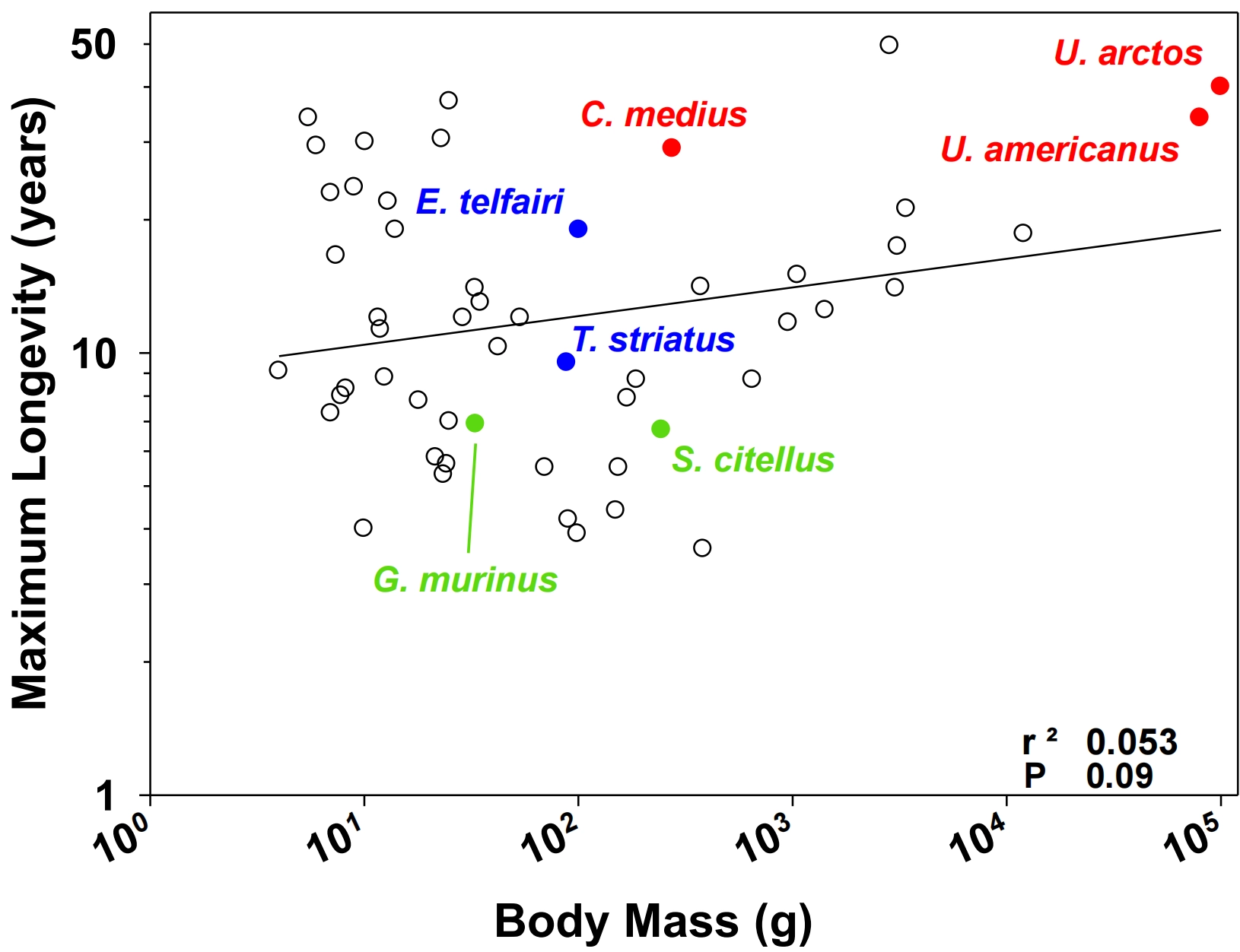
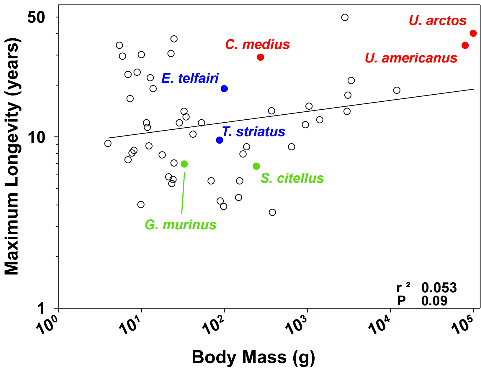
Shown is the relationship between the maximum longevity (in years) from the AnAge database and body mass in grams (average of values found in AnAge[20] and Ruf and Geiser[1]) for mammalian hibernating species according to Ruf and Geiser[1]. Bats and marsupials were excluded because both groups are established outliers in the body size-longevity relationship (see above). We could observe that similar values of maximum longevity (MaxL) can occur in hibernating species with markedly different BM. For example, as shown in red, the fat-tailed dwarf lemur (Cheirogaleus medius; BM ≈ 275 g, MaxL = 29 years) exhibits a longevity comparable to that of the American black bear (Ursus americanus; BM ≈ 80 kg, MaxL = 34 years) and the brown bear (Ursus arctos; BM ≈ 100 kg, MaxL = 40 years), despite BM differing by more than two orders of magnitude. Similarly, the European ground squirrel (Spermophilus citellus; BM ≈ 250 g, MaxL = 6.7 years) and the woodland dormouse (Graphiurus murinus; BM ≈ 35 g, MaxL = 6.9 years), displayed in green, have nearly identical longevities despite a sevenfold difference in BM. Conversely, maximum longevity can vary substantially among hibernating species with similar BM, as illustrated in blue by the Eastern chipmunk (Tamias striatus; BM ≈ 90 g, MaxL = 9.5 years) and the lesser hedgehog tenrec (Echinops telfairi; BM ≈ 100 g, MaxL = 19 years).
Taken together, these observations indicate that body size alone does not fully account for the variation in longevity observed among hibernating mammals. Instead, the weak relationship between body mass and longevity suggests that lifespan is influenced by physiological mechanisms extending beyond those captured by allometric scaling. Figure 1, however, does not demonstrate that hibernation itself accounts for the observed variation in longevity. Differences in torpor depth and duration, arousal duration and frequency, total time spent in metabolic depression and the various interconnected physiological features of hibernation may contribute to variation in longevity among hibernating species. The following section examines the physiological, cellular, and molecular mechanisms through which these traits may modulate fundamental ageing processes.
3. Hibernation as a Naturally Evolved Maintenance Program Relevant to Biological Ageing
Biological ageing can be defined as the progressive loss of physiological functions with advancing age, accompanied by increased mortality risk and ultimately leading to death[21]. To facilitate a better understanding of the molecular, cellular, and systemic mechanisms underlying ageing, twelve hallmarks of ageing have been proposed[22]. These hallmarks include genomic instability (accumulation of mutations and defects in DNA repair), telomere attrition (shortening of chromosomal ends leading to cellular senescence or apoptosis), epigenetic alterations (changes in DNA methylation, histone modifications, and dysregulation of non-coding RNAs), and loss of proteostasis (accumulation of misfolded or aggregated proteins). They also encompass disabled macroautophagy (age-related decline in autophagy resulting in the accumulation of cellular waste), deregulated nutrient sensing (altered signaling pathways such as insulin-like growth factor 1 (IGF-1) and mechanistic target of rapamycin (mTOR) affecting growth, metabolism, and longevity), mitochondrial dysfunction (declining mitochondrial function, increased reactive oxygen species production, and mitochondrial DNA mutations), and cellular senescence (accumulation of senescent cells secreting pro-inflammatory factors known as the senescence-associated secretory phenotype (SASP)). Finally, the framework includes stem cell exhaustion (reduced tissue regenerative capacity), altered intercellular communication (hormonal, neuroendocrine, and extracellular matrix imbalances), chronic inflammation (inflammaging, characterized by elevated levels of pro-inflammatory cytokines), and dysbiosis (microbiota imbalance affecting immunity, metabolism, and cognition).
Although the hallmarks of ageing have been primarily characterized in traditional laboratory models, growing evidence suggests that hibernating mammals naturally modulate several of these processes, and we propose here a hypothetical framework in which hibernation may offer insights into the mechanisms underlying healthy ageing (Figure 2). During hibernation, the marked suppression of energetically costly processes substantially reduces metabolic demand. The energetic resources spared may then become available to maintain essential cellular and tissue functions. Whether this energetic reallocation simply results from reduced metabolic demand or involves specific regulatory mechanisms that actively direct these resources toward particular maintenance pathways remains unclear. For example, the profound metabolic depression that occurs during torpor is associated with a marked reduction in mitochondrial respiration and whole-body energy expenditure, which is predicted to limit the production of reactive oxygen species (ROS) and may thereby attenuate oxidative damage, a major contributor to genomic instability and mitochondrial dysfunction. Consistent with this hypothesis, hibernators exhibit remarkable mitochondrial plasticity and enhanced antioxidant defenses that protect cellular components during hibernation[23-25]. Hibernation has also been linked to alterations in nutrient-sensing pathways, including, for example, insulin/IGF-1[26,27] and mTOR[28,29] signaling, which are known regulators of metabolism, growth, and longevity. Furthermore, the prolonged fasting periods associated with hibernation require efficient maintenance of proteostasis[10] and activation of autophagy-related processes[29], potentially limiting the accumulation of damaged proteins and cellular debris. At the organismal level, hibernators exhibit a marked suppression of immune and inflammatory functions during torpor, including reduced leukocyte numbers, cytokine production, and innate immune responses[30,31], which may limit the persistent inflammatory burden associated with ageing. Moreover, accumulating evidence indicates that torpor engages molecular pathways associated with stem cell quiescence and cellular maintenance, potentially contributing to the preservation of tissue regenerative capacity despite prolonged periods of inactivity[29]. Together, these observations suggest that hibernation is associated with coordinated modulation of multiple processes relevant to ageing, spanning energy metabolism, cellular maintenance, stress resistance, and tissue homeostasis. The following sections focus on four interconnected biological processes for which current evidence is the strongest.
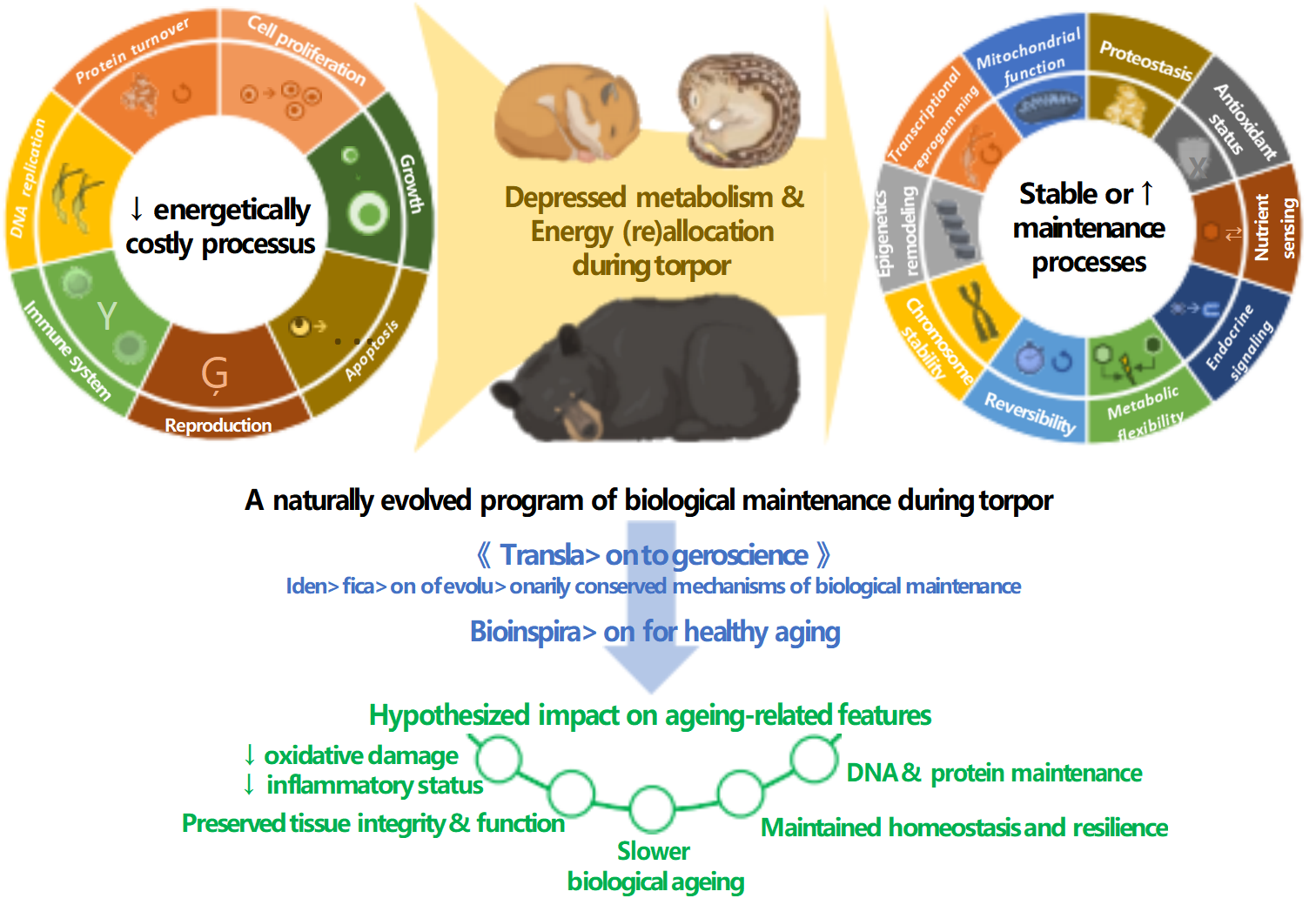
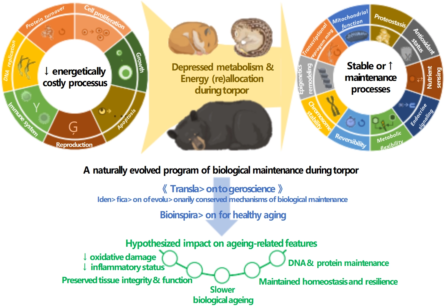
Figure 2. Proposed framework for hibernation as a natural blueprint for healthy ageing.
During hibernation, energy is hypothesized to be reallocated away from energetically costly processes, including protein synthesis, immune function, DNA replication, cell proliferation, growth and reproduction, and apoptosis, toward biological maintenance pathways that preserve cellular and tissue integrity. These include mitochondrial maintenance, proteostasis, antioxidant defenses and redox homeostasis, nutrient sensing and metabolic flexibility, endocrine regulation, telomere maintenance (mostly due to lengthening after emergence), and epigenetic remodeling with transcriptional reprogramming. While some physiological responses could be deleterious if maintained over the long run, their reversibility contributes to overall somatic maintenance. This coordinated redistribution of energetic resources is proposed to reduce the accumulation of molecular and cellular damage while maintaining tissue function, homeostasis, and physiological resilience, thereby contributing to slower biological ageing and the exceptional longevity observed in several hibernating species. Comparative investigation of these naturally evolved adaptive mechanisms may facilitate the identification of evolutionarily conserved pathways of biological maintenance, providing opportunities for bioinspired strategies to promote healthy human ageing.
3.1 Metabolic suppression and mitochondrial homeostasis
The reversible metabolic depression found in mammals during hibernation is not merely a passive consequence of hypothermia, as metabolic rate begins to decrease before any drop in body temperature, indicating that metabolic suppression is actively orchestrated by endogenous circannual programs[32]. One of the major consequences of this seasonal reprogramming that occurs during the pre-hibernation period and continues throughout hibernation is a profound redistribution of energy utilization. Carbohydrate metabolism is progressively replaced by lipid oxidation, while energetically expensive processes such as protein synthesis, cell proliferation, and anabolic pathways are markedly reduced[10,11,33]. In bears, for instance, this metabolic shift is accompanied by extensive remodeling of circulating lipids and adipose tissue metabolism[34], allowing prolonged reliance on lipid-derived fuels while minimizing protein catabolism despite several months of complete fasting[10]. Moreover, a marked repression of the eicosanoid pathway has been reported in bear tissues during hibernation[35], which may contribute to a coordinated downregulation of inflammatory responses and thermoregulation, while also reducing energy expenditure. Remarkably, despite pronounced seasonal lipid mobilization and the massive accumulation of lipid stores, no signs of the atherogenic profiles typically associated with chronic lipid overload in humans have been observed in hibernating bears[36]. This preservation of cardiovascular health is accompanied by enhanced antioxidant defenses, lower levels of inflammatory lipid mediators, and an increased proportion of the cardioprotective high-density lipoprotein 2b (HDL2b) lipoprotein[36].
Numerous studies have demonstrated reversible, tissue- and species-specific changes in mitochondrial function during hibernation[25,37,38]. In brown bears, winter skeletal muscle exhibits reduced respiratory capacity that is largely explained by lower mitochondrial content rather than intrinsic mitochondrial dysfunction, suggesting a controlled reduction in oxidative capacity while maintaining mitochondrial quality and ATP homeostasis[23]. Beyond energy production, mitochondrial remodeling also contributes to minimizing oxidative damage. Repeated torpor-arousal cycles expose tissues to rapid increases in oxygen consumption that would normally generate substantial oxidative stress. However, small hibernators display remarkable tolerance to these transitions. Numerous studies report maintenance or anticipatory upregulation of antioxidant defenses, including superoxide dismutase, catalase, glutathione peroxidases, peroxiredoxins, and glutathione metabolism, before or during torpor[24,39-41]. These adaptations are consistent with the concept of “preparation for oxidative stress” initially proposed as a phenomenon favoring survival under stress such as exposure to hypoxia, freezing, dehydration, or estivation[42]. The limitation of ROS production likely contributes to the remarkable tolerance of hibernators to ischemia-reperfusion injury, a phenomenon that has been particularly well documented in the brain and is also observed in other organs including the heart and kidney[43-46].
Thus, metabolic suppression in hibernators is an actively regulated process that reduces energetic demand, while preserving mitochondrial integrity and preparing tissues for the oxidative challenges of arousal. These adaptations provide the metabolic foundation for the cellular quality-control mechanisms discussed below.
3.2 Cellular quality control and preservation of tissue integrity
Prolonged metabolic suppression markedly reduces energetic demand and oxidative stress (see above), but this reduction alone probably cannot fully prevent the continuous production of damaged proteins, dysfunctional organelles, and other altered macromolecules. However, without efficient quality-control systems, these lesions would inevitably compromise cellular function. This has been particularly discussed for humans during fasting and physical inactivity[47,48]. In non-hibernating mammals, similar conditions rapidly induce skeletal muscle atrophy, cardiac remodeling, bone loss, and metabolic dysfunction; however, hibernators remarkably avoid many of the detrimental consequences typically associated with impaired cellular quality control. They emerge from hibernation in a healthy physiological state characterized by preserved muscle and bone mass, as well as intact cardiovascular and renal function[12,46]. These observations suggest that highly efficient cellular quality-control mechanisms continuously preserve proteome integrity and tissue homeostasis throughout the hibernation cycle[10,11].
Proteostasis constitutes a central component of this maintenance program. The mechanisms underlying body protein sparing and reduction of protein turnover during hibernation have been reviewed elsewhere[10]. Notably, the coordinated downregulation of anabolic and catabolic processes is accompanied by the maintenance or selective regulation of cellular stress- and energy-sensing pathways, including AMP-activated kinase (AMPK), forkhead box protein O (FOXO) transcription factors, and silent information regulator 1 (SIRT1)[49], thereby supporting concurrent metabolic adaptation and stress resistance[11,50]. Importantly, proteomic studies in bears indicate that the reduction in translation is not indiscriminate but is accompanied by selective preservation or increased abundance of proteins involved, e.g., antioxidant defense and carbohydrate metabolism, illustrating that hibernation reshapes rather than simply suppresses the cellular proteome[10,24,33]. Maintaining protein quality also requires the efficient recognition and elimination of damaged cellular components. Overall, the ubiquitin-proteasome[51-53] and autophagy[29,54-56] systems undergo species- and tissue-specific regulation during hibernation, with evidence suggesting maintenance of their functionality, thereby preserving tissue integrity and cellular homeostasis. Molecular chaperones and heat shock proteins provide an additional level of protection by preserving protein folding and preventing irreversible aggregation during repeated torpor-arousal cycles. They are differentially regulated in a tissue-specific manner during hibernation, reflecting, in some cases, enhanced cellular protection against protein damage, such as, for example, in bear muscle[24]. Together with antioxidant defenses (see above), these chaperones probably also facilitate the recovery of protein conformation following arousal and reduce the accumulation of misfolded proteins that would otherwise trigger proteotoxic stress.
This coordinated regulation illustrates that hibernators rely not on a single protective mechanism but on an integrated network of surveillance and recycling pathways. By maintaining protein quality, eliminating damaged cellular components, and preserving the functional integrity of skeletal muscle and other organs, hibernators largely avoid the progressive loss of proteostasis that characterizes normal ageing.
3.3 Nutrient sensing, endocrine plasticity and immune remodeling
The metabolic and cellular adaptations described in the previous sections cannot be achieved through cell-autonomous mechanisms alone but instead would require an integrated regulatory network involving nutrient-sensing pathways, endocrine signaling and immune function, capable of coordinating fuel utilization, tissue maintenance and stress resistance according to seasonal energetic constraints. During hibernation, markedly reduced circulating insulin concentrations together with a reversible state of peripheral insulin resistance[27,57] allow glucose to be spared for obligate glucose-dependent tissues while promoting lipolysis, hepatic ketogenesis and fatty acid oxidation in peripheral organs[10,11]. Importantly, this seasonal insulin resistance does not develop into a pathological state as observed in obesity and type 2 diabetes, as suggested by restoration of insulin sensitivity after emergence[27,57]. Rather than triggering glucotoxicity, oxidative stress and chronic inflammation, it represents a tightly regulated physiological adaptation that is rapidly reversed following arousal or spring emergence. At the cellular level, the metabolic shift is orchestrated by highly conserved nutrient-sensing pathways, i.e. AMPK and FOXO (see above), together with context-dependent regulation of mammalian target of rapamycin complex 1 (mTORC1), a major regulator of anabolic processes. In many seasonal hibernators, mTORC1 activity is reduced during torpor, consistent with the suppression of anabolic metabolism and reduced protein synthesis[28]. By contrast, studies in hibernating brown bears have reported increased phosphorylation of S6K1[58], suggesting that components of the mTORC1 pathway may remain active or even be enhanced during torpor. Another study found higher skeletal muscle S6K1 phosphorylation in black bears one week after hibernation compared to before hibernation, although phosphorylation during hibernation was not assessed[55]. If S6K1 phosphorylation also increases during hibernation in black bears, as reported in brown bears, its elevated levels one week after hibernation could reflect a prolonged alteration in S6K1 phosphorylation dynamics. The apparent divergence between variation of mTORC1 and S6K1 raises the possibility that mTORC1 signaling may be differentially coupled to whole-body metabolic suppression across hibernating species. In bears, sustained mTORC1 signaling could potentially contribute to tissue maintenance and remodeling despite overall metabolic depression. However, S6K1 phosphorylation alone is not sufficient to establish sustained functional activation of the mTORC1 pathway. A dedicated comparative study integrating measurements of mTORC1 signaling activity, protein synthesis, nutrient availability, and tissue turnover across hibernators with contrasting body sizes and energetic strategies would help determine whether this divergence reflects a genuine adaptation for balancing energy conservation with tissue maintenance during prolonged metabolic suppression, or instead arises from differences in sampling time, torpor depth, or tissue-specificity. This question is also relevant to the broader relationship between mTOR signaling and longevity, as chronic activation of mTOR is increasingly recognized as a major driver of ageing and reduced lifespan[59,60]. Hibernation may thus provide a unique physiological context in which mTOR signaling is uncoupled from its usual pro-ageing consequences, potentially through tight temporal, tissue-specific regulation.
Seasonal endocrine remodeling during hibernation does not involve isolated hormonal fluctuations, but coordinated adjustments across multiple endocrine axes that collectively reprioritize physiological functions, with different species achieving similar physiological outcomes through distinct regulatory strategies. For example, whereas bears generally exhibit reduced circulating triiodothyronine (T3) and thyroxine (T4) concentrations consistent with metabolic suppression and reduced thermogenesis[61,62], several small hibernating rodents display maintained or even elevated thyroid hormone levels during torpor. Whether these concentrations are buffered by altered thyroid hormone availability or contribute to sustaining protein turnover, lipid metabolism, or arousal energetics remains unresolved. Similar flexibility is observed in nutrient-regulating hormones. In addition to insulin (see above), decreased IGF-1 signaling limits anabolic investment during hibernation[63,64]. Seasonal regulation of reproductive hormones[65-67], glucocorticoids[68,69], and potentially other unidentified mediators[70] supports skeletal muscle maintenance despite prolonged inactivity. Across taxa, these diverse hormonal adaptations converge toward common outcomes: metabolic suppression, efficient fuel allocation, and preservation of tissue integrity.
Immune function undergoes equally profound and species-specific remodeling. Although hibernation is often associated with a suppression of innate immune responses in small hibernating mammals[31,71] and brown bears (U. arctos)[72,73], immune function may be differentially regulated across species, with American black bears (U. americanus) maintaining immune competence during hibernation[74]. Moreover, as already mentioned above, downregulation of inflammatory responses is expected to result from the repression of the eicosanoid pathway in hibernating bears[35]. These adaptations illustrate how brown bears have evolved mechanisms that dissociate physiological insulin resistance and hyperlipidemia from the inflammatory and vascular complications that characterize human metabolic disease[27,35,36]. Whether this conclusion can be generalized to other hibernators remains unknown.
Collectively, these observations indicate that nutrient sensing, endocrine plasticity, and immune remodeling act as interconnected components of integrated seasonal physiological regulation. Conserved nutrient-sensing pathways coordinate cellular metabolism, endocrine networks synchronize whole-body physiological priorities, and immune remodeling minimizes inflammatory damage while preserving host defense. Understanding more precisely how hibernators maintain this finely coordinated systems-level regulation without progressing toward chronic metabolic dysfunction or inflammaging may provide valuable insights for developing interventions aimed at promoting healthy ageing and preventing age-associated diseases.
3.4 Telomere dynamics and epigenetic regulation
3.4.1 Telomeres: Guarding the ends
Telomeres contribute to the maintenance of genome integrity and are increasingly recognized as key mediators of life-history trade-offs across living organisms[75]. They consist of non-coding repetitive DNA sequences located at the ends of eukaryotic chromosomes that prevent the progressive loss of coding DNA during DNA replication and cell division. Telomeres indeed generally shorten with each round of cell division, a process accelerated by oxidative stress, which damages DNA and other macromolecules when ROS production exceeds antioxidant defenses[76]. However, specific mechanisms, including telomerase activation[76] and alternative lengthening of telomeres (ALT)[77], can maintain or restore telomere length by elongating telomeric DNA. Consequently, telomere length is widely regarded as a marker of somatic maintenance, reflecting the balance between cumulative cellular damage and the mechanisms that preserve genomic integrity.
Studies in hibernators indicate that telomere dynamics are far more complex than the progressive shortening typically associated with ageing. Initially, hibernation was associated with telomere shortening in several mammalian species, notably small-bodied hibernators that undergo periodic arousals during the winter[78-80]. However, telomere lengthening has been observed upon emergence from hibernation[78] or during the subsequent active season[80], suggesting that telomere maintenance mechanisms can be reactivated following hibernation. This seasonal telomere recovery might reflect the dependence of telomere maintenance on energy availability, consistent with regulation by circannual rhythms[81].
The energetic conditions experienced during hibernation also appear to shape telomere dynamics. In hibernating garden dormice provided with food throughout winter, telomere length gradually increased over the course of the hibernation season[82]. Likewise, telomere lengthening during hibernation was negatively correlated with time spent in euthermia, suggesting that energy-efficient torpor phases may preserve telomeric integrity, whereas metabolically costly arousals accelerate telomere attrition[79,82]. Consistent with this interpretation, further research on garden dormice revealed that DNA strand breaks accumulate during torpor bouts, although the extent of DNA damage was inversely related to body temperature[83].
Recent findings in the fat-tailed dwarf lemur (C. medius), a tropical hibernating primate, further challenge the assumption that telomere attrition is an inevitable consequence of metabolic stress[84,85]. Under captive conditions, food-deprived individuals undergoing deep torpor-arousal cycles and substantial body mass loss exhibited telomere elongation during hibernation, followed by shortening upon emergence. By contrast, daily fed lemurs display shallower torpor and little change in telomere length, suggesting that torpor depth and energy availability jointly shape telomere dynamics. A recent study further demonstrated that telomerase activity is downregulated in the liver of garden dormice during the pre-hibernation period but remains elevated throughout hibernation, consistent with the maintenance of telomere integrity during torpor[86].
Together, these findings suggest that telomere dynamics during hibernation reflect a balance between torpor depth, energy availability, oxidative stress, and arousal-associated damage. While the notion of seasonal telomere maintenance, with damage during arousal and recovery during torpor, remains partly speculative, it is increasingly supported by experimental data[81]. Nevertheless, evidence remains sparse as to whether seasonal telomere preservation or elongation is mediated by telomerase activity or other DNA-maintenance or repair mechanisms.
3.4.2 Epigenetic adaptations to torpor as a model for healthy ageing
Epigenetics refers to the ensemble of molecular and cellular mechanisms that modulate gene expression without altering the nucleotide sequence of DNA, thereby contributing to phenotypic variation[87]. In its broadest sense, epigenetic regulation includes DNA methylation, post-translational histone modifications, chromatin remodeling, and the activity of regulatory non-coding RNAs, which collectively influence genome function and cellular phenotype[88]. With ageing, these epigenetic regulatory systems undergo progressive alterations that disrupt gene expression programs and cellular homeostasis, thereby promoting the loss of cellular identity, impaired tissue regeneration, cellular senescence, chronic low-grade inflammation, and increased susceptibility to age-related disease[89-91]. Interestingly, epigenetic modifications are reversible and highly responsive to lifestyle factors. Several interventions capable of modulating the epigenetic mechanisms underlying ageing have been identified. Beyond promising pharmacological therapies and other strategies involving CRISPR-based technologies, microbiota transplantation, and immunotherapy, efforts to reduce biological age primarily rely on lifestyle interventions, notably caloric restriction, regular physical activity, stress management, and sufficient high-quality sleep[91,92]. In this context, hibernation represents a particularly intriguing physiological model. As mentioned above, torpid animals display adaptations that are likely to modulate several hallmarks of ageing, suggesting the involvement of adaptive molecular and epigenetic mechanisms that contribute to the preservation of cellular function and tissue integrity.
DNA methylation, histone modifications, and non-coding RNAs allow animals to rapidly and reversibly modulate their biological functions in response to seasonal fluctuations in environmental conditions[93]. Accumulating evidence indicates that multiple layers of epigenetic control act in a coordinated and reversible manner to regulate global gene expression during torpor, therefore being involved in the molecular reprogramming underlying transitions between active euthermic and hypometabolic states in hibernators. Global histone modifications during hibernation are generally consistent with reduced transcriptional activity. For example, in skeletal muscle and brown adipose tissue of the thirteen-lined ground squirrel (Ictidomys tridecemlineatus), decreased histone H3 phosphorylation and acetylation, together with increased abundance and activity of histone deacetylases (HDAC)1 and HDAC4[94,95], as well as small ubiquitin-related modifier (SUMO)ylation-mediated regulation[96], are expected to promote chromatin compaction and transcriptional repression. Similarly, differential modulation of histone H3/H4 acetylation and coordinated regulation of histone acetyltransferase and deacetylase systems in the liver of the Syrian hamster (Mesocricetus auratus) indicate active and reversible chromatin remodeling during torpor[97]. Moreover, torpor-associated changes in miRNA expression have been reported across multiple tissues and taxa[14,98,99]. Collectively, predicted miRNA targets point to metabolic suppression, repression of anabolic and other energy-demanding cellular processes, and tissue-specific protective responses. These include reduced fatty acid oxidation and preservation of skeletal muscle integrity in the brown bear (U. arctos)[100] and little brown bat (M. lucifugus)[101], coordination of cardiac hypertrophy, skeletal muscle atrophy and hepatic metabolic remodelling in the thirteen-lined ground squirrel (I. tridecemlineatus)[102], regulation of energy metabolism, the cell-cycle and chromatin-associated pathways in the liver of the garden dormouse (Eliomys quercinus)[103], regulation of heart and skeletal muscle metabolism and of neuroprotective pathways in the brain of the Australian central bearded dragon (Pogona vitticeps)[104], and regulation of mitochondrial function and metabolic flexibility, i.e. the capacity to adjust substrate use to changes in substrate availability, by mitomiRs in the liver of the thirteen-lined ground squirrel (I. tridecemlineatus)[105]. Seasonal changes in DNA methylation have also been documented, likely contributing to the regulation of gene expression during hibernation. Increased global DNA methylation occurs during torpor in the liver of the greater horseshoe bat (Rhinolophus ferrumequinum), together with elevated expression of DNA methyltransferases 3B (DNMT3B) and ZBTB4[106], and in brown adipose tissue of the thirteen-lined ground squirrel (I. tridecemlineatus)[95], whereas no change has been detected in the liver of the Syrian hamster (M. auratus)[97]. More detailed analyses further reveal locus-specific regulation: in wing tissue of the big brown bat (Eptesicus fuscus), hibernation is associated with slower epigenetic ageing, and widespread methylation changes affecting genes involved in metabolism, transcriptional regulation and innate immunity, predominantly within quiescent chromatin states[107]. Taken together, these findings indicate that hibernation involves coordinated epigenetic remodeling through histone modifications, miRNAs and DNA methylation. Understanding how hibernators repeatedly engage these protective programs may provide insights into biological resilience, healthy ageing and strategies to mitigate age-related decline in humans.
The biomarkers of ageing currently described in the literature encompass functional, physiological, and molecular dimensions. They include measures of physical and cognitive performance, circulating markers of inflammation, metabolism, and endocrine function, as well as molecular indicators of biological age such as telomere length, DNA damage and repair, DNA methylation profiles, circulating microRNAs, and signatures derived from multi-omics approaches[108-111]. Age-dependent changes in DNA methylation patterns have enabled the development of epigenetic clocks, which are currently among the most robust and widely used biomarkers of biological ageing[91]. In yellow-bellied marmots (Marmota flaviventris), hibernation was associated with a marked slowing of epigenetic ageing, with near-stabilization of DNA methylation age during winter dormancy and resumed age acceleration during the active season[112]. This seasonal pattern is consistent with an effect of metabolic suppression on epigenetic ageing trajectories, although whether it translates into slower biological ageing remains to be established.
Together, these findings raise the possibility that hibernation may influence biological ageing, but this hypothesis requires validation using complementary measures of ageing-related damage and functional healthspan. The relative contributions of hypometabolism, reduced body temperature, fasting, torpor architecture and epigenetic regulation remain unresolved.
4. Transferability & Future Directions
Collectively, the evidence reviewed here supports the view that hibernation slows the accumulation of molecular and cellular damage through integrated regulation of metabolism, mitochondrial function, proteostasis, endocrine signaling and immune homeostasis. Across the mechanisms reviewed herein, several common principles emerge despite substantial interspecific diversity (Figure 2):
● Several protective responses are anticipatory, being initiated before or at the onset of torpor.
● Physiological states that resemble human pathology, including insulin resistance, hyperlipidemia, immune suppression, reduced mitochondrial activity, and transcriptional repression, remain tightly controlled, seasonal, and reversible.
● The profound reduction in metabolic demand may create an energetic context that we hypothesize could favor the maintenance of essential cellular processes, including antioxidant defense, proteostasis, autophagy, DNA repair, and tissue integrity.
Whether energy is actively redirected toward the maintenance processes, rather than simply conserved through reduced demand, remains an important area for future investigations.
The evidence linking hibernation to slower biological ageing remains largely associative. Many findings derive from small samples, heterogeneous species, cross-sectional seasonal comparisons, or molecular signatures that have not been functionally validated. A central priority is therefore to establish causal links between specific features of hibernation and healthspan preservation. Particular attention should be paid to transitions into and out of torpor.
Although metabolic depression may reduce ATP turnover and molecular damage during torpor, rapid rewarming, reperfusion, and metabolic reactivation may transiently increase oxidative and cellular stress. Repeated sampling across preparation for torpor, deep torpor, torpor arousal, and seasonal recovery will therefore be essential to determine the temporal sequence of damage, protection, and repair.
Importantly, the different mechanisms associated with hibernation are not equally mature for translation. We propose three research priorities. First, experimentally induced torpor-like states in non-hibernating mammals will provide a direct way to test whether components of the hibernation phenotype causally modify biological ageing. Second, circulating or secreted factors derived from hibernators will offer an opportunity to identify potential protective signals that may be transferable without reproducing torpor per se. Third, continued comparative investigation of natural hibernators remains essential for identifying evolutionarily conserved mechanisms of biological maintenance that should be prioritized for future testing of cause-and-effect relationships.
4.1 Induction of torpor-like states as a proof of transferability
The clearest proof-of-concept that hibernation-associated benefits can be induced in non-hibernators comes from experimentally induced torpor-like states in mice. Activation of hypothalamic neuronal circuits capable of inducing a torpor-like phenotype was shown to slow epigenetic ageing across multiple tissues and improve healthspan[113]. Importantly, decreased core body temperature emerged as the primary driver of this effect, providing evidence that at least one component of the hibernation phenotype can influence ageing-related outcomes outside hibernators.
Future similar studies are now needed to disentangle the respective contributions of hypothermia, fasting, reduced physical activity and other factors by manipulating these components independently or in combination. Caloric restriction, for example, is known to extend lifespan in a wide range of animal species, including non-human animals[114]. In addition, human studies of sustained caloric restriction suggest favorable effects on some, but not all, biomarkers or hallmarks of ageing[115-117]. Future studies should determine which components of the torpor-like phenotype are necessary or sufficient for the observed geroprotective effects and whether combinations of these components produce additive or synergistic responses. Importantly, outcomes should progressively extend beyond hallmarks and epigenetic age to functional measures of ageing, including tissue function, physiological resilience, morbidity, healthspan and ultimately lifespan where feasible.
4.2 Use of circulating factors to transfer torpor-like characteristics
A second experimental strategy is to identify circulating factors that contribute to tissue protection during hibernation. Serum collected from hibernating bears has already been shown to inhibit proteolysis and increase protein content, as well as regulate the transforming growth factor-β (TGF-β)/bone morphogenetic proteins (BMP) balance, in cultured human muscle cells[70,118], demonstrating that at least part of the hibernation phenotype can be transferred across species through circulating signals. Systemic fractionation and multi-omic profiling of hibernator serum, followed by functional screening in human cells and non-hibernating animal models, could identify proteins, metabolites, lipids, extracellular vesicles, or combinations of circulating factors responsible for these effects. Similar approaches could be extended beyond skeletal muscle to identify circulating signals involved in mitochondrial protection, inflammation, bone preservation, or other ageing-relevant processes. Such approaches may ultimately be more readily translatable than inducing whole-body torpor because they aim to reproduce discrete protective mechanisms rather than the complete physiological state.
4.3 Deepening our understanding of how hibernation influences biological ageing
Another priority is to establish more directly how hibernation influences the trajectory of biological ageing itself. Longitudinal studies should determine whether variation in torpor depth, duration, arousal frequency, and cumulative time spent in metabolic depression predicts trajectories of biological age, physiological function, healthspan and lifespan.
A separate but complementary priority is to determine how these protective capacities change with advancing age. Longitudinal studies are again required to establish whether older hibernators retain the ability to preserve muscle, bone, cardiovascular, metabolic, cognitive, and immune function and to identify the mechanisms underlying this resilience. Linking age-related changes in hibernation architecture to molecular markers of maintenance and functional resilience will be critical for determining whether the mechanisms that protect hibernators remain effective throughout life.
4.3.1 Telomere and genome maintenance
Future studies should notably determine whether the observed preservation or elongation of telomeres involves changes in telomerase activity, alternative lengthening of telomeres, or both, and establish how these pathways vary across tissues and successive phases of the hibernation cycle. This work should subsequently be broadened to DNA-damage sensing and repair to determine whether hibernators merely limit genome damage or also possess enhanced mechanisms for restoring genome integrity during torpor, arousal, or seasonal recovery.
4.3.2 Cellular senescence
The trajectory of cellular senescence represents another particularly important but underexplored dimension of hibernation biology. Current evidence is insufficient to determine whether hibernators accumulate fewer senescent cells than non-hibernating mammals. Among the major upstream drivers of cellular senescence, including mitochondrial dysfunction, oxidative stress, chronic inflammation, loss of proteostasis, and metabolic dysregulation[119,120], several appear to be attenuated or tightly regulated during hibernation[10,23,24]. Hibernators may therefore possess mechanisms that limit the accumulation of damage normally leading to irreversible cell-cycle arrest, or alternatively that actively eliminate or reprogram senescent cells once they arise. Addressing these hypotheses will require systematic characterization of senescence across tissues and successive phases of the hibernation cycle using complementary markers of cell-cycle arrest, persistent DNA damage markers, DNA repair capacity, and components of the senescence-associated secretory phenotype, combined with single-cell transcriptomics, spatial omics, cytogenetics, and epigenetic profiling. Such integrative analyses would provide an unprecedented opportunity to investigate not only the presence of senescent cells but also their tissue specificity, temporal dynamics, and potential contribution to seasonal physiological remodelling. Another particularly promising avenue concerns the dynamics of senescent-cell turnover. Rather than completely preventing senescence, hibernators may transiently generate senescent cells during torpor and subsequently eliminate or reprogram them during interbout arousals or seasonal transitions. This hypothesis is consistent with the increasingly recognized beneficial roles of transient cellular senescence in tissue remodelling, regeneration, and wound repair[121]. It is also possible that hibernators can efficiently clear senescent cells when needed, potentially through mechanisms involving their immune system. Determining whether hibernation relies on coordinated cycles of senescence induction and clearance could fundamentally reshape our understanding of how these animals preserve tissue function despite experiencing repeated episodes of profound physiological stress.
4.3.3 Epigenetic programs and cross-species conservation
A related objective is to determine whether the epigenetic programs naturally engaged during torpor can be safely activated, wholly or in part, in non-hibernating species. Integrating epigenetic clocks, multi-omics approaches, and functional ageing biomarkers will help identify evolutionarily conserved molecular signatures associated with torpor-induced resilience and tissue preservation. The availability of a comprehensive open-access atlas of human age-related epigenetic changes across multiple organs[122] now offers an unprecedented framework for cross-species comparisons, enabling the identification of epigenetic pathways shared between hibernation and healthy human ageing. Particular attention should be devoted to identifying which longevity- and resilience-associated pathways are conserved across hibernators and remain inducible in non-hibernating species.
4.3.4 Comparative studies
Comparative studies across species will also be essential. Small hibernators undergo profound hypothermia and repeated interbout arousals, whereas larger hibernators such as bears markedly suppress metabolism while maintaining relatively high body temperatures. These contrasting strategies offer an opportunity to distinguish the respective contributions of hypometabolism, hypothermia, body size, and torpor architecture to tissue preservation and healthy ageing. Progress will require standardized physiological phenotyping, longitudinal sampling, improved genomic resources, and integrated multi-omics analyses across complementary species.
4.4 Roadmap
Together, these three research priorities cited above in Section 4.1, Section 4.2 and Section 4.3 provide a roadmap for hibernation-inspired geroscience. Studies in hibernators should continue to identify mechanisms associated with biological maintenance and determine how these mechanisms influence ageing. The immediate translational objective should not be to reproduce whole-body hibernation in humans, but rather to identify discrete protective modules that can be activated independently, including anticipatory antioxidant responses, mitochondrial quality control, proteostasis, autophagy, DNA repair, metabolic flexibility, inflammation resolution, and reversible epigenetic regulation. Candidate mechanisms can then be tested causally by inducing torpor-like states or transferring circulating factors in non-hibernating models. Ultimately, mechanisms that are both protective and evolutionarily conserved can be prioritized for testing in human cells and controlled human experimental models. This staged progression offers a path for moving the field from association toward intervention, while potentially revealing new biomarkers of biological resilience and therapeutic targets for delaying biological ageing and improving healthspan.
5. Conclusion
Hibernation should not simply be viewed as a state of metabolic depression, but we hypothesize that it may rather be considered as a naturally evolved maintenance program that transiently prioritizes biological maintenance over processes of growth and reproduction, thereby limiting the accumulation of biological damage associated with ageing (Figure 2). If this hypothesis is ultimately validated, hibernation could provide a compelling naturally evolved model of long-term biological maintenance, offering a unique opportunity to investigate mechanisms that may promote healthy ageing and inform research in geroscience. By bringing together expertise in geroscience and comparative physiology (or ecophysiology), this perspective paves the way for an exciting new era of interdisciplinary research at the intersection of hibernation and healthy ageing.
Acknowledgements
The authors used ChatGPT and Grammarly to assist with English-language editing. The authors thoroughly reviewed and take full responsibility for the final content.
Authors contribution
Giroud S, Bergouignan A, Bertile F: Conceptualization, writing-original draft.
Conflicts of interest
The authors declare no conflicts of interest.
Ethical approval
Not applicable.
Consent to participate
Not applicable.
Consent for publication
Not applicable.
Availability of data and materials
Not applicable.
Funding
None.
Copyright
© The Author(s) 2026.
References
-
2. Klug BJ, Brigham RM. Changes to metabolism and cell physiology that enable mammalian hibernation. Springer Sci Rev. 2015;3(1):39-56.[DOI]
-
3. Jastroch M, Giroud S, Barrett P, Geiser F, Heldmaier G, Herwig A. Seasonal control of mammalian energy balance: Recent advances in the understanding of daily torpor and hibernation. J Neuroendocrinology. 2016.[DOI]
-
5. Fowler PA, Racey PA. Overwintering strategies of the badger, Meles meles, at 57 °N. J Zool. 1988;214(4):635-651.[DOI]
-
6. Evans AL, Singh NJ, Friebe A, Arnemo JM, Laske TG, Fröbert O, et al. Drivers of hibernation in the brown bear. Front Zool. 2016;13(1):7.[DOI]
-
12. Fröbert O, Frøbert AM, Kindberg J, Arnemo JM, Overgaard MT. The brown bear as a translational model for sedentary lifestyle-related diseases. J Intern Med. 2020;287(3):263-270.[DOI]
-
14. Al-Attar R, Storey KB. Suspended in time: Molecular responses to hibernation also promote longevity. Exp Gerontol. 2020;134:110889.[DOI]
-
16. Drew KL. Hibernation-inspired innovations in biomedicine: Addressing aging, chronic diseases, and critical care. Can J Zool. 2026;104:1-12.[DOI]
-
17. Austad SN. Comparative aging and life histories in mammals. Exp Gerontol. 1997;32(1-2):23-38.[DOI]
-
18. Kuparinen A, Yeung E, Hutchings JA. Correlation between body size and longevity: New analysis and data covering six taxonomic classes of vertebrates. Acta Oecologica. 2023;119:103917.[DOI]
-
19. Austad SN, Fischer KE. Mammalian aging, metabolism, and ecology: Evidence from the bats and marsupials. J Gerontol. 1991;46(2):B47-B53.[DOI]
-
20. de Magalhães JP, Abidi Z, Dos Santos GA, Avelar RA, Barardo D, Chatsirisupachai K, et al. Human Ageing Genomic Resources: Updates on key databases in ageing research. Nucleic Acids Res. 2024;52(D1):D900-D908.[DOI]
-
21. Kirkwood TBL, Austad SN. Why do we age? Nature. 2000;408(6809):233-238.[DOI]
-
22. López-Otín C, Blasco MA, Partridge L, Serrano M, Kroemer G. Hallmarks of aging: An expanding universe. Cell. 2023;186(2):243-278.[DOI]
-
30. Reitsema VA, Oosterhof MM, Henning RH, Bouma HR. Phase specific suppression of neutrophil function in hibernating Syrian hamster. Dev Comp Immunol. 2021;119:104024.[DOI]
-
32. Ortmann S, Heldmaier G. Regulation of body temperature and energy requirements of hibernating Alpine marmots (Marmota marmota). Am J Physiol Regul Integr Comp Physiol. 2000;278(3):R698-R704.[DOI]
-
36. Giroud S, Chery I, Arrivé M, Prost M, Zumsteg J, Heintz D, et al. Hibernating brown bears are protected against atherogenic dyslipidemia. Sci Rep. 2021;11:18723.[DOI]
-
43. Dave KR, Christian SL, Perez-Pinzon MA, Drew KL. Neuroprotection: Lessons from hibernators. Comp Biochem Physiol Part B Biochem Mol Biol. 2012;162(1-3):1-9.[DOI]
-
57. Weitten M, Robin JP, Oudart H, Pévet P, Habold C. Hormonal changes and energy substrate availability during the hibernation cycle of Syrian hamsters. Horm Behav. 2013;64(4):611-617.[DOI]
-
63. Schmidt KE, Kelley KM. Down-regulation in the insulin-like growth factor (IGF) axis during hibernation in the golden-mantled ground squirrel, Spermophilus lateralis: IGF-I and the IGF-binding proteins (IGFBPs). J Exp Zool. 2001;289(1):66-73.[DOI]
-
67. Strauss A, Hoffmann IE, Vielgrader H, Millesi E. Testis development and testosterone secretion in captive European ground squirrels before, during, and after hibernation. Acta Theriol. 2008;53(1):47-56.[DOI]
-
69. Harlow HJ, Beck TDI, Walters LM, Greenhouse SS. Seasonal serum glucose, progesterone, and Cortisol levels of black bears (Ursus americanus). Can J Zool. 1990;68(1):183-187.[DOI]
-
70. Chanon S, Chazarin B, Toubhans B, Durand C, Chery I, Robert M, et al. Proteolysis inhibition by hibernating bear serum leads to increased protein content in human muscle cells. Sci Rep. 2018;8:5525.[DOI]
-
73. Sahdo B, Evans AL, Arnemo JM, Fröbert O, Särndahl E, Blanc S. Body temperature during hibernation is highly correlated with a decrease in circulating innate immune cells in the brown bear (Ursus arctos): A common feature among hibernators? Int J Med Sci. 2013;10(5):508-514.[DOI]
-
76. Chan SRWL, Blackburn EH. Telomeres and telomerase. Phil Trans R Soc Lond B. 2004;359(1441):109-121.[DOI]
-
81. Redon L, Constant T, Smith S, Habold C, Giroud S. Understanding seasonal telomere length dynamics in hibernating species. J Therm Biol. 2024;123:103913.[DOI]
-
83. de Wit L, Hamberg MR, Ross AM, Goris M, Lie FF, Ruf T, et al. Temperature effects on DNA damage during hibernation. Physiol Biochem Zool. 2023;96(2):144-152.[DOI]
-
84. Blanco MB, Smith DL, Greene LK, Yoder AD, Ehmke EE, Lin J, et al. Telomere dynamics during hibernation in a tropical primate. J Comp Physiol B. 2024;194(2):213-219.[DOI]
-
90. Greer EL, Lee SS, Prahlad V. Chromatin and epigenetics in aging biology. Genetics. 2025;230:iyaf055.[DOI]
-
92. von Hattburg AT. Epigenetics and life extension: The role of epigenetic modifications in ageing and reversing biological age through lifestyle interventions. Am J Biomed Sci Res. 2025;25(6):769-772.[DOI]
-
93. Fishman B, Tauber E. Epigenetics and seasonal timing in animals: A concise review. J Comp Physiol A. 2024;210(4):565-574.[DOI]
-
97. Oosterhof MM, Coussement L, van Pijkeren A, Kwiatkowski M, Zwinderman MRH, Dekker FJ, et al. Changes in histone lysine acetylation, but not DNA methylation during facultative hibernation in Syrian hamster liver. Epigenet Commun. 2024;4(1):1.[DOI]
-
101. Kornfeld SF, Biggar KK, Storey KB. Differential expression of mature microRNAs involved in muscle maintenance of hibernating little brown bats, myotis lucifugus: A model of muscle atrophy resistance. Genom Proteom Bioinform. 2012;10(5):295-301.[DOI]
-
108. Lara J, Cooper R, Nissan J, Ginty AT, Khaw KT, Deary IJ, et al. A proposed panel of biomarkers of healthy ageing. BMC Med. 2015;13(1):222.[DOI]
-
115. Das JK, Banskota N, Candia J, Griswold ME, Orenduff M, de Cabo R, et al. Calorie restriction modulates the transcription of genes related to stress response and longevity in human muscle: The CALERIE study. Aging Cell. 2023;22(12):e13963.[DOI]
-
118. Richard C, Pourpe C, Fourneaux G, Cueff G, Parry L, Coudy-Gandilhon C, et al. Hibernating brown bear serum modulates the balance of TGF-β and BMP pathways in human muscle cells. Int J Biochem Cell Biol. 2025;189:106864.[DOI]
Copyright
© The Author(s) 2027. This is an Open Access article licensed under a Creative Commons Attribution 4.0 International License (https://creativecommons.org/licenses/by/4.0/), which permits unrestricted use, sharing, adaptation, distribution and reproduction in any medium or format, for any purpose, even commercially, as long as you give appropriate credit to the original author(s) and the source, provide a link to the Creative Commons license, and indicate if changes were made.
Publisher’s Note
Science Exploration remains a neutral stance on jurisdictional claims in published
maps
and institutional affiliations. The views expressed in this article are solely those
of
the author(s) and do not reflect the opinions of the Editors or the publisher.
Share And Cite


Science Exploration Style
Giroud S, Bergouignan A, Bertile F. Hibernation: An underexplored natural model of biological maintenance for advancing geroscience. Geromedicine. 2027;3:202648. https://doi.org/10.70401/Geromedicine.2026.0041
Tips
Copy completed.
Submit a Manuscript
Author Instructions
Cite this Article
Article Metrics
0
View
0
Download
Cited
Article Updates

- Abstract
- Keywords
- 1. Introduction
- 2. Body Size Does Not Fully Explain Variation in Longevity Among Hibernators
- 3. Hibernation as a Naturally Evolved Maintenance Program Relevant to Biological Ageing
- 4. Transferability & Future Directions
- 5. Conclusion
- Acknowledgements
- Authors contribution
- Conflicts of interest
- Ethical approval
- Consent to participate
- Consent for publication
- Availability of data and materials
- Funding
- References
- Copyright
 Get citation
Science Exploration Style

Giroud S, Bergouignan A, Bertile F. Hibernation: An underexplored natural model of biological maintenance for advancing geroscience. Geromedicine. 2027;3:202648. https://doi.org/10.70401/Geromedicine.2026.0041
copy
Share Link
copy